. 2023 Dec 22:21:eAI0534.
doi: 10.31744/einstein_journal/2023AI0534. eCollection 2023.

Stenosis of the left pulmonary veins after atrial fibrillation catheter ablation

Affiliations

Stenosis of the left pulmonary veins after atrial fibrillation catheter ablation

Tainá Estruzani et al. Einstein (Sao Paulo). .
No abstract available


Figures

Figure 1
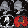
Figure 1. Computed tomography pulmonary angiogram demonstrates a focal reduction in the caliber of both the ostia of the left upper pulmonary vein (purple circle in image A) and left lower pulmonary vein (orange circle in image B); occlusion of pulmonary venous branches upstream of the stenotic point is observed (red circle in image C) along with corresponding segmental pulmonary hypoperfusion within the territory drained by the stenosed veins is evident in the iodine map images (image D)
Figure 2
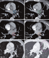
Figure 2. Control computed tomography pulmonary angiogram. The initial examination (images in column E) and a follow-up study after 3 months (images in column F) illustrate the progressive stenosis of the left inferior pulmonary vein (orange circle in image F2) and more extensive stenosis in the superior venous branch (green circle in image F1), which is now occluded. This occlusion is associated with the presence of a pulmonary infarction area (pink circle in image F3)
Figure 3
Figure 3. Stent placement within the upper pulmonary vein ostia (G1), and lower pulmonary vein ostia (H1). After 6 months, a subsequent computed tomography pulmonary angiogram examination reveals restenosis in the left upper pulmonary vein (pink circle in image G2) while demonstrating a patent inferior pulmonary vein with preserved caliber (blue circle in image H2)

References

    1. Lacomis JM, Goitein O, Deible C, Schwartzman D. CT of the pulmonary veins. J Thorac Imaging . 2007;22(1):63–76. Review. - PubMed
    1. Raeisi-Giglou P, Wazni OM, Saliba WI, Barakat A, Tarakji KG, Rickard J, et al. Outcomes and management of patients with severe pulmonary vein stenosis from prior atrial fibrillation ablation. Circ Arrhythm Electrophysiol . 2018;11(5):e006001. - PubMed
    1. Scanavacca MI, Sosa E. Catheter ablation of atrial fibrillation. Techniques and results. Arq Bras Cardiol . 2005;85(4):295–301. - PubMed
    1. Sarabanda AV, Beck LC, Ferreira LG, Gali WL, Melo F, Netto, Monte GU. Tratamento de estenose de veia pulmonar após ablação percutânea de fibrilação atrial. Arq Bras Cardiol . 2010;94(1):e7–e10. - PubMed
    1. Fender EA, Widmer RJ, Hodge DO, Cooper GM, Monahan KH, Peterson LA, et al. Severe pulmonary vein stenosis resulting from ablation for atrial fibrillation: presentation, management, and clinical outcomes. Circulation . 2016;134(23):1812–1821. - PubMed

MeSH terms