

Expression of influenza A virus glycan receptor candidates in mallard, chicken, and tufted duck
- PMID: 38127648
- PMCID: PMC10987293
- DOI: 10.1093/glycob/cwad098
Expression of influenza A virus glycan receptor candidates in mallard, chicken, and tufted duck
Abstract
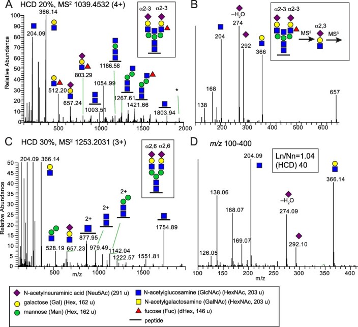
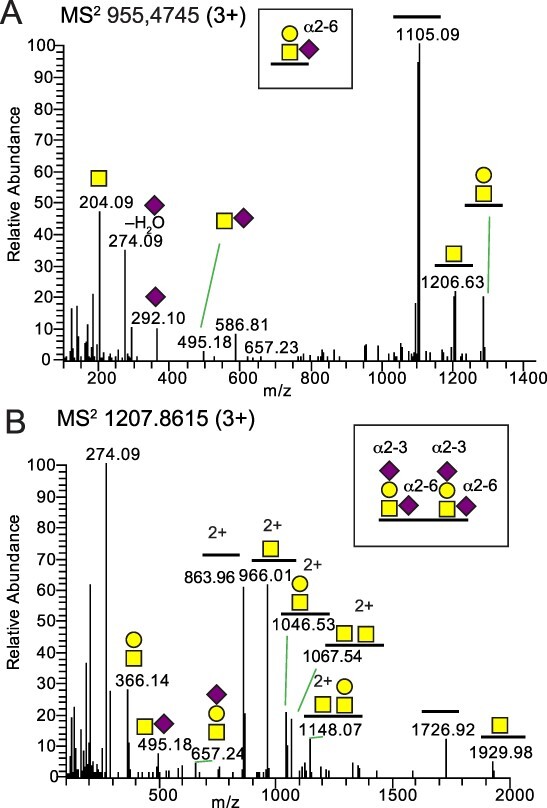
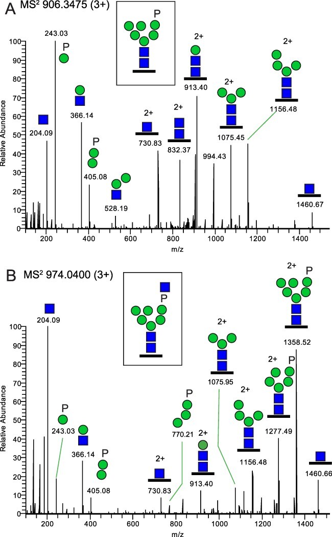
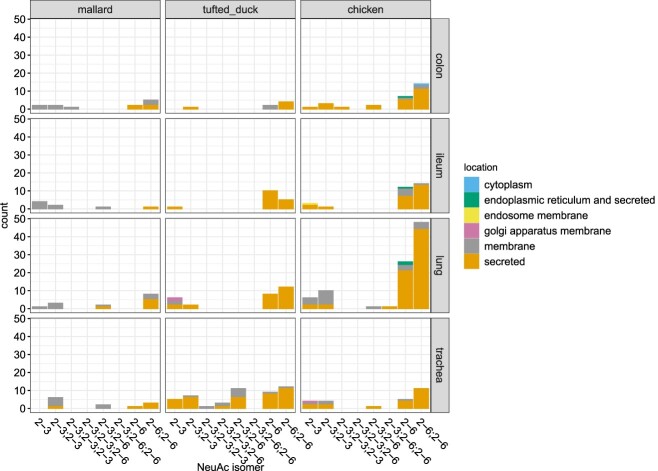
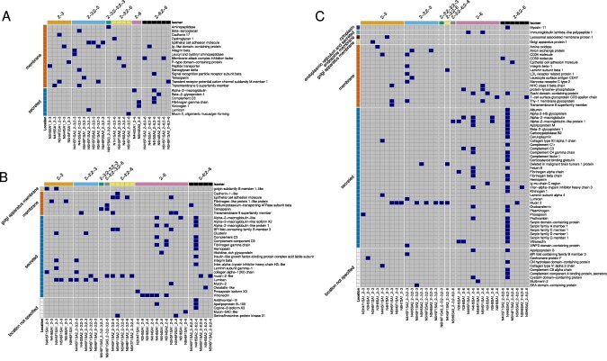
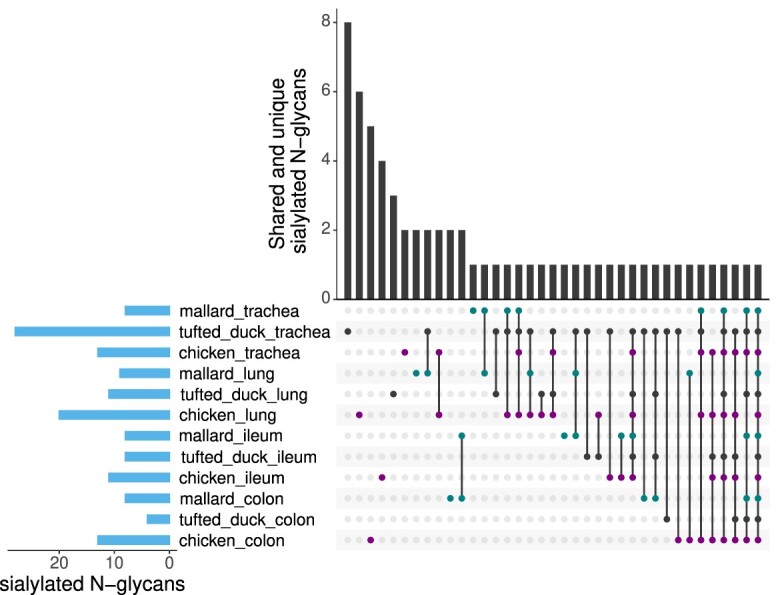
Influenza A virus (IAV) pandemics result from interspecies transmission events within the avian reservoir and further into mammals including humans. Receptor incompatibility due to differently expressed glycan structures between species has been suggested to limit zoonotic IAV transmission from the wild bird reservoir as well as between different bird species. Using glycoproteomics, we have studied the repertoires of expressed glycan structures with focus on putative sialic acid-containing glycan receptors for IAV in mallard, chicken and tufted duck; three bird species with different roles in the zoonotic ecology of IAV. The methodology used pinpoints specific glycan structures to specific glycosylation sites of identified glycoproteins and was also used to successfully discriminate α2-3- from α2-6-linked terminal sialic acids by careful analysis of oxonium ions released from glycopeptides in tandem MS/MS (MS2), and MS/MS/MS (MS3). Our analysis clearly demonstrated that all three bird species can produce complex N-glycans including α2-3-linked sialyl Lewis structures, as well as both N- and O- glycans terminated with both α2-3- and α2-6-linked Neu5Ac. We also found the recently identified putative IAV receptor structures, Man-6P N-glycopeptides, in all tissues of the three bird species. Furthermore, we found many similarities in the repertoires of expressed receptors both between the bird species investigated and to previously published data from pigs and humans. Our findings of sialylated glycan structures, previously anticipated to be mammalian specific, in all three bird species may have major implications for our understanding of the role of receptor incompatibility in interspecies transmission of IAV.
Keywords: birds; glycoproteomics; influenza virus; interspecies transmission; sialic acid receptor.
© The Author(s) 2023. Published by Oxford University Press. All rights reserved. For permissions, please e-mail: journals.permissions@oup.com.
Figures
Similar articles
-
Complex N-glycans are important for interspecies transmission of H7 influenza A viruses.J Virol. 2024 Apr 16;98(4):e0194123. doi: 10.1128/jvi.01941-23. Epub 2024 Mar 12. J Virol. 2024. PMID: 38470143 Free PMC article.
-
Host Range of Influenza A Virus H1 to H16 in Eurasian Ducks Based on Tissue and Receptor Binding Studies.J Virol. 2021 Feb 24;95(6):e01873-20. doi: 10.1128/JVI.01873-20. Print 2021 Feb 24. J Virol. 2021. PMID: 33361418 Free PMC article.
-
The Sialyl Lewis X Glycan Receptor Facilitates Infection of Subtype H7 Avian Influenza A Viruses.J Virol. 2022 Oct 12;96(19):e0134422. doi: 10.1128/jvi.01344-22. Epub 2022 Sep 20. J Virol. 2022. PMID: 36125302 Free PMC article.
-
Roles of Glycans and Non-glycans on the Epithelium and in the Immune System in H1-H18 Influenza A Virus Infections.Methods Mol Biol. 2022;2556:205-242. doi: 10.1007/978-1-0716-2635-1_16. Methods Mol Biol. 2022. PMID: 36175637 Review.
-
Glycans as receptors for influenza pathogenesis.Glycoconj J. 2010 Aug;27(6):561-70. doi: 10.1007/s10719-010-9303-4. Epub 2010 Aug 24. Glycoconj J. 2010. PMID: 20734133 Free PMC article. Review.
Cited by
-
Comparative IP-MS Reveals HSPA5 and HSPA8 Interacting with Hemagglutinin Protein to Promote the Replication of Influenza A Virus.Pathogens. 2025 May 27;14(6):535. doi: 10.3390/pathogens14060535. Pathogens. 2025. PMID: 40559543 Free PMC article.
-
Evaluating the epizootic and zoonotic threat of an H7N9 low-pathogenicity avian influenza virus (LPAIV) variant associated with enhanced pathogenicity in turkeys.J Gen Virol. 2024 Jul;105(7):002008. doi: 10.1099/jgv.0.002008. J Gen Virol. 2024. PMID: 38980150 Free PMC article.
References
-
- Altschul SF, Gish W, Miller W, Myers EW, Lipman DJ. Basic local alignment search tool. J Mol Biol. 1990:215(3):403–410. - PubMed
-
- Arike L, Seiman A, van der Post S, Rodriguez Piñeiro AM, Ermund A, Schütte A, Bäckhed F, Johansson MEV, Hansson GC. Protein turnover in epithelial cells and mucus along the gastrointestinal tract is coordinated by the spatial location and microbiota. Cell Rep. 2020:30(4):1077–1087.e3. - PMC - PubMed
-
- Bewley CA. Illuminating the switch in influenza viruses. Nat Biotechnol. 2008:26(1):60–62. - PubMed
