. 2023 Dec 21;18(12):e0292455.
doi: 10.1371/journal.pone.0292455. eCollection 2023.

Evaluation of anticancer potential of tetracene-5,12-dione (A01) and pyrimidine-2,4-dione (A02) via caspase 3 and lactate dehydrogenase cytotoxicity investigations

Affiliations

Evaluation of anticancer potential of tetracene-5,12-dione (A01) and pyrimidine-2,4-dione (A02) via caspase 3 and lactate dehydrogenase cytotoxicity investigations

Mubashir Aziz et al. PLoS One. .

Abstract

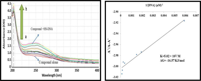
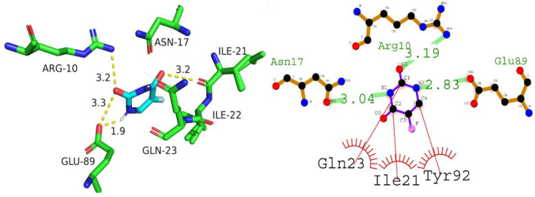
Cancer stands as a significant global cause of mortality, predominantly arising from the dysregulation of key enzymes and DNA. One strategic avenue in developing new anticancer agents involves targeting specific proteins within the cancer pathway. Amidst ongoing efforts to enhance the efficacy of anticancer drugs, a range of crucial medications currently interact with DNA at the molecular level, exerting profound biological effects. Our study is driven by the objective to comprehensively explore the potential of two compounds: (7S,9S)-7-[(2R,4S,5S,6S)-4-amino-5-hydroxy-6-methyloxan-2-yl]oxy-6,9,11-trihydroxy-9-(2-hydroxyacetyl)-4-methoxy-8,10-dihydro-7H-tetracene-5,12-dione (A01) and 5-fluoro-1H-pyrimidine-2,4-dione (A02). These compounds have demonstrated marked efficacy against breast and cervical cancer cell lines, positioning them as promising anticancer candidates. In our investigation, A01 has emerged as a particularly potent candidate, with its potential bolstered by corroborative evidence from lactate dehydrogenase release and caspase-3 activity assays. On the other hand, A02 has exhibited remarkable anticancer potential. To further elucidate their molecular mechanisms and interactions, we employed computational techniques, including molecular docking and molecular dynamics simulations. Notably, our computational analyses suggest that the A01-DNA complex predominantly interacts via the minor groove, imparting significant insights into its mechanism of action. While earlier studies have also highlighted the anticancer activity of A01, our research contributes by providing a deeper understanding of its binding mechanisms through computational investigations. This knowledge holds potential for designing more effective drugs that target cancer-associated proteins. These findings lay a robust groundwork for future inquiries and propose that derivatives of A01 could be synthesized as potent bioactive agents for cancer treatment. By elucidating the distinctive aspects of our study's outcomes, we address the concern of distinguishing our findings from those of prior research.


Conflict of interest statement

The authors have declared that no competing interests exist.

Figures

Fig 1
Fig 1. Already reported Pyrazole derivatives alongside compound A01 and A02 [28, 30, 31].
Fig 2
Fig 2
The representation of % Lactate dehydrogenase (LDH) release at different concentration of A01 against HeLa (A), MDA-MB-231 (B) and MCF-7 cells using LDH Assay Kit (Cytotoxicity) (ab65393). Data represent the mean ± SD (n = 3). * p < 0.05 compares the treated cell with control cell.
Fig 3
Fig 3. Detection of Caspase-3 activity was detected in HeLa, MDA-MB231 and MCF-7 cells treated at two different concentrations, respectively, for 24 h using Caspase-3 Assay Kit (Fluorometric:ab39383).
Data represent the mean ± SD (n = 3). * p < 0.05 compares the treated cell with control cell.
Fig 4
Fig 4
A)UV-visible spectrum responses of A01 in the absence and presence of various concentrations of HS-DNA as was highlighted above. The pointed end of the arrow represents an increase in the amount of DNA present. (B) Ao–A/Ao vs. 1/[DNA] is the graph that is used to calculate the binding constant.
Fig 5
Fig 5
A) UV-visible spectrum responses of A02 in the absence and presence of various concentrations of HS-DNA as was highlighted above. The pointed end of the arrow represents an increase in the amount of DNA present. (B) Ao–A/Ao vs. 1/[DNA] is the graph that is used to calculate the binding constant.
Fig 6
Fig 6
The optimized structure A01 (right) and A02 (left) alongside electrostatic potential map (ESP).
Fig 7
Fig 7
HOMO/LUMO orbitals of A01 (up) and A02 (down).
Fig 8
Fig 8. Predicted 2D and 3D binding interactions of compound A01 with caspase 3.
Fig 9
Fig 9. Putative 2D and 3D binding mode of compound A02 against Caspase-3.
Fig 10
Fig 10. Putative 2D and 3D interaction of A01 (A01) with NF-κB.
Fig 11
Fig 11. Illustration of 2D and 3D interaction of A02 against NF-κB.
Fig 12
Fig 12. The putative binding mode of compound A01 against P53.
Fig 13
Fig 13. The putative binding mode of compound A02 against P53.
Fig 14
Fig 14. Intercalation of DNA groove by compound A01.
Fig 15
Fig 15. Intercalation of DNA groove by compound A02.
Fig 16
Fig 16. Representation of RMSD pattern for apo protein NF-κB (red colored trajectory) and liganded protein (NF-κB -A01 complex, represented by green colored trajectory).
Fig 17
Fig 17. The amino acid residues wise fluctuation (RMSF) for c alpha atoms of NF-κB (apo protein) and liganded protein (NF-κB -A01 complex).
Fig 18
Fig 18. The RMSD pattern for the apo protein, P53 (represented by the blue trajectory), and the liganded protein, P53-A02 complex (represented by the brown trajectory), is depicted in the illustration.
Fig 19
Fig 19. The amino acid residues wise fluctuation (RMSF) of Apo protein (P53) and liganded protein (P53-A02 complex).
Fig 20
Fig 20. The evolution of RMSD trajectories for sole ligand molecules.
The Blue colored trajectory is for compound A01 whereas orange colored trajectory is depicting the RMSD for compound A02.
Fig 21
Fig 21. Superimposed structures of the NF-κB-A01 complex and the P53-A02 complex were observed during MD simulation.
(A) Overlay frames of the NF-κB-A01 complex, where the blue-colored ligand indicates the conformation observed during the first frame, while the pink-colored ligand indicates the conformation at the final frame of the MD simulation. (B) Superimposed conformations of the P53-A02 complex observed during the initial and final frames. The green-colored ligand indicates the conformation of A02 in the first frame, whereas the blue-colored ligand indicates the conformation during the final frame.

References

    1. Feng Y, Panwar N, Tng DJH, Tjin SC, Wang K, Yong K-T. The application of mesoporous silica nanoparticle family in cancer theranostics. Coordination chemistry reviews. 2016;319:86–109.
    1. Qi F, Li A, Inagaki Y, Gao J, Li J, Kokudo N, et al. Chinese herbal medicines as adjuvant treatment during chemoor radio-therapy for cancer. Bioscience trends. 2010;4(6). - PubMed
    1. Jabłońska-Trypuć A, Matejczyk M, Rosochacki S. Matrix metalloproteinases (MMPs), the main extracellular matrix (ECM) enzymes in collagen degradation, as a target for anticancer drugs. Journal of enzyme inhibition medicinal chemistry. 2016;31(sup1):177–83. doi: 10.3109/14756366.2016.1161620 - DOI - PubMed
    1. Koutras A, Ntounis T, Fasoulakis Z, Papalios T, Pittokopitou S, Prokopakis I, et al. Cancer Treatment and Immunotherapy during Pregnancy. Pharmaceutics. 2022;14(10):2080. doi: 10.3390/pharmaceutics14102080 - DOI - PMC - PubMed
    1. Wagner JM, Karnitz LM. Cisplatin-induced DNA damage activates replication checkpoint signaling components that differentially affect tumor cell survival. Molecular pharmacology. 2009;76(1):208–14. doi: 10.1124/mol.109.055178 - DOI - PMC - PubMed