

Contrast enhanced computed tomography of small ruminants: Caprine and ovine
- PMID: 38127918
- PMCID: PMC10735035
- DOI: 10.1371/journal.pone.0287529
Contrast enhanced computed tomography of small ruminants: Caprine and ovine
Abstract
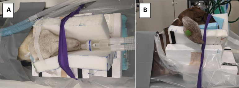
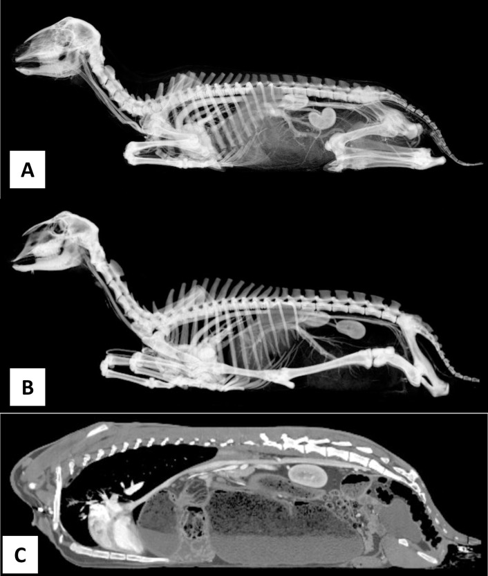
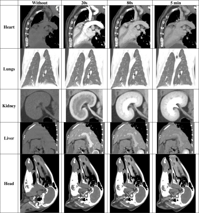
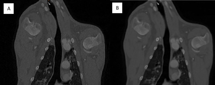
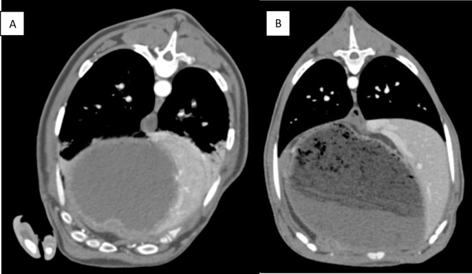
The use of small ruminants, mainly sheep and goats, is increasing in biomedical research. Small ruminants are a desirable animal model due to their human-like anatomy and physiology. However, the large variability between studies and lack of baseline data on these animals creates a barrier to further research. This knowledge gap includes a lack of computed tomography (CT) scans for healthy subjects. Full body, contrast enhanced CT scans of caprine and ovine subjects were acquired for subsequent modeling studies. Scans were acquired from an ovine specimen (male, Khatadin, 30-35 kg) and caprine specimen (female, Nubian 30-35 kg). Scans were acquired with and without contrast. Contrast enhanced scans utilized 1.7 mL/kg of contrast administered at 2 mL/s and scans were acquired 20 seconds, 80 seconds, and 5 minutes post-contrast. Scans were taken at 100 kV and 400 mA. Each scan was reconstructed using a bone window and a soft tissue window. Sixteen full body image data sets are presented (2 specimens by 4 contrast levels by 2 reconstruction windows) and are available for download through the form located at: https://redcap.link/COScanData. Scans showed that the post-contrast timing and scan reconstruction method affected structural visualization. The data are intended for further biomedical research on ruminants related to computational model development, device prototyping, comparative diagnostics, intervention planning, and other forms of translational research.
Copyright: This is an open access article, free of all copyright, and may be freely reproduced, distributed, transmitted, modified, built upon, or otherwise used by anyone for any lawful purpose. The work is made available under the Creative Commons CC0 public domain dedication.
Conflict of interest statement
The authors have declared that no competing interests exist.
Figures
Similar articles
-
Differential diagnosis of theileriosis through blood smear examination and polymerase chain reaction in small ruminants from Pakistan.Open Vet J. 2023 Jun;13(6):697-704. doi: 10.5455/OVJ.2023.v13.i6.4. Epub 2023 Jun 6. Open Vet J. 2023. PMID: 37545708 Free PMC article.
-
Seroprevalence and associated risk factors of peste des petits ruminants among ovine and caprine in selected districts of Afar region, Ethiopia.BMC Vet Res. 2022 Dec 9;18(1):429. doi: 10.1186/s12917-022-03528-6. BMC Vet Res. 2022. PMID: 36494681 Free PMC article.
-
Identification of Main Brucella species Implicated in Ovine and Caprine Abortion Cases by Molecular and Classical Methods.Arch Razi Inst. 2021 Mar;76(1):51-60. doi: 10.22092/ari.2019.128003.1398. Epub 2021 Mar 1. Arch Razi Inst. 2021. PMID: 33818957 Free PMC article.
-
Proteomics and protein analyses of ovine and caprine body fluids: current studies and future promises.Curr Protein Pept Sci. 2014 Feb;15(1):45-55. doi: 10.2174/1389203715666140221113544. Curr Protein Pept Sci. 2014. PMID: 24555895 Review.
-
Imaging of the Urinary and Reproductive Tract in Small Ruminants.Vet Clin North Am Food Anim Pract. 2021 Mar;37(1):75-92. doi: 10.1016/j.cvfa.2020.10.002. Epub 2020 Dec 24. Vet Clin North Am Food Anim Pract. 2021. PMID: 33358313 Review.
References
-
- Fulton L. K., Clarke M. S. & Farris H. E. The Goat as a Model for Biomedical Research and Teaching. ILAR Journal 36, 21–29 (1994).
-
- Salerno C. T., Droel J. & Bianco R. W. Current state of in vivo preclinical heart valve evaluation. Journal of Heart Valve Disease 7, 158–162 (1998). - PubMed
MeSH terms
LinkOut - more resources
Full Text Sources
Miscellaneous
