. 2023 Dec 21;19(12):e1011220.
doi: 10.1371/journal.ppat.1011220. eCollection 2023 Dec.

FLAgellum Member 8 modulates extravascular distribution of African trypanosomes

Affiliations

FLAgellum Member 8 modulates extravascular distribution of African trypanosomes

Estefanía Calvo-Alvarez et al. PLoS Pathog. .

Abstract

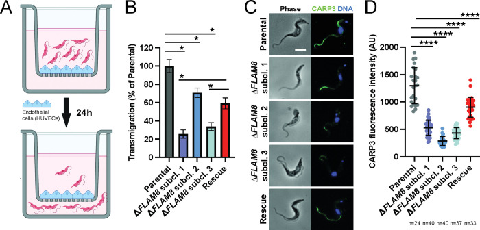
In the mammalian host, the biology of tissue-dwelling Trypanosoma brucei parasites is not completely understood, especially the mechanisms involved in their extravascular colonization. The trypanosome flagellum is an essential organelle in multiple aspects of the parasites' development. The flagellar protein termed FLAgellar Member 8 (FLAM8) acts as a docking platform for a pool of cyclic AMP response protein 3 (CARP3) that is involved in signaling. FLAM8 exhibits a stage-specific distribution suggesting specific functions in the mammalian and vector stages of the parasite. Analyses of knockdown and knockout trypanosomes in their mammalian forms demonstrated that FLAM8 is not essential in vitro for survival, growth, motility and stumpy differentiation. Functional investigations in experimental infections showed that FLAM8-deprived trypanosomes can establish and maintain an infection in the blood circulation and differentiate into insect transmissible forms. However, quantitative bioluminescence imaging and gene expression analysis revealed that FLAM8-null parasites exhibit a significantly impaired dissemination in the extravascular compartment, that is restored by the addition of a single rescue copy of FLAM8. In vitro trans-endothelial migration assays revealed significant defects in trypanosomes lacking FLAM8. FLAM8 is the first flagellar component shown to modulate T. brucei distribution in the host tissues, possibly through sensing functions, contributing to the maintenance of extravascular parasite populations in mammalian anatomical niches, especially in the skin.


Conflict of interest statement

The authors have declared that no competing interests exist.

Figures

Fig 1
Fig 1. Characterization of the FLAM8::mNG FLAM8RNAi strain in vitro and in vivo functional investigation in the mammalian host.
A) Expression of FLAM8 mRNA assessed by RT-PCR by the comparative ΔΔCT method in control, non-induced and induced FLAM8::mNG FLAM8RNAi parasites (72 h). B) Immunofluorescence pictures of non-induced (upper panels) and induced (bottom panels) FLAM8::mNG FLAM8RNAi BSF during 72 h. Methanol-fixed trypanosomes were stained with an anti-mNG antibody (green) and DAPI for DNA content (blue). The scale bars represent 5 μm. C) Growth curves of control, non-induced and induced FLAM8::mNG FLAM8RNAi BSF parasites. All cell lines received 1 μg tetracycline for 6 days. Control parasites are Lister 427 “Single Marker” BSF parasites that do not bear the pZJM-FLAM8 plasmid for RNAi silencing. Results represent the mean (± standard deviation, SD) of three independent experiments. D) Groups of 4 BALB/c mice were injected IP with either control, non-induced or induced FLAM8RNAi BSF trypanosomes. One PBS-injected BALB/c animal was used as negative control. Representative normalized in vivo images of the bioluminescence radiance signal (in photons / second / cm2 / steradian) emitted from BALB/c mice infected with control, non-induced and induced FLAM8::mNG FLAM8RNAi parasites 4 days post-infection (non-infected technical control mice were negative for bioluminescence, not shown). RNAi silencing of FLAM8 was maintained in vivo by the addition of doxycycline in sugared drinking water 48 h prior infection and until the end of the experiment. E) Number of parasites in the blood (intravascular, IV) of infected BALB/c mice during the course of the infection (5 days) counted from a tail bleed using a cytometer. F) Number of parasites in the extravascular compartment (extravascular, EV) of the same infected mice as in E). G) Dissemination of control, non-induced and induced FLAM8::mNG FLAM8RNAi parasites, measured over the entire animal body (in cm2) through the total bioluminescent surface, during the entire infection course. Results represent means ± standard deviation (SD).
Fig 2
Fig 2. Characterization of ΔFLAM8 null mutants in vitro.
A) Immunofluorescence pictures of parental, ΔFLAM8 knockout and rescue pleomorphic BSF parasites labelled with the anti-FLAM8 (green) and mAb25 (axoneme in red) antibodies, DAPI staining for DNA content (blue). Scale bars show 5 μm. B) Quantification of the total fluorescence intensity of FLAM8 along the entire flagellum length in methanol-fixed parental, ΔFLAM8 knockout and rescue parasites. C) Growth curve of one parental, three ΔFLAM8 subclones and one rescue pleomorphic BSF trypanosome cell lines over 6 consecutive days. D) Measurements of the flagellum length based on the axonemal marker mAb25 profiles in parental, three ΔFLAM8 subclones and rescue parasites. No statistical differences were found. E-H) Motility tracking analysis showing the average speeds (E and G) and linearity (F and H) of BSF cell lines in matrix-dependent culture medium containing 0.5% (E-F) or 1.1% (G-H) methylcellulose. No statistical differences were observed. The number of parasites considered for quantifications (N) is indicated under and above graphs in (B) and (D-H), respectively. Results represent the mean ± standard deviation (SD) of three independent experiments. Statistical tests included one-way ANOVA and Tukey’s ad-hoc post-tests for multiple comparisons.
Fig 3
Fig 3. Functional investigations on the ΔFLAM8 null mutants in vivo in the mammalian host.
Groups of 3 BALB/c mice were injected IP with either one parental, three ΔFLAM8 null subclones or one rescue strains. One PBS-injected BALB/c animal was used as negative control. A) Normalized in vivo images of the bioluminescence radiance intensity (in photons / second / cm2 / steradian) emitted 8 days post-infection in BALB/c mice infected with parental, three ΔFLAM8 subclones or rescue parasites (non-infected control mice C- were negative for bioluminescence). B) Total number of parasites in the blood of infected mice (intravascular, IV) daily counted from tail bleeds using a cytometer over 4 weeks. Statistically significant differences (p<0.01) are indicated with one, two or three asterisks (*, **, ***) representing differences between the parental strain and one, two or three ΔFLAM8 subclones, respectively. C) Total number of extravascular (EV) trypanosomes in the same mice. Statistically significant differences between the parental strain and ΔFLAM8 subclones are indicated as in B). D-E) Variations of the total numbers of intravascular (IV as in B) and extravascular (EV as in C) parasite populations in the same mice plotted as fold differences to values obtained with mice infected with parental trypanosomes at the same time point. F) Dissemination of the parental, three ΔFLAM8 subclones and rescue parasite strains, measured over the entire animal body (in cm2) through the total surface of bioluminescent signal, during the entire infection course. Statistically significant differences between the parental strain and ΔFLAM8 subclones are indicated as described above. Results represent means ± standard deviation (SD). Statistical tests included two-way ANOVA and Tukey’s ad-hoc post-tests for multiple comparisons in B-F. Detailed individual data are provided in S4 Fig.
Fig 4
Fig 4. The absence of FLAM8 reduces extravascular trypanosome dissemination.
In a second experimental infection, groups of 3 BALB/c animals were injected IP with either parental, three ΔFLAM8 null subclones or rescue strains, while one PBS-injected BALB/c animal was used as negative control. At day 24 post-infection, animals were euthanized, and the gut, kidney, liver, lung, skin, spleen and testes were collected to quantify the parasite proportion in each organ. A) Total number of intravascular parasites (IV) during the infection period. B) Total number of extravascular (EV) trypanosomes in the same mice. C) Dissemination of the parental, three ΔFLAM8 subclones and rescue parasite strains, measured over the entire animal body (in cm2) through the total surface of bioluminescent signal, during the entire infection course. Statistically significant differences (p<0.01) are indicated with one or three asterisks (*, ***) representing differences between the parental strain and one or three ΔFLAM8 subclones, respectively. Detailed individual data are provided in S5 Fig D) Delta number of parasites per dissected organs and strains. The total number of parasites in each sample was calculated per mg of tissues by using a Tubulin RT-qPCR standard curve. To better compare the variations of the parasite populations in each compartment between strains, the Delta number of parasites was calculated as the difference between the number of parasites in each tissue sample of a given mouse and the number of parasites in the blood sample from the same mouse. E) Delta number of parasites per strain calculated as the average of values for individual organs shown in D. Statistical differences (p<0.0001) according to one-way ANOVA and Dunnett’s comparison tests. Detailed individual data are provided in S1 Table and S6 Fig.
Fig 5
Fig 5. Parasite differentiation is not impacted in FLAM8-depleted trypanosomes.
A) Representative immunofluorescence pictures of stumpy parasites after in vitro differentiation from proliferative slenders of parental (left panel), three ΔFLAM8 subclones (middle panels) and rescue (right panel) parasite strains upon in vitro treatment with a nucleotide 5’-AMP analog. Methanol-fixed parasites were labelled with the anti-PAD1 antibody (red), and DAPI staining for DNA content (blue). Scale bars show 5 μm. B) Quantification of the proportion of stumpy trypanosomes in all pleomorphic cell lines after in vitro differentiation. The number of parasites considered for quantification (n) is indicated above the graph. No significant differences were found (one-way ANOVA and Tukey’s comparisons test). Results represent the mean ± standard deviation (SD) of three independent experiments. C) Selected immunofluorescence images of freshly in vitro differentiated procyclic cells. Trypanosomes were doubly labelled with anti-FLAM8 (green) and mAb25 (axoneme in magenta), DAPI staining showing DNA content in blue. Scale bars show 5 μm. D) Upon differentiation, procyclic forms (PCF) of parental, three ΔFLAM8 subclones and rescue parasite strains were equally diluted, and their in vitro growth assessed. Statistical differences (p<0.01) were observed only when comparing parental and KO 2 PCF trypanosomes (one-way ANOVA and Tukey’s comparison test). Results represent the mean ± standard deviation (SD) of four independent experiments. E) Average relative proportions of PAD1 expression levels quantified by RT-qPCR on blood and dissected organs at day 24 of the second in vivo challenge. Tubulin expression was used to normalize the PAD1 mRNA levels in all parental-, ΔFLAM8- and rescue-infected mice samples. F) Representative immunofluorescence images of naturally differentiated stumpy trypanosomes from parental, one selected ΔFLAM8 subclone and rescue strains isolated from mouse blood during the first peak of parasitemia (first experimental in vivo infection). Parasites were labelled with anti-PAD1 antibody (red), and DAPI staining for DNA content (blue). Scale bars show 5 μm.
Fig 6
Fig 6. Parasites lacking FLAM8 are impaired in transmigrating through endothelial cells in vitro.
A) Schematic representation of the trans-endothelial migration assay, showing the transwell system (Boyden chamber) containing the upper and the lower compartments, the monolayer of endothelial HUVEC cells and slender trypanosomes seeded on the top of the chamber for 24 hours. B) After this period, parasites within the upper and lower chambers were counted and values further used to calculate the proportion of parental, three ΔFLAM8 subclones and rescue parasites that migrated through the endothelial monolayer into the lower compartment. Trypanosome transmigration was compared between the FLAM8 mutant cell lines and the parental reference line (100% of trans-endothelial migration) using the Generalised Linear Model function in R with a Gaussian family function and proportion of transmigration as the dependent variable. A probability value of p<0.001 was considered significant (*). Error bars show SD. C) Representative immunofluorescence images of cultured slender trypanosomes from parental, ΔFLAM8 subclone and rescue strains. Methanol-fixed parasites were labelled with anti-CARP3 antibody (green intensity normalized), and DAPI staining for DNA content (blue). Scale bars show 5 μm. D) Quantification of CARP3 fluorescence signal in normalized images from methanol-fixed parental, ΔFLAM8 subclones and rescue trypanosomes, expressed as the ratio between the fluorescent CARP3 signal intensity along the entire flagellum divided by the length of individual flagella. The number of parasites considered for quantification (n) is indicated under the graph. Statistical differences (p<0.0001) according to one-way ANOVA and Dunnett’s comparison test. Results represent the mean ± standard deviation (SD) of two independent experiments.

Similar articles

Cited by

References

    1. MacGregor P, Szoor B, Savill NJ, Matthews KR. Trypanosomal immune evasion, chronicity and transmission: an elegant balancing act. Nat Rev Microbiol. 2012;10(6):431–8. Epub 2012/05/01. doi: 10.1038/nrmicro2779 . - DOI - PMC - PubMed
    1. Smith TK, Bringaud F, Nolan DP, Figueiredo LM. Metabolic reprogramming during the Trypanosoma brucei life cycle. F1000Res. 2017;6. Epub 2017/06/20. doi: 10.12688/f1000research.10342.2 ; PubMed Central PMCID: PMC5461901. - DOI - PMC - PubMed
    1. Rotureau B, Van Den Abbeele J. Through the dark continent: African trypanosome development in the tsetse fly. Frontiers in cellular and infection microbiology. 2013;3:53. Epub 2013/09/26. doi: 10.3389/fcimb.2013.00053 ; PubMed Central PMCID: PMC3776139. - DOI - PMC - PubMed
    1. Capewell P, Cren-Travaille C, Marchesi F, Johnston P, Clucas C, Benson RA, et al.. The skin is a significant but overlooked anatomical reservoir for vector-borne African trypanosomes. eLife. 2016;5. doi: 10.7554/eLife.17716 ; PubMed Central PMCID: PMC5065312. - DOI - PMC - PubMed
    1. Caljon G, Van Reet N, De Trez C, Vermeersch M, Perez-Morga D, Van Den Abbeele J. The Dermis as a Delivery Site of Trypanosoma brucei for Tsetse Flies. PLoS Pathog. 2016;12(7):e1005744. doi: 10.1371/journal.ppat.1005744 ; PubMed Central PMCID: PMC4956260. - DOI - PMC - PubMed

Substances