. 2023 Dec 21;18(12):e0295434.
doi: 10.1371/journal.pone.0295434. eCollection 2023.

Five-year clinical outcomes of 107 consecutive DMEK surgeries

Affiliations

Five-year clinical outcomes of 107 consecutive DMEK surgeries

Pierre Bichet et al. PLoS One. .

Abstract

Purpose: The long-term clinical outcomes, postoperative complications, and graft survival of Descemet-membrane endothelial keratoplasty (DMEK) remain poorly understood. We retrospectively assessed these variables in all consecutive eyes that underwent DMEK for any indication in 2014-2018. The findings were compared to the long-term DMEK studies of five other groups (3-10-year follow-up).

Methods: Patients underwent ophthalmological tests preoperatively, at 1, 3, 6, and 12 postoperative months, and then annually. Five-year graft survival was determined by Kaplan-Meier estimator. Change in best-corrected visual acuity (BCVA), endothelial-cell density (ECD), and central-corneal thickness (CCT) at each timepoint was determined.

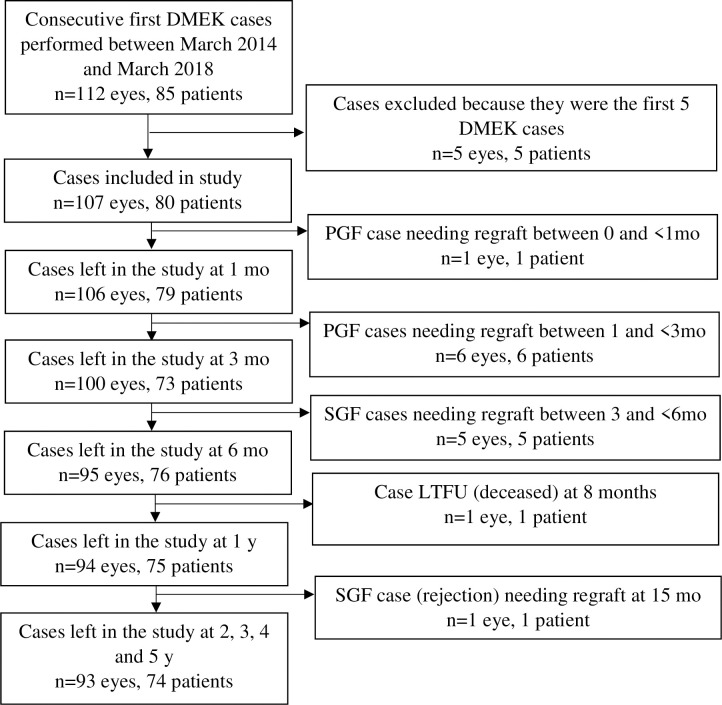
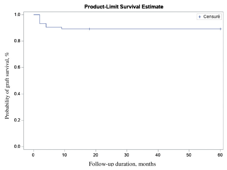
Results: 107 eyes (80 patients; 72 years old; 67% female) underwent first-time DMEK for uncomplicated Fuchs endothelial corneal dystrophy (94% of eyes), pseudophakic bullous keratopathy (3%), and regraft after previous keratoplasty (3%). The most common complication was graft detachment requiring rebubbling (18%). Thirteen grafts (12%) failed at ≤15 months. Cumulative 5-year graft-survival probability was 88% (95% confidence intervals = 79-94%). BCVA improved from 0.6 logMAR preoperatively to 0.05 logMAR at 1 year (p<0.0001) and then remained stable. Donor ECD dropped by 47% at 6 postoperative months and then continued to decrease by 4.0%/year. Five-year endothelial-cell loss was 65% (from 2550 to 900 cells/mm2). CCT dropped from 618 to 551 μm at 5 years (p<0.0001). These findings are generally consistent with previous long-term DMEK studies.

Conclusions: DMEK has low complication and high graft-survival rates and excellent clinical outcomes that persist up to 5 years post-surgery. DMEK seems to be a safe and effective treatment in the long term.


Conflict of interest statement

The authors have declared that no competing interests exist.

Figures

Fig 1
Fig 1. Patient distribution during the 5-year study period.
LTFU, loss to follow-up; mo, months; PGF, primary graft failure (≤3 months); SGF, secondary graft failure (>3 months); y, years.
Fig 2
Fig 2. Kaplan-Meier analysis of the DMEK graft survival probability of the whole cohort over 5 years (n = 107).
Fig 3
Fig 3
Average change over time in BCVA expressed as logMAR (A) and decimal categories (B) in the whole cohort (n = 100). Seven eyes with preoperative conditions that could affect visual acuity recovery were excluded from this analysis. (A) shows the median (orange line), interquartile range (green boxes), and maximum and minimum data as box-and-whisker plots.
Fig 4
Fig 4. Average change in graft ECD and endothelial cell loss over time in the whole cohort (n = 107).
(A) shows the median (black line), interquartile range (orange boxes), and maximum and minimum data as box-and-whisker plots. (B) shows median endothelial cell loss.
Fig 5
Fig 5
Comparison of our study (n = 107) with the literature in terms of (A) the percentage of original cohort eyes that did not attend follow-up and (B) graft survival. The study of Zwingelberg et al. [23] is not shown in (A) because only the patients who completed 3 years of follow-up were examined. The percentages in (A) are based on eye numbers in survival analyses [–19] or the stated number of eyes at each follow-up [,–22]; thus, they include graft failures and loss to follow-up. * Studies on the Melles-group cohort; ** studies on the Kruse-group cohort.
Fig 6
Fig 6. Comparison of our study (n = 100) with the literature in terms of long-term visual outcomes of DMEK.
The percentage of the original cohort that was available at each timepoint is indicated. * studies on the Melles-group cohort; ** studies on the Kruse-group cohort. All visual acuity analyses excluded eyes with preoperative conditions that could interfere with visual acuity recovery.
Fig 7
Fig 7. Comparison of our study (n = 107) with the literature in terms of the long-term graft endothelial cell changes after DMEK.
The percentage of the original cohort that was available at each timepoint is indicated. * studies on the Melles-group cohort; ** studies on the Kruse-group cohort.
Fig 8
Fig 8. Comparison of our study (n = 97) with the literature in terms of the long-term CCT changes after DMEK.
The percentage of the original cohort that was available at each timepoint is indicated. * studies on the Melles-group cohort; ** studies on the Kruse-group cohort.

References

    1. Melles GRJ, Wijdh RHJ, Nieuwendaal CP. A technique to excise the descemet membrane from a recipient cornea (descemetorhexis). Cornea. 2004;23: 286–288. doi: 10.1097/00003226-200404000-00011 - DOI - PubMed
    1. Melles GRJ, Ong TS, Ververs B, van der Wees J. Descemet membrane endothelial keratoplasty (DMEK). Cornea. 2006;25: 987–990. doi: 10.1097/01.ico.0000248385.16896.34 - DOI - PubMed
    1. Vedana G, Villarreal G, Jun AS. Fuchs endothelial corneal dystrophy: Current perspectives. Clinical Ophthalmology. 2016;10: 321–330. doi: 10.2147/OPTH.S83467 - DOI - PMC - PubMed
    1. Fuchs E. Dystrophia epithelialis corneae. Graefes Arhiv für Ophthalmologie. 1910;76: 478–508.
    1. Sarnicola C, Farooq A, Colby K. Fuchs Endothelial Corneal Dystrophy: Update on Pathogenesis and Future Directions. Eye Contact Lens. 2019;45: 1–10. doi: 10.1097/ICL.0000000000000469 - DOI - PubMed