. 2024 Mar:159:105471.
doi: 10.1016/j.yhbeh.2023.105471. Epub 2023 Dec 21.

Beyond the binary: Characterizing the relationships between sex and neuropeptide receptor binding density measures in the rat brain

Affiliations

Beyond the binary: Characterizing the relationships between sex and neuropeptide receptor binding density measures in the rat brain

Daphna Joel et al. Horm Behav. 2024 Mar.

Abstract

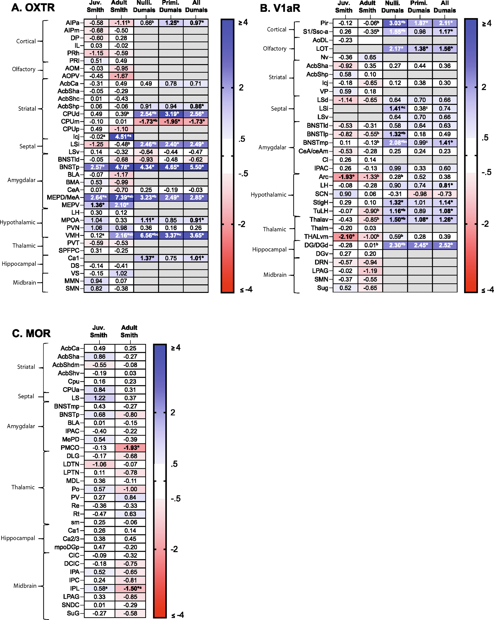
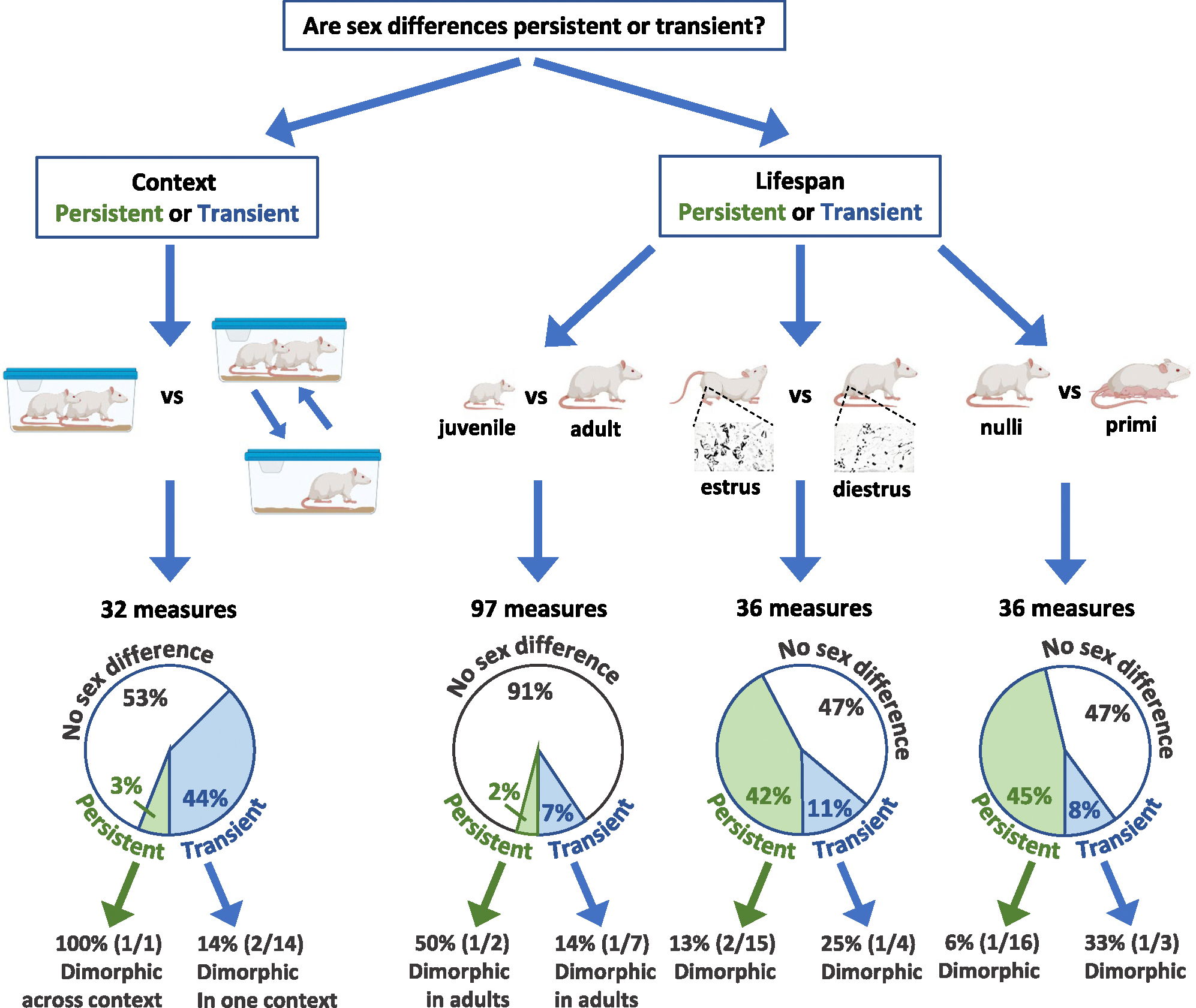
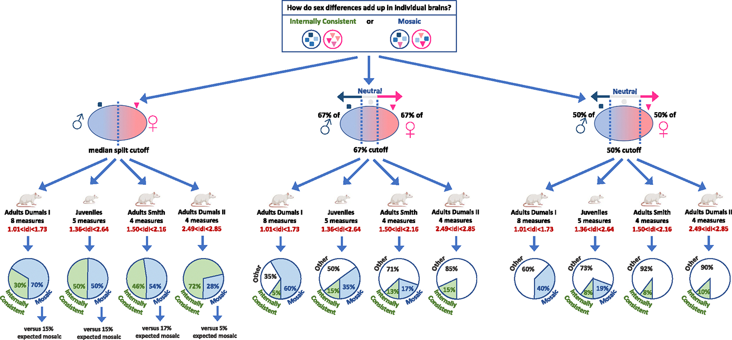
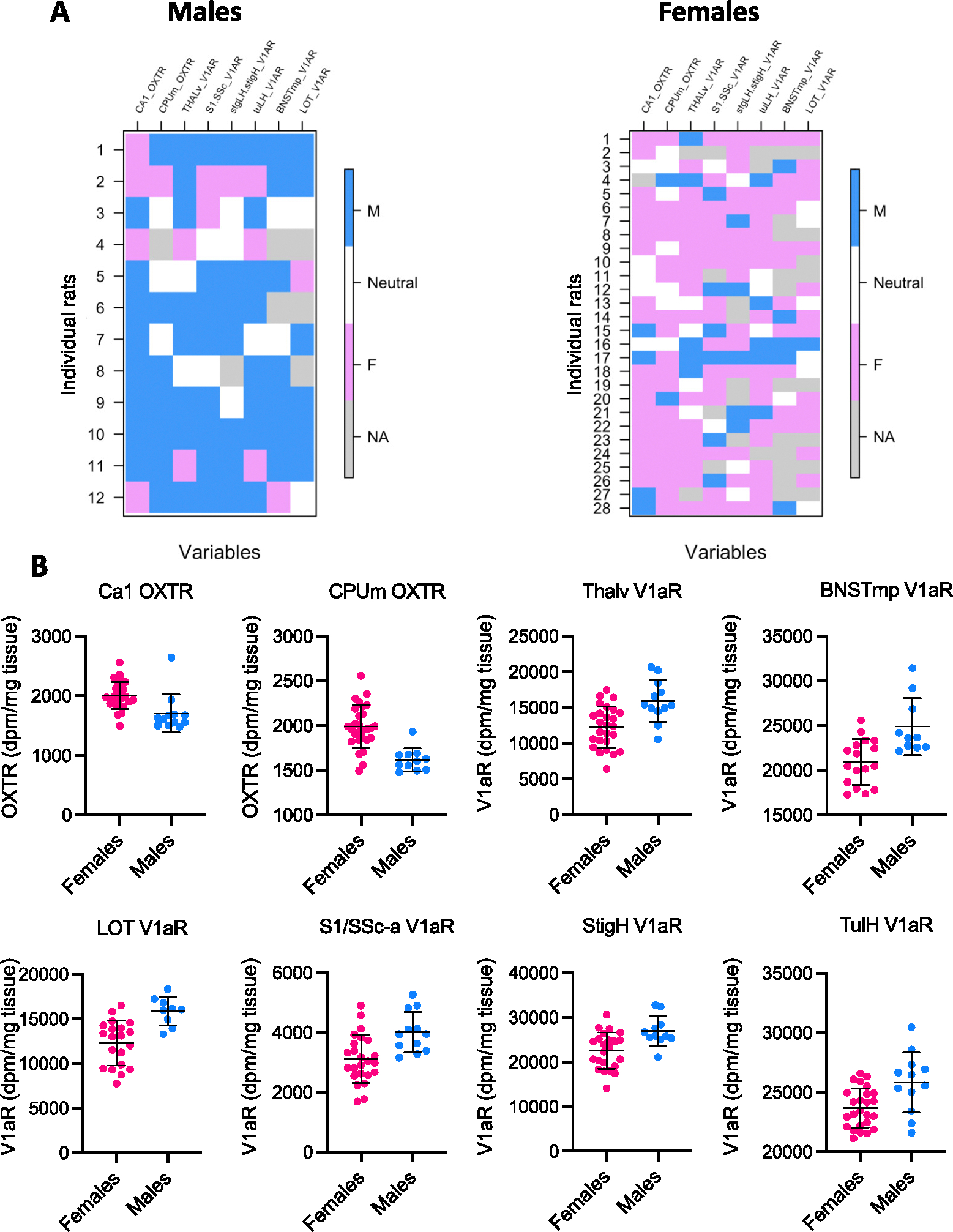
Sex differences exist in numerous parameters of the brain. Yet, sex-related factors are part of a large set of variables that interact to affect many aspects of brain structure and function. This raises questions regarding how to interpret findings of sex differences at the level of single brain measures and the brain as a whole. In the present study, we reanalyzed two datasets consisting of measures of oxytocin, vasopressin V1a, and mu opioid receptor binding densities in multiple brain regions in rats. At the level of single brain measures, we found that sex differences were rarely dimorphic and were largely persistent across estrous stage and parental status but not across age or context. At the level of aggregates of brain measures showing sex differences, we tested whether individual brains are 'mosaics' of female-typical and male-typical measures or are internally consistent, having either only female-typical or only male-typical measures. We found mosaicism for measures showing overlap between females and males. Mosaicism was higher a) with a larger number of measures, b) with smaller effect sizes of the sex difference in these measures, and c) in rats with more diverse life experiences. Together, these results highlight the limitations of the binary framework for interpreting sex effects on the brain and suggest two complementary pathways to studying the contribution of sex to brain function: (1) focusing on measures showing dimorphic and persistent sex differences and (2) exploring the relations between specific brain mosaics and specific endpoints.

Keywords: Age; Estrous cycle; Juvenile; Mosaics; Opioids; Oxytocin; Primiparous; Sex differences; Sex similarities; Vasopressin.


Conflict of interest statement

Declaration of competing interest None.

Figures

Fig. 1.
Fig. 1.
Heatmaps depicting sex difference size and persistency across conditions. Heatmaps depict Cohen’s d values for all sex differences in Smith Juvenile (Juv. Smith), Smith Adult, Dumais Adult with only nulliparous females (Nulli. Dumais), Dumais Adult with only primiparous females (Primi Dumais), and Dumais Adult with all females (All Dumais) in all brain regions for (A) OXTR, (B) V1aR, and (C) MOR. Measures for which the average score is higher in males compared to females and Cohen’s d > 0.5 are colored in blue shades (hue represents the effect size, darker = larger, up to Cohen’s d = 4); measures for which the average score is lower in males compared to females and Cohen’s d < −0.5 are colored in red shades. Significant sex differences are bolded and marked with an asterisk. a: significantly different effect size across age (Juv. Smith vs Adult Smith), b: significantly different effect size across studies (Adult Smith vs. Nulli. Dumais), c: significantly different effect size with parity (Nulli. Dumais vs Primi. Dumais). Definitions for all abbreviations can be found in the main text.
Fig. 2.
Fig. 2.
Persistency of sex differences in rat brain measures is variable across context and lifespan conditions. Context: 32 measures were assessed in sexually naïve rats from the Smith’s (left) and Dumais’ (right) cohorts. Rats in Dumais’ cohort were exposed to 4 × 24 h social isolation with a subsequent 4-min exposure to a juvenile rat. Significant sex differences in at least one of the cohorts were found in 15/32 measures; in one of these (1/15), the sex difference was persistent, that is, significant in both cohorts and of the same magnitude. This sex difference (1/1) was also sexually dimorphic in the two cohorts. Among the 14 non-persistent sex differences, two (2/14) were sexually dimorphic in one of the cohorts. Lifespan: Age: 97 measures were assessed in juvenile (5-week-old) and sexually naïve adult (12-week-old) rats from the Smith’s cohort. Significant sex differences in at least one age group were found in 9/97 measures. In two of these (2/9), the sex difference was persistent, that is, significant in both age groups and of the same magnitude, and in one of these (1/2), the sex difference was sexually dimorphic in adults. Among the 7 non-persistent sex differences, one (1/7) was sexually dimorphic in adults. Estrous stage: 36 measures were assessed in adult male and female rats (Dumais’ cohort) in which the females were either in estrus (estrus/proestrus) or in diestrus (diestrus/metestrus). Significant sex differences in at least one of the groups were found in 19/36 measures. Of these, 15 (15/19) were persistent across estrous stage, and two of these (2/15) were sexually dimorphic in at least one estrous stage. Among the 4 non-persistent sex differences, one (1/4) was sexually dimorphic in one estrous stage. Parental status: 36 measures were assessed in adult male and female rats (Dumais’ cohort) in which the females were either nulliparous or primiparous. Significant sex differences in at least one of the groups were found in 19/36 measures. Of these, 16 (16/19) were persistent across parental status, and one of these (1/16) was sexually dimorphic under one parental status. Among the 3 non-persistent sex differences, one (1/3) was sexually dimorphic under one parental status. The rat images were created with BioRender.com.
Fig. 3.
Fig. 3.
Analyses of internal consistency versus mosaic across rat brain measures in individual brains. The results of the mosaic analysis of four sets of brain measures: Adults Dumais I (8 measures), Juveniles (Smith’s cohort; 5 measures), Adults Smith (4 measures), Adults Dumais II (4 measures), with three definitions of female-typical (F), male-typical (M) (and neutral (N), were relevant) ranges of scores: median split, 67 % cutoff, and 50 % cutoff. The pie charts present the percent of internally consistent brains (containing either only F or only M measures), mosaic brains (containing both F and M measures), or, for the 67 % and 50 % cutoff analyses, ‘other’ (brains containing either only N measures, or a combination of N and M measures or of N and F measures). For the median split analysis, the number of expected mosaics under the assumption of internal consistency is reported. The rat images were created with BioRender.com.
Fig. 4.
Fig. 4.
Mosaic analysis and individual scores on the eight brain measures of the Dumais I dataset using the 67 % cutoff. A. Visual representation of the mosaic analysis of the Dumais I dataset using the 67 % cutoff. Each vertical column represents one brain measure, and each horizontal row represents one rat depicting whether a rat’s score is ‘male-typical’ (blue), ‘neutral’ (white), or ‘female-typical’ (pink; grey indicates missing data = NA). B. The individual scores grouped by sex on each of the eight brain measures included in the mosaic analysis presented in A. The mean and standard deviation of the mean score of females and males are also presented.

Similar articles

Cited by

References

    1. Albers HE, 2015. Jan. Species, sex and individual differences in the vasotocin/vasopressin system: relationship to neurochemical signaling in the social behavior neural network. Front. Neuroendocrinol. 36, 49–71. - PMC - PubMed
    1. Arnold AP, Chen X, 2009. What does the “four core genotypes” mouse model tell us about sex differences in the brain and other tissues? Front. Neuroendocrinol. 30 (1), 1–9. - PMC - PubMed
    1. Arsenijevic Y, Dreifuss JJ, Vallet P, Marguerat A, Tribollet E, 1995. Nov 6. Reduced binding of oxytocin in the rat brain during aging. Brain Res. 698 (1–2), 275–279. - PubMed
    1. Bale TL, Dorsa DM, Johnston CA, 1995. Jul. Oxytocin receptor mRNA expression in the ventromedial hypothalamus during the estrous cycle. J. Neurosci. 15 (7 Pt 1), 5058–5064. - PMC - PubMed
    1. Baxter A, Anderson M, Seelke AM, Kinnally EL, Freeman SM, Bales KL, 2020. Oct 14. Oxytocin receptor binding in the titi monkey hippocampal formation is associated with parental status and partner affiliation. Sci. Rep. 10 (1), 17301. - PMC - PubMed

Publication types