. 2023 Dec 21;14(1):8512.
doi: 10.1038/s41467-023-44181-3.

Unique adipose tissue invariant natural killer T cell subpopulations control adipocyte turnover in mice

Affiliations

Unique adipose tissue invariant natural killer T cell subpopulations control adipocyte turnover in mice

Sang Mun Han et al. Nat Commun. .

Abstract

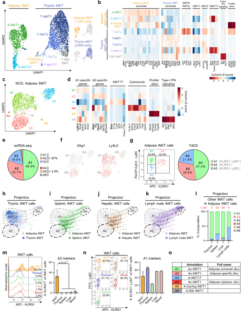
Adipose tissue invariant natural killer T (iNKT) cells are a crucial cell type for adipose tissue homeostasis in obese animals. However, heterogeneity of adipose iNKT cells and their function in adipocyte turnover are not thoroughly understood. Here, we investigate transcriptional heterogeneity in adipose iNKT cells and their hierarchy using single-cell RNA sequencing in lean and obese mice. We report that distinct subpopulations of adipose iNKT cells modulate adipose tissue homeostasis through adipocyte death and birth. We identify KLRG1+ iNKT cells as a unique iNKT cell subpopulation in adipose tissue. Adoptive transfer experiments showed that KLRG1+ iNKT cells are selectively generated within adipose tissue microenvironment and differentiate into a CX3CR1+ cytotoxic subpopulation in obese mice. In addition, CX3CR1+ iNKT cells specifically kill enlarged and inflamed adipocytes and recruit macrophages through CCL5. Furthermore, adipose iNKT17 cells have the potential to secrete AREG, and AREG is involved in stimulating adipose stem cell proliferation. Collectively, our data suggest that each adipose iNKT cell subpopulation plays key roles in the control of adipocyte turnover via interaction with adipocytes, adipose stem cells, and macrophages in adipose tissue.


Conflict of interest statement

The authors declare no competing interests.

Figures

Fig. 1
Fig. 1. scRNA-seq analysis reveals distinct subpopulations of adipose iNKT cells.
a Unsupervised clustering of TCRβint/CD1d.PBS57 tetramer+ iNKT cells from 16-week-old male mice. 1,287 cells from epididymal white adipose tissue (WAT) and 2,630 cells from thymus of 16-week-old male mice on a uniform manifold approximation and projection (UMAP) plot. b Heatmap showing the expression levels of tissue-selective genes and iNKT cell subtype markers. c Unsupervised clustering of adipose iNKT cells from 16-week-old male mice on a UMAP plot. d Heatmap showing the expression levels of subpopulation marker genes. e The ratio of each adipose iNKT cell subpopulation in scRNA-seq data. f Gene expression levels of Klrg1 and Ly6c2 in adipose iNKT cells. g Representative FACS plot and proportion of major adipose iNKT cell subpopulations from 16-week-old male mice (n = 5). hk Projection plots of iNKT cells from other organs to adipose iNKT cells. iNKT cells from other organs are projected to average of similar adipose iNKT cells’ coordinates. Adipose iNKT cells are shown in gray, and iNKT cells from other organs are shown in color. Thymic, splenic, hepatic, and lymph node iNKT cells were used. scRNA-seq data of splenic, hepatic, and lymph node iNKT cells were obtained from GSE161495. l Result of reference mapping of iNKT cells from other organs on adipose iNKT cells. The ratio of other iNKT cells mapped to specific adipose iNKT cell subpopulations was depicted as bar graphs and the percentage of cells mapped to A2 subpopulation was indicated on each bar graph. m Representative FACS plots and proportion of KLRG1+ iNKT cells from 10-week-old male mice (n = 6). n Representative FACS plots and proportion of KLRG1Ly6C+ cells from 10-week-old male mice (n = 3). o Annotation table of each adipose iNKT cell subpopulation. Data are represented as mean ± SD. One-way ANOVA (m).
Fig. 2
Fig. 2. Adipose tissue microenvironment generates As-iNKT1 cells.
a, b Representative FACS plots and proportion of iNKT cells (a) and As-iNKT1 cells (b) from male mice WAT (2w, 4w, 12w (n = 4), and 8w (n = 3)). c Experimental scheme for adoptive transfer of primary iNKT cells established from spleen. d Representative FACS plot and proportion of CD45.1+ iNKT cells among adipose iNKT cells (3w (n = 4) and 8w (n = 3)). e Experimental scheme for adoptive transfer of primary iNKT cells. f Representative FACS plot and proportion of KLRG1+ cells among primary iNKT cells (n = 4) and infiltrated CD45.1+ iNKT cells (n = 5). g Representative FACS plot and proportion of Ki-67+ cells from 8-week-old male mice (n = 4). h mRNA levels in adipocyte fraction of WAT from male mice (n = 4). p-values; versus 4-week-old. i, j Representative FACS plot and proportion of KLRG1+ cells among CD45.1+ iNKT cells infiltrated into WAT of WT or CD1d KO mice (n = 3) (i) and among DN32.D3 iNKT hybridoma cells after culture with Control media (Ctrl), primary adipocyte-conditioned media (AD CM), or primary hepatocyte-conditioned media (Hep CM) (n = 3 biologically independent samples) (j). k mRNA levels in DN32.D3 cells after culture with Ctrl (n = 6), AD CM (n = 5), or Hep CM (n = 6). l Heatmap showing the expression levels of cytokine genes. m Intracellular cytokine staining of As-iNKT1, Au-iNKT1, and A-iNKT17 cells from WT mice stimulated with PMA/Ionomycin. Representative FACS plots and proportion of cytokine-positive cells among each adipose iNKT cell subpopulation (TNFα, IL-17A (n = 4), and IFNγ (n = 5)). n Representative FACS plot and proportion of eGFP+ cells among As-iNKT1, Au-iNKT1, and A-iNKT17 cells from IL-4/GFP enhanced transcript (4Get) mice (n = 6 biologically independent mice). Connected dots represent paired cell populations in a single stromal vascular fraction (SVF) (m and n). Data are represented as mean ± SD. n.s., non-significant. One-way ANOVA (a, b, f, h, j, and k). Paired one-way ANOVA (m and n). Two-tailed unpaired Student’s t test (d and i). Two-tailed paired Student’s t test (g). As-iNKT1; Adipose specific iNKT1, Au-iNKT1; Adipose universal iNKT1.
Fig. 3
Fig. 3. In obesity, As-iNKT1 cells give rise to Ac-iNKT1 cells.
a Unsupervised clustering of TCRβint/CD1d.PBS57 tetramer+ iNKT cells from WAT of NCD-, 1-week, or 8-week HFD-fed mice. 1,287 cells from NCD-, 1,800 cells from 1-week, and 2,400 cells from 8-week HFD-fed mice on a UMAP plot. b Heatmap showing the expression levels of subpopulation marker genes. c Proportion of each adipose iNKT cell subpopulation in scRNA-seq data. d Representative FACS plots and proportion of each subpopulation among total adipose iNKT cells in NCD- (n = 7) or 8-week HFD-fed mice (n = 8). e Gene expression levels of Nr4a1, Hmgb2, Trbv13-1, Cd160, and Klrk1 in adipose iNKT cell subpopulations under each condition. p-values were adjusted for multiple comparisons using Bonferroni correction. f In silico pseudotime analysis of adipose iNKT1 cells. g Clonotype overlap analysis of adipose iNKT cell subpopulations. h Representative clones showing clonotype overlapping between As-iNKT1, Ac-iNKT1, and A-Cycling iNKT1 cells. i Experimental scheme for adoptive transfer of CD45.1+ Au-iNKT1 and As-iNKT1 cells. iNKT cells were sorted from CD45.1 mice 1-week after α-GC injection and injected into each WAT fat pad of 16-week HFD-fed CD45.2 Jα18 KO mice. j, k Representative FACS plots and composition of injected CD45.1+ donor iNKT cells in recipient mice after 3 weeks (Before injection (n = 2), Au-iNKT1 post injection (n = 2), and As-iNKT1 post injection (n = 3)). l Schematic diagram of iNKT1 differentiation process in adipose tissue. m CDR3β amino acid sequences of most prevalent CDR3β length of each adipose iNKT1 cell subpopulation. Data were collected from 8-week-HFD condition. Data are represented as mean ± SD. n.s., non-significant. Two-tailed unpaired Student’s t test (d, e, j, and k). Au-iNKT1; Adipose universal iNKT1, As-iNKT1; Adipose specific iNKT1. Ac-iNKT1; Adipose cytotoxic iNKT1.
Fig. 4
Fig. 4. Ac-iNKT1 cells kill hypertrophic and inflammatory adipocytes and recruit macrophages by secreting CCL5.
a KEGG pathway analysis of Ac-iNKT1 high-differentially expressed genes (DEGs) compared to As-iNKT1 cells (P < 0.05). b Expression levels of cytotoxic marker genes in adipose iNKT cell subpopulations. c Experimental design for coculture of each iNKT cell subpopulation with hypertrophic adipocytes. d Representative images of coculture between PA-treated 3T3-L1 adipocytes and iNKT cell subpopulations. Arrows; PI+ dead adipocytes. Scale bars, 50 μm. e, f Proportion of PI+ adipocytes among large or small adipocytes ((-) (n = 7), As-iNKT1 (n = 6), and Ac-iNKT1 (n = 7)) (e) and among total adipocytes (n = 12 biologically independent cells) (f). Cells in (d) were counted by using microscope (e and f). g Experimental scheme for injection of each iNKT cell subpopulation. iNKT cells were sorted from 8-week HFD-fed WT male mice 1-week after α-GC injection and injected into HFD-fed Jα18 KO mice. h Representative histological images of PBS or iNKT cell subpopulation-injected WAT fat pads. Arrows; CLS. Scale bars, 100 μm. i Quantification of the number of CLS (Ac-iNKT1/PBS (n = 17), Ac-iNKT1/+iNKT (n = 24), As-iNKT1/PBS (n = 22), As-iNKT1/+iNKT (n = 23), Au-iNKT1/PBS (n = 14), Au-iNKT1/+iNKT (n = 12), A-iNKT17/PBS (n = 18), and A-iNKT17/+iNKT (n = 19)). j Heatmap showing the expression levels of macrophage-recruiting cytokines in adipose iNKT cell subpopulations. k Ccl5 mRNA level in in vivo-expanded iNKT cell subpopulations sorted from 10-week-old male mice (n = 5 biologically independent samples). l Ccr5 expression level in immune cells from WAT (GSE182233). m Left: representative images of monocyte infiltration with or without CCL5 peptides. Right: quantification of the number of infiltrated THP1 cells (n = 5 biologically independent samples). Scale bars, 100 μm. Data are represented as mean ± SD except (i) represented as box and whiskers plot. In (i), the lower, central, and upper line in each box represents the first (Q1), the second (median), and the third quartile (Q3), respectively. The whiskers extend from the box to the minimum and maximum observations, respectively. n.s., non-significant. One-way ANOVA (f and k). Two-way ANOVA (e and i). Two-tailed unpaired Student’s t test (m). One-tailed Fisher’s exact test (a).
Fig. 5
Fig. 5. A-iNKT17 cells stimulate adipose stem cell proliferation by secreting amphiregulin.
a Representative FACS plots and proportion of Ki-67+ among ASCs (CD31-/CD45-/PDGFRα+) from WT WAT 4-days after vehicle (n = 4) or α-GC injection (n = 5). b The total number of ASCs from WT WAT 1-week after vehicle (n = 4) or α-GC injection (n = 5). c Representative FACS plots and proportion of each adipose iNKT cell subpopulation among total adipose iNKT cells 4-days after vehicle or α-GC injection (n = 5 biologically independent mice). d Representative FACS plots and proportion of Ki-67+ among ASCs from WAT of 12-week HFD-fed Jα18 KO mice with or without A-iNKT17 cell injection (n = 4 biologically independent mice). Mice were sacrificed 4 days after injection. e Heatmap showing the expression levels of growth factors in adipose iNKT cell subpopulations. f Areg mRNA level in in vivo-expanded iNKT cell subpopulations sorted from 10-week-old male mice (n = 5 biologically independent samples). g Areg mRNA level in WAT SVFs from 8-week HFD-fed WT (n = 6) and Jα18 KO mice (n = 5). h Representative FACS plots and proportion of Ki-67+ among ASCs from WT WAT. Mice were injected with vehicle, AREG 0.1 μg (n = 4), or AREG 1 μg (n = 3). i Experimental scheme using C57BL/6J adipocyte lineage-tracing male mice. j Representative microscopic images of vehicle or AREG injected WAT 4 weeks after injection. Asterisks indicate perilipin+/YFP new adipocytes. Scale bars, 100 μm. k Proportion of YFP new adipocytes among total adipocytes from vehicle (n = 4) or AREG injected mice (n = 6). l Quantification of lipid droplet (LD) size of vehicle (n = 489) or AREG injected WAT (n = 527) in (j). Data are represented as mean ± SD except (l) represented as box and whiskers plot. In (l), the lower, central, and upper line represents the first (Q1), the second (median), and the third quartile (Q3), respectively. The whiskers extend from the box to the 2.5 and 97.5 percentile, respectively. n.s., non-significant. One-way ANOVA (f and h). Two-tailed unpaired Student’s t test (ac, g, k, and l). Two-tailed paired Student’s t test (d).
Fig. 6
Fig. 6. Proposed Model.
Adipose iNKT cells are composed of adipose-specific (As)-iNKT1 cells which possess distinct transcriptome profiles including KLRG1. As-iNKT1 cells are differentiated from adipose-universal (Au)-iNKT1 cells under adipose tissue microenvironment. In obesity, some As-iNKT1 cells differentiate into adipose-cytotoxic (Ac)-iNKT1 cells. Ac-iNKT1 cells are highly cytotoxic and they selectively kill large and inflamed adipocytes. Also, they recruit macrophages around dead adipocytes by secreting CCL5. Adipose iNKT17 cells stimulate the proliferation of adipose stem cells by amphiregulin secretion, helping adipose tissue regeneration.

References

    1. Sun K, Kusminski CM, Scherer PE. Adipose tissue remodeling and obesity. J. Clin. Invest. 2011;121:2094–2101. doi: 10.1172/JCI45887. - DOI - PMC - PubMed
    1. Choe SS, Huh JY, Hwang IJ, Kim JI, Kim JB. Adipose Tissue Remodeling: Its Role in Energy Metabolism and Metabolic Disorders. Front Endocrinol. (Lausanne) 2016;7:30. doi: 10.3389/fendo.2016.00030. - DOI - PMC - PubMed
    1. Sakers A, De Siqueira MK, Seale P, Villanueva CJ. Adipose-tissue plasticity in health and disease. Cell. 2022;185:419–446. doi: 10.1016/j.cell.2021.12.016. - DOI - PMC - PubMed
    1. Rosen ED, Spiegelman BM. What we talk about when we talk about fat. Cell. 2014;156:20–44. doi: 10.1016/j.cell.2013.12.012. - DOI - PMC - PubMed
    1. Reilly SM, Saltiel AR. Adapting to obesity with adipose tissue inflammation. Nat. Rev. Endocrinol. 2017;13:633–643. doi: 10.1038/nrendo.2017.90. - DOI - PubMed

Publication types

LinkOut - more resources