Review
. 2023 Dec 21;9(1):73.
doi: 10.1038/s41572-023-00484-9.

Central neuropathic pain

Affiliations
Review

Central neuropathic pain

Jan Rosner et al. Nat Rev Dis Primers. .

Abstract

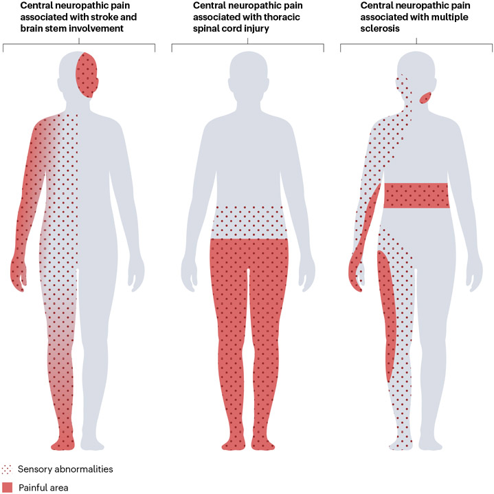
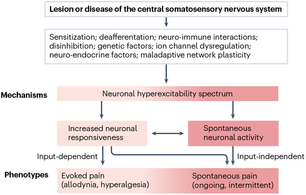
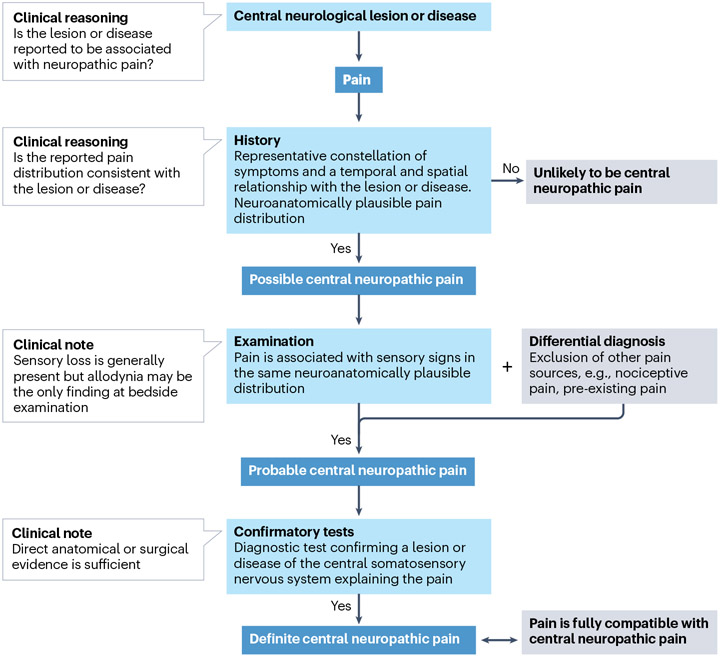
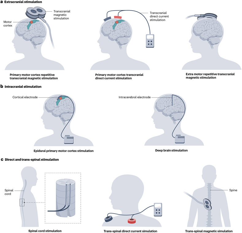
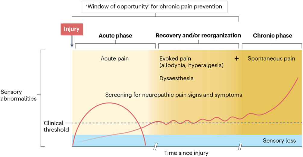
Central neuropathic pain arises from a lesion or disease of the central somatosensory nervous system such as brain injury, spinal cord injury, stroke, multiple sclerosis or related neuroinflammatory conditions. The incidence of central neuropathic pain differs based on its underlying cause. Individuals with spinal cord injury are at the highest risk; however, central post-stroke pain is the most prevalent form of central neuropathic pain worldwide. The mechanisms that underlie central neuropathic pain are not fully understood, but the pathophysiology likely involves intricate interactions and maladaptive plasticity within spinal circuits and brain circuits associated with nociception and antinociception coupled with neuronal hyperexcitability. Modulation of neuronal activity, neuron-glia and neuro-immune interactions and targeting pain-related alterations in brain connectivity, represent potential therapeutic approaches. Current evidence-based pharmacological treatments include antidepressants and gabapentinoids as first-line options. Non-pharmacological pain management options include self-management strategies, exercise and neuromodulation. A comprehensive pain history and clinical examination form the foundation of central neuropathic pain classification, identification of potential risk factors and stratification of patients for clinical trials. Advanced neurophysiological and neuroimaging techniques hold promise to improve the understanding of mechanisms that underlie central neuropathic pain and as predictive biomarkers of treatment outcome.


Figures

Fig. 1 ∣
Fig. 1 ∣. Distribution of sensory abnormalities and central neuropathic pain.
In a ventral view of the human body, the distribution of pain and sensory abnormalities associated with central neuropathic pain conditions are presented. Sensory abnormalities and pain are present contralateral to the lesion in stroke but may affect the ipsilateral face in the case of brainstem lesions. After spinal cord injury, the location of pain and sensory abnormalities can be at or below the lesion level. In multiple sclerosis, the location varies depending on the site of the central nervous system lesions, for example, facial pain in trigeminal neuralgia or segmental belt-like pain in the case of spinal cord involvement. Sensory abnormalities (dotted area) often extend beyond the painful area (red). The locations of pain and sensory abnormalities can vary substantially between individuals, with the possibility of uni- or bilateral distribution. Thus, these illustrations aim to depict examples of neuroanatomically plausible locations rather than typical presentations. The proximal to-distal gradient in pain intensity as demonstrated in the left body chart, wherein pain intensity gradually increases towards the distal extremity, is observed in some patients.
Fig. 2 ∣
Fig. 2 ∣. From sensory-discriminative mechanisms to the clinical pain phenotype.
A lesion or disease of the central somatosensory nervous system initiates a pathophysiological cascade that involves various mechanisms potentially dependent on injury type and with largely unknown interactions. These mechanisms may, to some extent, converge to modify neuronal excitability. Dynamic changes in the response properties and excitability of central somatosensory neurons can result in two types of pain — spontaneous pain and evoked pain (allodynia, hyperalgesia) and/or spontaneous or evoked dysaesthesia (not shown). Evoked pain depends on preserved afferent input (input-dependent). Spontaneous pain may arise independently of afferent input or may be (partially) maintained by continuous exteroceptive and/or interoceptive input, which may otherwise not be consciously perceived by the patient.
Fig. 3 ∣
Fig. 3 ∣. Molecular mechanisms in spinal and supraspinal neuronal circuitry.
A lesion or disease in the central nervous system (CNS) causes both direct and secondary changes in the integrity and plasticity of spinal or supraspinal circuitry. On the left, in a limited view of the nociceptive system, two main pathways of the ascending somatosensory nervous system related to sensory-discriminative aspects of pain are displayed. The figure on the right highlights important molecular mechanisms that shape inhibition and excitation within spinal and supraspinal circuits related to the sensory-discriminative aspects of central neuropathic pain (CNP). Dysfunction of local inhibition (γ-aminobutyric acid (GABA) and glycine) and descending inhibition owing to damage can lead to loss of inhibitory control (disinhibition). Altered synaptic plasticity, notably N-methyl-d-aspartate (NMDA)-dependent long-term potentiation, may increase the sensitivity of the nociceptive system to subthreshold input. Such activity-dependent plasticity may be triggered or facilitated by spontaneous discharges arising from neuronal damage or altered inhibitory tone. Non-neuronal cells, including astrocytes and microglia, have important roles — impaired astrocytic glutamate buffering may result in excitotoxicity, whereas microglial activation via messengers such as ATP can affect neuronal function, potentially mediated by brain-derived neurotrophic factor (BDNF)-induced downregulation of the potassium chloride co-transporter KCC2. AMPAR, α-amino-3-hydroxy-5-methyl-4-isoxazolepropionic acid receptor; CatS, cathepsin S; CX3CR/L1, C-X3-C motif chemokine receptor/ligand 1; DRG, dorsal root ganglion; EAAT2, excitatory amino acid transporter 2; GlyR, glycine receptor; NMDAR, N-methyl-d-aspartate receptor; TRKB, tyrosine receptor kinase B.
Fig. 4 ∣
Fig. 4 ∣. The thermal grill illusion — a model for central neuropathic pain?
One hypothesis to explain thermal grill illusions suggests that the integration of simultaneous innocuous warm and cool input to thermoreceptive spinothalamic neurons (COLD), which show selective activation by Aδ-fibres mediating cooling, results in diminished COLD neuron activity. Consequently, this reduction induces a central disinhibition, or ‘unmasking’ of polymodal nociceptive neurons that respond to nociceptive heat, pinch and cold (HPC),. Conceptually, a lesion or disease that affects COLD and HPC neurons differentially could also generate an imbalance, leading to the release of HPC pathway activity,. According to this thermal grill illusion model, this release manifests as a burning, painful or unpleasant sensation, which resembles the spontaneous neuropathic pain phenotype of patients with central neuropathic pain. The thermal grill illusion may facilitate our understanding of the pathophysiology of central neuropathic pain (CNP) and has been explored in CNP after multiple sclerosis and non-neuropathic conditions,.
Fig. 5 ∣
Fig. 5 ∣. Grading system for central neuropathic pain.
A grading system is used to attain different levels of diagnostic certainty for central neuropathic pain (CNP). If reported symptoms are temporally associated with the lesion or disease and align with the anticipated neuroanatomical pain pattern, the presence of neuropathic pain is possible. To advance to ‘probable’, a neurological examination needs to detect somatosensory abnormalities such as loss of sensation or hypersensitivity. Importantly, differential diagnoses such as musculoskeletal pain must be ruled out at this stage. Once a lesion within the central somatosensory nervous system is confirmed and other types of pain excluded, the definite CNP level is reached.
Fig. 6 ∣
Fig. 6 ∣. Neuromodulatory techniques for central neuropathic pain.
Various modulation techniques using extracranial stimulation (panel a), intracranial stimulation (panel b) and direct and trans-spinal stimulation (panel c). The schematic shows neuromodulation of primary motor cortex (left figures, panels a and b) and deep brain regions (right figures, panels a and b).
Fig. 7 ∣
Fig. 7 ∣. Progression and monitoring of central neuropathic pain.
In the immediate aftermath of a central nervous system lesion, acute pain can occur owing to changes induced by the trauma or disease. Neurological function is compromised, leading to sensory impairments as the primary somatosensory manifestation. As time progresses, individuals may experience heightened sensitivity to sensory stimuli, which can progress to the emergence of spontaneous central neuropathic pain (CNP), along with evoked pain. This phase is particularly important for implementing preventive treatments and clinical monitoring, emphasizing the need for screening for abnormal sensory signs. Once spontaneous CNP has developed (with possible concurrent evoked pain), the focus shifts to achieving an optimal (differential) diagnosis and implementing effective pain management strategies. Note that this is only one example for a patient trajectory; not all patients experience evoked pain, and many other trajectories exist.

References

    1. Jensen TS et al. A new definition of neuropathic pain. Pain 152, 2204–2205 (2011). - PubMed
    1. Scholz J. et al. The IASP classification of chronic pain for ICD-11: chronic neuropathic pain. Pain 160, 53–59 (2019).

      This paper is an overview of conditions included in the International Classification of Diseases 11th Revision classification of chronic neuropathic pain, including CNP.

    1. Borsook D. Neurological diseases and pain. Brain 135, 320–344 (2012). - PMC - PubMed
    1. Widerström-Noga E, Loeser JD, Jensen TS & Finnerup NB AAPT diagnostic criteria for central neuropathic pain. J. Pain 18, 1417–1426 (2017). - PubMed
    1. Widerström-Noga E, Felix ER, Adcock JP, Escalona M & Tibbett J Multidimensional neuropathic pain phenotypes after spinal cord injury. J. Neurotrauma 33, 482–492 (2016). - PubMed

Substances

LinkOut - more resources