. 2023 Dec 20;13(1):22782.
doi: 10.1038/s41598-023-49578-0.

Inhibition of Drp1 orchestrates the responsiveness of breast cancer cells to paclitaxel but insignificantly relieves paclitaxel-related ovarian damage in mice

Affiliations

Inhibition of Drp1 orchestrates the responsiveness of breast cancer cells to paclitaxel but insignificantly relieves paclitaxel-related ovarian damage in mice

Adel I Alalawy et al. Sci Rep. .

Erratum in

Abstract

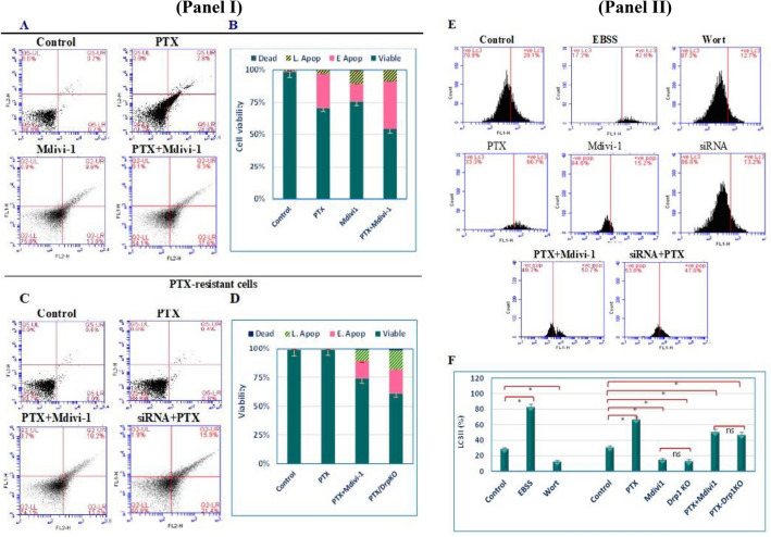
Chemoresistance and chemotherapy-related ovarian damage are well-reported in breast cancer (BC) young patients. Herein, the inhibition of the mitochondrial fission was invested to explore its chemosensitizing role in Paclitaxel (PTX)-resistant cells, and its ability to restore the ovarian integrity in mice receiving PTX or cisplatin chemotherapy. To establish these aims, PTX-resistance was generated in BC cells, which were treated with PTX in combination with Drp1 deficiency, via mdivi-1, or Drp1-specific siRNA transfection. Furthermore, the alterations in the ovarian structure and the endocrine-related hormones were explored in mice receiving repetitive doses of PTX or cisplatin. We found that combining PTX with mdivi-1 improved cell responsiveness to PTX, induced apoptosis- and autophagy-mediated cell death, and relieved cellular oxidative stress. Additionally, the expression of PCNA1 and cyclin B1 genes were downregulated, meanwhile, p53, p21, and mitochondrial fusion proteins (Mfu1&Mfu2) were increased. The in vivo investigations in mice demonstrated that PTX induced gonadotoxic damage similar to cisplatin, whereas dual treatment of mice with PTX+ mdivi-1 failed to restore their normal follicular count and the circulating levels of E2 and AMH hormones. These results suggested that combining Drp1 inhibition with PTX resensitized breast cancer cells to PTX but failed to offer enough protection against chemotherapy-related gonadotoxicity.


Conflict of interest statement

The authors declare no competing interests.

Figures

Figure 1
Figure 1
The cytotoxic effect of PTX is enhanced by Drp1 deficiency in breast cancer cells. The cytotoxicity of PTX was evaluated in PTX-sensitive and PTX-resistant cells by MTT assay, where the IC50 of mdivi-1 in PTX-sensitive cells was 59.5 µM (A). PTX-sensitive cells (green dashed line) and resistant cells (red dashed line) were incubated with a concentration range of PTX. Also, resistant cells were incubated with different concentrations of PTX combined with the IC50 of mdivi-1 (blue dashed line) and the percent of viable cells was determined and normalized to the control cells (B). Also, the percentage of cell viability was determined in PTX-resistant cells exposed to concentration range of PTX after their transfection Drp1-specific siRNA (C). The IC50 of PTX in PTX-sensitive cells is 85 nM, where for PTX+mdivi-1 is 132 uM. D through F are representative photomicrographs of PTX-sensative cells treated with PTX, PTX-resistant cells treated with PTX, and PTX-resistant cells breated with both PTX and mdivi-1, respectively (Magnification 20X). Dots represent the mean (± SD) of viable cells of n = 5–6 readings. (*) and (#): refer to significant changes in cell viability of PTX+mdivi-1 versus PTX, P < 0.05 and P < 0.001, respectively. “D-F” are representative light micrographs of PTX-regular or resistant cells treated with PTX or cells dually treated with PTX+Mdivi1, respectively.
Figure 2
Figure 2
Drp1 deficiency induced apoptosis and autophagy in PTX-resistant cells. Regular cells (IA) were left untreated, treated with the IC50 concentration (85 nM) of PTX, the IC50 concentration (59.5 µM) of mdivi-1, or PTX+mdivi-1. The apoptotic effect in PTX or mdivi-1 treated cells was enhanced in when cells were cotreated with both drugs. Resistant cells (IC) were left untreated, treated with PTX, PTX+mdivi-1, or PTX after they were transfected with Drp1-specific siRNA. Apoptosis was significantly developed in PTX cells cotreated with Drp1 inhibitor or transfected with siRNA (D). “E” depicts the level autophagy marker (LC3II) in cells left untreated, transiently incubated in EBSS, treated with Wort, PTX, mdivi-1, transfected with siRNA, PTX+mdivi-1, or treated with PTX, after siRNA transfection. Resistant cells responded to the autophagy inducer EBSS or repressor (Wort). PTX induced a twofold increase in the LC3II (P < 0.001). Drp1 inhibition (or silencing), insignificantly, repressed autophagy (P > 0.05) compared to its basal level. However, combined treatments led to a significant increase in the LC3II. Bars (B, D and F) represent the mean of three independent experiments (± SD), (*): refers to a significant difference between the indicated cells versus the corresponding untreated cells. “ns”: insignificant.
Figure 3
Figure 3
Expression of apoptosis-related genes (p21 and p53), cell cycle-related genes (cyclin B1 and PCNA1), MDR1, and Drp1. RNAs were isolated form untreated or treated cells, reverse transcribed, and then used as templates in qRT-PCR reactions. In PTX-resistant cells, the expression of p53 and p21 increased (A), meanwhile cyclin B1 and PCNA1 were downregulated (B) relative to the untreated resistant cells. MDR1 expression was higher in resistant cells compared to sensitive cells (Control-S), and decreased in dually-treated resistant cells compared to the corresponding untreated cells. No significant difference was seen between normal and resistant cells; however it decreased in dully treated resistant cells.
Figure 4
Figure 4
Computational modeling of the molecular interactions of paclitaxel, and mdivi-1 with mitochondrial redox-related enzymes. The 2D interaction diagrams demonstrate the interaction of ligands (PTX or mdivi-1) with mitochondrial ATP synthase, SOD or Thioredoxin reductase 1. The hot spots in protein domains, the superimposition of the docking pose, and the co-crystallized ligands in the active sites in the enzyme active site are shown. The docking score is shown next each protein.
Figure 5
Figure 5
Changes in the mitochondrial morphology, and mitochondrial dynamic-related proteins. Electronmicrographs of the mitochondrial morphological changes in PTX-resistant cells (A), cells treated with mdivi-1, PTX, or PTX+mdivi-1. (B) depicts immunoblotting of the mitochondrial-related protein (Drp1), and fusion proteins (Mfu1 & Mfu2) and the corresponding band intensities (C). Drp1 expression was normally expressed in PTX-resistant cells, similar to regular cells. Mdivi-1, alone or combined with PTX did not affect the expression of Drp1, whereas it is downregulated in cells treated with PTX after siRNA transfection. Mfu1&Mfu2 were significantly upregulated in cell treated with mdivi-1 or PTX after siRNA transfection. (D) shows the in vitro cell migration of cells treated with PTX, Mdivi-1, mdivi-1+PTX, or cells transfected with siRNA and then were treated with PTX. *, **, *** refer to P < 0.05, P < 0.01, P < 0.001, compared to the untreated cells.
Figure 6
Figure 6
Change in mice body weight, ovarian weight, follicular count, and E2 and AMH levels. Mice body weight (A) after various treatments were compared to the initial body weight (Day 0), ovarian weights of treated groups were compared to the corresponding control group (B). Differential follicular count demonstrated massive decrease of the count of various types (C). Both E2 (D) and AMH (E) decreased in PTX or cisplatin groups, whereas cotreatment with mdivi-1 did not show significant improvements compared to PTX or cisplatin groups. Data are shown as bars or dots indicating the mean (± SD). Abbreviations: E2: estradiol, AMH: antimullerian hormone.
Figure 7
Figure 7
Representative hematoxylin and eosin-stained ovarian sections recovered from healthy mice (A), mice intraperitoneally injected with PTX (B), Cisplatin (C), PTX+mdivi-1 (D), or Cis+mdivi-1 (E). Both PTX and Cisplatin reduced the size of ovaries, the number of healthy follicles. Cotreatment with mdivi-1did not exert a significant repair.

References

    1. Yu R, Lendahl U, Nistér M, Zhao J. Regulation of mammalian mitochondrial dynamics: Opportunities and challenges. Front. Endocrinol. 2020;11:374. doi: 10.3389/fendo.2020.00374. - DOI - PMC - PubMed
    1. Kashatus DF, Lim KH, Brady DC, Pershing NL, Cox AD, Counter CM. RALA and RALBP1 regulate mitochondrial fission at mitosis. Nat. Cell Biol. 2011;13(9):1108–1115. doi: 10.1038/ncb2310. - DOI - PMC - PubMed
    1. Hall AR, Burke N, Dongworth RK, Hausenloy DJ. Mitochondrial fusion and fission proteins: Novel therapeutic targets for combating cardiovascular disease. Br. J. Pharmacol. 2014;171(8):1890–1906. doi: 10.1111/bph.12516. - DOI - PMC - PubMed
    1. Danino D, Hinshaw JE. Dynamin family of mechanoenzymes. Curr. Opin. Cell Biol. 2001;13(4):454–460. doi: 10.1016/S0955-0674(00)00236-2. - DOI - PubMed
    1. Ingerman E, Perkins EM, Marino M, Mears JA, McCaffery JM, Hinshaw JE, Nunnari J. Dnm1 forms a spiral that is structurally tailored to fit mitochondria. J. Cell Biol. 2005;170(7):1021–1027. doi: 10.1083/jcb.200506078. - DOI - PMC - PubMed