. 2023 Dec 21;6(1):1289.
doi: 10.1038/s42003-023-05682-5.

Atomistic ensemble of active SHP2 phosphatase

Affiliations

Atomistic ensemble of active SHP2 phosphatase

Massimiliano Anselmi et al. Commun Biol. .

Abstract

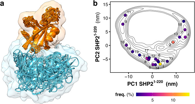
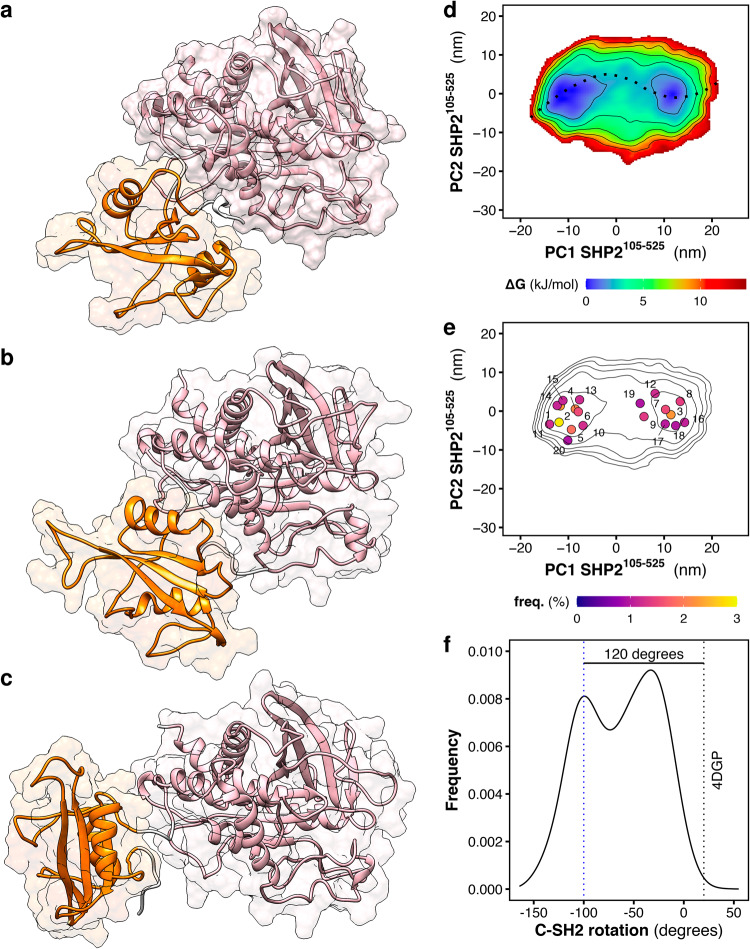
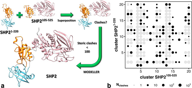
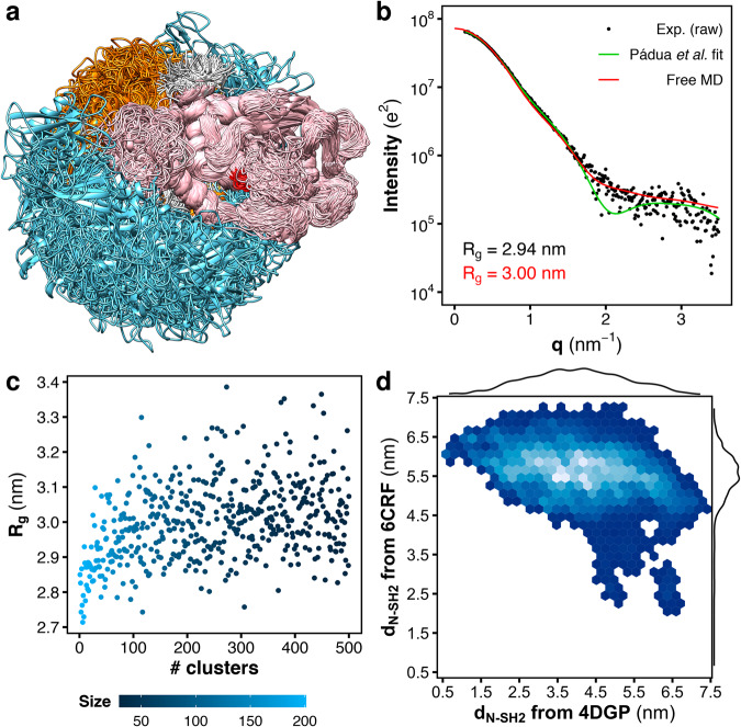
SHP2 phosphatase plays an important role in regulating several intracellular signaling pathways. Pathogenic mutations of SHP2 cause developmental disorders and are linked to hematological malignancies and cancer. SHP2 comprises two tandemly-arranged SH2 domains, a catalytic PTP domain, and a disordered C-terminal tail. Under physiological, non-stimulating conditions, the catalytic site of PTP is occluded by the N-SH2 domain, so that the basal activity of SHP2 is low. Whereas the autoinhibited structure of SHP2 has been known for two decades, its active, open structure still represents a conundrum. Since the oncogenic mutant SHP2E76K almost completely populates the active, open state, this mutant has been extensively studied as a model for activated SHP2. By molecular dynamics simulations and accurate explicit-solvent SAXS curve predictions, we present the heterogeneous atomistic ensemble of constitutively active SHP2E76K in solution, encompassing a set of conformational arrangements and radii of gyration in agreement with experimental SAXS data.


Conflict of interest statement

The authors declare no competing interests.

Figures

Fig. 1
Fig. 1. Crystal structures and SAXS curves from closed SHP2 and open SHP2E76K.
a Crystal structure of autoinhibited wild-type SHP2 (SHP2wt). In the autoinhibited state (PDB ID 4DGP), SHP2wt adopts a closed conformation; the N-SH2 domain (cyan cartoon) blocks the catalytic site (red) of the PTP domain (pink) with the blocking loop (blue). The N-SH2 domain is connected to PTP in tandem with the homologous C-SH2 domain (orange). b Crystal structure of the constitutively active SHP2E76K mutant. In the active state, SHP2E76K adopts an open conformation with the catalytic pocket exposed to the solvent. The crystal structure of SHP2E76K (PDB ID 6CRF) reveals a 120° rotation of the C-SH2 domain and the relocation of the N-SH2 domain to a PTP surface opposite the catalytic site. c Small-angle X-ray scattering (SAXS) curve calculated from the solution ensemble of the autoinhibited wild-type SHP2 (unrestrained MD simulation from PDB ID 4DGP, red line) is compared with the experimental curve (black dots), reported as raw data, and with the single-structure model fitting by Pádua et al. (green line). d The SAXS curve calculated from the crystal structure of the constitutively active SHP2E76K mutant (restrained MD simulation from PDB ID 6CRF, red line) is compared with the experimental curve (black dots), reported as raw data, and with the single-structure model fitting by Pádua et al. (green line), Experimental, model-estimated, and calculated radii of gyration (Rg) are reported in each panel in black, green, and red font, respectively.
Fig. 2
Fig. 2. Structural dynamics and free energy landscape of tandem SH2 domains.
a Overlay of the representative structures of the tandem SH2 obtained from MD simulations, as taken from ref. . N-SH2 and C-SH2 are depicted as ribbons and colored respectively in cyan and orange. The structures were superimposed at the C-SH2 domain. b Free energy landscape along the principal components PC1 and PC2 of the tandem SH2 (SHP21–220) as obtained from MD simulations. The projection of the central structure of the twenty most populated clusters are reported as dots in the essential plane.
Fig. 3
Fig. 3. Structural dynamics and free energy landscape of truncated SHP2.
Simulation snapshots of truncated SHP2 ΔN-SH2 (SHP2105–525): (a) conformation adopted by the C-SH2−PTP moiety in autoinhibited SHP2, (b, c) the two most populated conformations adopted by ∆N-SH2 during the simulations. C-SH2 and PTP are depicted as ribbons and colored respectively in orange and pink. d Free energy landscape along the principal components PC1 and PC2 of truncated SHP2 ΔN-SH2 as obtained from MD simulations. The free-energy minimum pathway, reported over the essential plane as a dashed line, corresponds to the roto-translation of the C-SH2 domain relative to PTP (see also Movie S1). e Free energy landscape as contour plot together with the central structures of the twenty most populated clusters. f Distribution of the dihedral angle representing the rotation of the C-SH2 domain relative to PTP. The value of the dihedral angle in autoinhibited SHP2 (PDB ID 4DGP) is reported as a dashed vertical black line. The blue line indicates the value of the dihedral angle that corresponds to the structure depicted in (c).
Fig. 4
Fig. 4. Homology modeling workflow for generating SHP2E76K conformations.
a Scheme representing the homology modeling flowchart used for the generation of the open conformations of SHP2E76K used as seeds in MD simulations. Representative structures of the tandem SH2 (SHP21–220) and of truncated SHP2 ∆N-SH2 (SHP2105–525) were superposed at the C-SH2 domain. After the superposition, the number of clashes raising between PTP and N-SH2 was determined. If the number of clashes did not exceed 100, the representative structure pair was further used as a template for the following modeling of the full-length SHP2E76K (SHP21–529). b Number of clashes between PTP and N-SH2 for each pair of representative structures of the tandem SH2 (SHP21–220) and of truncated SHP2 ∆N-SH2 (SHP2105–525). Among the 400 possible pairs, 181 representative structure pairs had no clashes (void spaces), 275 pairs had fewer than 100 clashes (void spaces and black dots), 125 pairs had at least 100 clashes (gray dots) and were discarded from homology modeling.
Fig. 5
Fig. 5. Heterogeneity of the SHP2E76K structural ensemble is supported by MD simulations and SAXS.
a Overlay of representative structures of SHP2E76K as obtained from MD simulations. N-SH2, C-SH2, and PTP are depicted as ribbons and colored respectively in cyan, orange, and pink. The catalytic site is highlighted in red. The structures were superposed at the PTP domain. b Small-angle X-ray scattering (SAXS) curve calculated from the solution ensemble of SHP2E76K (red line) is compared with the experimental curve (black dots), reported as raw data, and with the single-structure model by Pádua et al. (green line). Radii of gyration (Rg) from the MD ensemble and from experiment are reported with black and red font, respectively. c Rg values of SHP2E76K calculated from the ensembles of the first 500 most populated clusters via Guinier fit to calculated SAXS curves. The population of the clusters are reported in color scale. d Density map of the distance of the N-SH2 center-of-mass (dN-SH2) from the positions respectively occupied by the N-SH2 center-of-mass either in the crystal structure of autoinhibited SHP2 (4DGP) or in the crystal structure of open SHP2E76K (6CRF). The distributions of the distance dN-SH2 from 4DGP and from 6CRF are reported as marginal plots.
Fig. 6
Fig. 6. Comparison of the N-SH2 position in MD simulations and crystal structure.
a Three-dimensional density distribution of the center-of-mass position of the N-SH2 domain relative to PTP. The isovalues of the opaque dark cyan isosurface and of the transparent light cyan isosurface are respectively one tenth and one thousandth of the maximum density value. b Comparison of the crystal structure of open SHP2E76K with the most similar MD configuration. N-SH2, C-SH2, and PTP are depicted as ribbons and colored respectively in cyan, orange, and pink (see text labels). The catalytic site is highlighted in red. The inertia ellipsoid of each domain is reported as a transparent surface. The structures were superposed at the PTP domain.

References

    1. Tajan M, de Rocca Serra A, Valet P, Edouard T, Yart A. SHP2 sails from physiology to pathology. Eur. J. Med. Genet. 2015;58:509–25. doi: 10.1016/j.ejmg.2015.08.005. - DOI - PubMed
    1. Grossmann K. S., Rosário M., Birchmeier C., Birchmeier W. Chapter 2 - The Tyrosine Phosphatase Shp2 in Development and Cancer. In: Advances in Cancer Research (eds Vande Woude G. F., Klein G) (Academic Press, 2010). - PubMed
    1. Yuan X, Bu H, Zhou J, Yang C-Y, Zhang H. Recent advances of SHP2 inhibitors in cancer therapy: current development and clinical application. J. Med. Chem. 2020;63:11368–96. doi: 10.1021/acs.jmedchem.0c00249. - DOI - PubMed
    1. Dance M, Montagner A, Salles J-P, Yart A, Raynal P. The molecular functions of Shp2 in the Ras/Mitogen-activated protein kinase (ERK1/2) pathway. Cell. Signal. 2008;20:453–9. doi: 10.1016/j.cellsig.2007.10.002. - DOI - PubMed
    1. Digilio MC, et al. Grouping of multiple-Lentigines/LEOPARD and Noonan Syndromes on the PTPN11 Gene. Am. J. Hum. Genet. 2002;71:389–94. doi: 10.1086/341528. - DOI - PMC - PubMed

Publication types

MeSH terms

Substances