. 2023 Dec 21;21(1):365.
doi: 10.1186/s12964-023-01393-w.

Hyperglycemia-induced STING signaling activation leads to aortic endothelial injury in diabetes

Affiliations

Hyperglycemia-induced STING signaling activation leads to aortic endothelial injury in diabetes

Ying An et al. Cell Commun Signal. .

Abstract

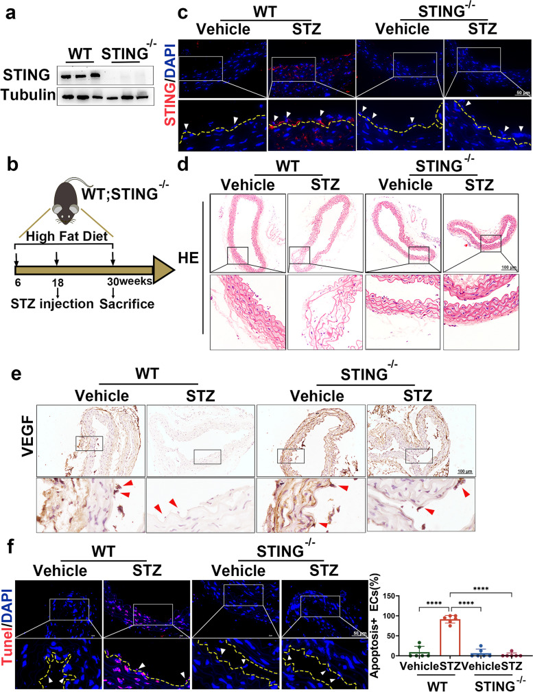
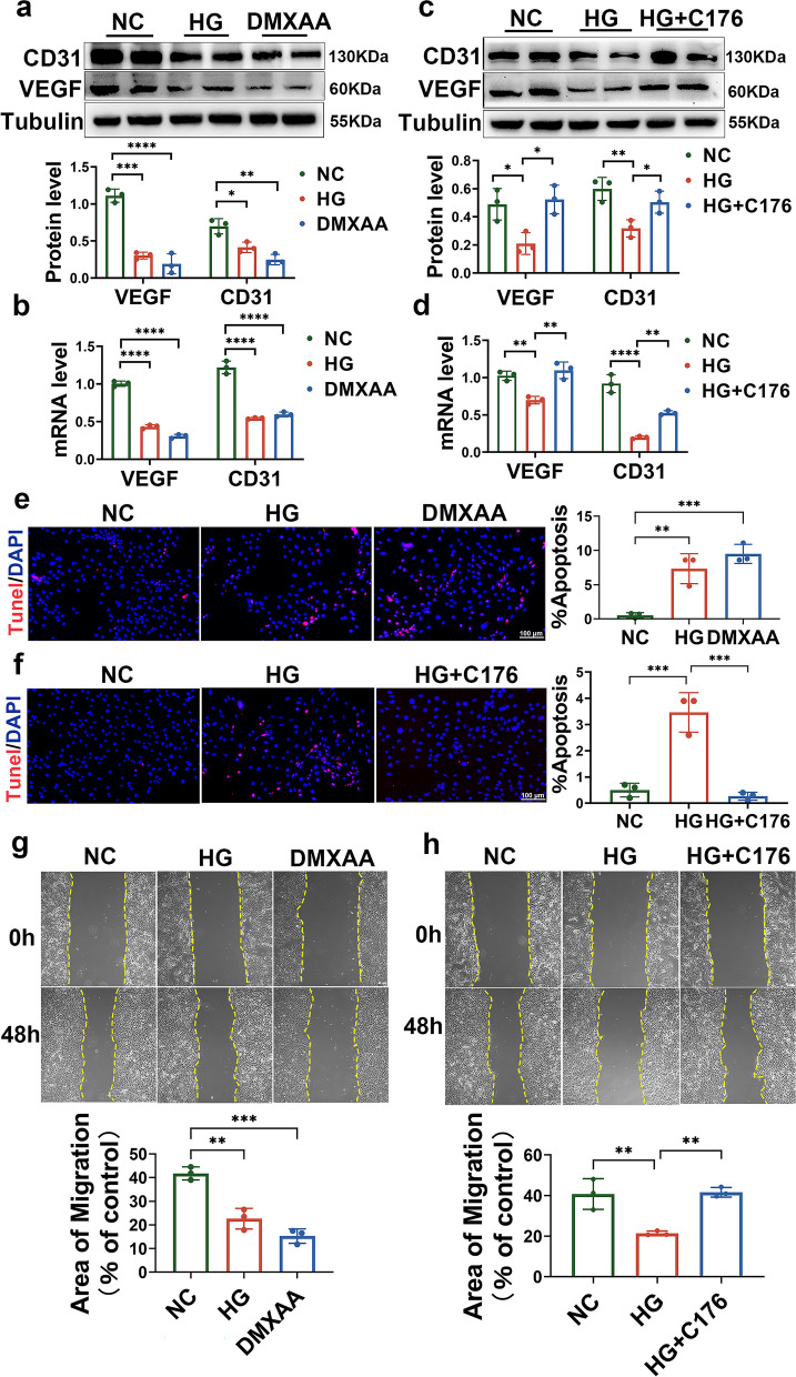
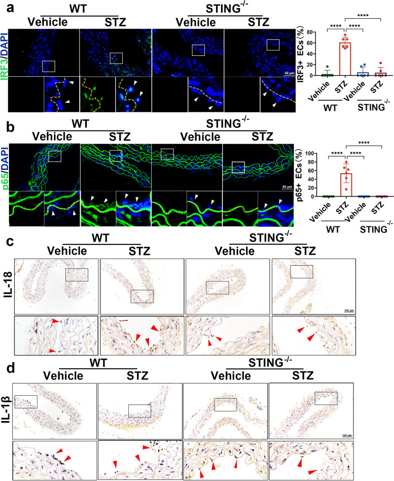
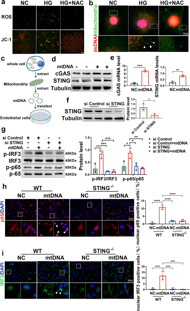
Hyperglycaemia-induced endothelial dysfunction is a key factor in the pathogenesis of diabetic microangiopathy and macroangiopathy. STING, which is a newly discovered regulator of innate immunity, has also been reported to play an important role in various metabolic diseases. However, the role of STING in diabetes-induced endothelial cell dysfunction is unknown. In this study, we established a diabetic macroangiopathy mouse model by streptozotocin (STZ) injection combined with high-fat diet (HFD) feeding and a glucotoxicity cell model in high glucose (HG)-treated rat aortic endothelial cells (RAECs). We found that STING expression was specifically increased in the endothelial cells of diabetic arteries, as well as in HG-treated RAECs. Moreover, genetic deletion of STING significantly ameliorated diabetes-induced endothelial cell dysfunction and apoptosis in vivo. Likewise, STING inhibition by C-176 reversed HG-induced migration dysfunction and apoptosis in RAECs, whereas STING activation by DMXAA resulted in migration dysfunction and apoptosis. Mechanistically, hyperglycaemia-induced oxidative stress promoted endothelial mitochondrial dysfunction and mtDNA release, which subsequently activated the cGAS-STING system and the cGAS-STING-dependent IRF3/NF-kB pathway, ultimately resulting in inflammation and apoptosis. In conclusion, our study identified a novel role of STING in diabetes-induced aortic endothelial cell injury and suggested that STING inhibition was a potential new therapeutic strategy for the treatment of diabetic macroangiopathy. Video Abstract.

Keywords: Aortic endothelial cells; Diabetes; Hyperglycemia; STING.


Conflict of interest statement

The authors declare no competing interests.

Figures

Fig. 1
Fig. 1
Expression of STING in the diabetic vasculature and high glucose-treated RAECs. a The mRNA levels of STING in the leukocytes from healthy controls (n = 6) and patients diagnosed with type 2 diabetes with macrovascular complications (DMCs; n = 6) based on the GSE160016 dataset. b Western blot analysis of STING protein levels in the aortas of vehicle- or STZ-injected mice. c qRT–PCR showing STING mRNA levels in the aortas of vehicle- and STZ-injected mice. d Representative images showing immunofluorescence staining of CD31 (green) and STING (red) in the aortas of vehicle- and STZ-injected mice. Bars: 50 μm. e Western blot analysis of STING protein levels in RAECs treated with HG (40 mmol/L). f qRT–PCR analysis showed the mRNA levels of STING in RAECs treated with HG (40 mmol/L). g Representative images of immunofluorescence staining of cGAS (green) in the aortas of vehicle- and STZ-injected mice. Bars: 50 μm. h Western blot analysis of cGAS protein levels in RAECs treated with HG (40 mmol/L). i qRT–PCR analysis of the mRNA levels of cGAS in RAECs treated with HG (40 mmol/L). j cGAMP levels were increased in the aortic endothelium of diabetic mice. k cGAMP levels were increased in HG-treated RAECs. The results are representative of three independent experiments. The data are presented as the mean ± SD. ∗P < 0.05; ∗∗P < 0.01; ∗∗∗P < 0.001
Fig. 2
Fig. 2
Suppressing STING ameliorates aortic endothelial cell injury in diabetes. a Representative Western blot analysis of STING expression levels in the aortas of WT and STING−/− mice. b Schematic representation of the diabetic mouse model induced by a high-fat diet combined with STZ. c Representative immunofluorescence staining of STING in the aortic endothelium of the indicated mice. The yellow dashed line depicts the aorta endothelium, and the arrow indicates endothelial cells. Bars: 50 μm. Representative images of aortic sections stained with H&E d, VEGF e and TUNEL f. The yellow dotted line indicates the vascular endodermis, and the arrow points to endothelial cells. Bars: 50 μm. The results are representative of three independent experiments. The data are presented as the mean ± SD. ∗P < 0.05; ∗∗P < 0.01; ∗∗∗P < 0.001
Fig. 3
Fig. 3
STING mediates high glucose-induced aortic endothelial cell dysfunction in vitro. a Representative Western blot analysis of VEGF and CD31 in RAECs treated with HG (40 mmol/L) and DMXAA (50 μg/ml). b qRT–PCR analysis of the expression of VEGF and CD31 in RAECs treated with HG and DMXAA. c-d RAECs were treated with HG in the absence or presence of C176 (5 nmol/L) for 72 h. Western blotting c and qRT–PCR d were performed to detect VEGF and CD31. e TUNEL staining showing the apoptotic levels of RAECs treated with HG and DMXAA. Bars: 100 μm. f TUNEL staining showing the apoptotic levels of HG-treated RAECs in the absence or presence of C176. Bars: 100 μm. g Representative images and quantification of migration of RAECs treated with HG and DMXAA. Bars: 50 μm. h Representative images and quantification of the migration of RAECs stimulated by HG in the absence or presence of C176. Bars: 50 μm. The results are representative of three independent experiments. The data are presented as the mean ± SD. ∗P < 0.05; ∗∗P < 0.01; ∗∗∗P < 0.001
Fig. 4
Fig. 4
Suppressing STING inhibits diabetes-induced inflammatory activation in aortic endothelial cells. a-b Representative immunofluorescence images and quantification of IRF3 a and p65 b in the aortic endothelium of mice in the indicated groups. The yellow dotted line indicates the aortic endothelium, and the arrow indicates endothelial cells. c-d Representative immunohistochemical staining of IL-18 and IL-1β in the aortas of mice in the indicated groups. The arrows indicate endothelial cells. The results are representative of three independent experiments. The data are presented as the mean ± SD. ∗P < 0.05; ∗∗P < 0.01; ∗∗∗P < 0.001
Fig. 5
Fig. 5
Suppressing STING inhibits HG-induced inflammatory activation in vitro. a Representative Western blot analysis and density quantification of phosphorylated IRF3, total IRF3, phosphorylated p65, total p65, IL-1β, and IL-18 in RAECs treated with HG and DMXAA. b RAECs were treated with HG in the absence or presence of C176 for 72 h. Western blot analysis and density quantification were performed to detect phosphorylated IRF3, total IRF3, phosphorylated p65, total p65, IL-1β and IL-18 in the indicated groups. c Schematic representation of primary aortic endothelial cell extraction. d-e Immunofluorescence staining showing the location of IRF3 and p65 in HG-treated primary aortic endothelial cells from WT and STING−/− mice. Bars: 50 μm. The results are representative of three independent experiments. The data are presented as the mean ± SD. ∗P < 0.05; ∗∗P < 0.01; ∗∗∗P < 0.001
Fig. 6
Fig. 6
HG-induced mtDNA release activates the STING signalling pathway in aortic endothelial cells. a The DCFH-DA probe was used to measure ROS levels in HG-induced primary aortic endothelial cells with or without NAC treatment (5 mmol/l) for 2 h. Bars: 50 μm. JC-1 staining revealed the mitochondrial membrane potential of HG-induced primary aortic endothelial cells with or without NAC treatment. Bars: 50 μm. b A coimmunostaining assay was used to detect the location of dsDNA (red) and mitofilin (green) in the cytoplasm of primary endothelial cells stimulated by high glucose with or without NAC treatment. Bars: 10 μm. c Flow chart of mtDNA extraction and transfection. d-e Western blotting and qRT–PCR analysis of the protein and mRNA levels of cGAS and STING in RAECs 24 h after transfection with 3 μg of mtDNA. f Representative Western blot showing STING in RAECs transfected with control siRNA or STING siRNA. g Western blot and density quantification showing the protein levels of phosphorylated IRF3, total IRF3, phosphorylated p65 and total p65 in RAECs transfected with control or STING siRNA in the absence or presence of mtDNA. h-i Immunofluorescence staining showing the location of IRF3 and p65 in mtDNA-treated primary aortic endothelial cells from WT and STING−/− mice. Bars: 50 μm. The results are representative of three independent experiments. The data are presented as the mean ± SD. ∗P < 0.05; ∗∗P < 0.01; ∗∗∗P < 0.001
Fig. 7
Fig. 7
Schematic illustration of the mechanism by which hyperglycaemia-induced activation of the cGAS-STING signalling pathway promotes aortic endothelial cell injury in diabetic macrovascular complications. Hyperglycaemia-induced mtROS production can cause mtDNA leakage into the cytoplasm, which is recognized by cGAS and subsequently activates STING by shifting from the endoplasmic reticulum to the Golgi apparatus. Activated STING promotes the phosphorylation and nuclear transfer of IRF3 and p65, which ultimately leads to inflammatory reactions and apoptosis in aortic endothelial cells in the context of diabetes

References

    1. Sun H, Saeedi P, Karuranga S, Pinkepank M, Ogurtsova K, Duncan BB, Stein C, Basit A, Chan JCN, Mbanya JC, Pavkov ME, Ramachandaran A, Wild SH, James S, Herman WH, Zhang P, Bommer C, Kuo S, Boyko EJ, Magliano DJ. Idf diabetes atlas: global, regional and country-level diabetes prevalence estimates for 2021 and projections for 2045. Diabetes Res Clin Pract. 2022;183:109119. doi: 10.1016/j.diabres.2021.109119. - DOI - PMC - PubMed
    1. Silvestre JS, Levy BI. Molecular basis of Angiopathy in diabetes mellitus. Circ Res. 2006;98(1):4–6. doi: 10.1161/01.RES.0000200396.90220.41. - DOI - PubMed
    1. Paul S, Ali A, Katare R. Molecular complexities underlying the vascular complications of diabetes mellitus - a comprehensive review. J Diabetes Complicat. 2020;34(8):107613. doi: 10.1016/j.jdiacomp.2020.107613. - DOI - PubMed
    1. Huynh DTN, Heo KS. Therapeutic targets for endothelial dysfunction in vascular diseases. Arch Pharm Res. 2019;42(10):848–861. doi: 10.1007/s12272-019-01180-7. - DOI - PubMed
    1. Sena CM, Pereira AM, Seica R. Endothelial dysfunction - a major mediator of diabetic vascular disease. Biochim Biophys Acta. 2013;1832(12):2216–2231. doi: 10.1016/j.bbadis.2013.08.006. - DOI - PubMed

Publication types

Substances