. 2023 Dec 21;21(1):366.
doi: 10.1186/s12964-023-01395-8.

Schistosome egg-derived extracellular vesicles deliver Sja-miR-71a inhibits host macrophage and neutrophil extracellular traps via targeting Sema4D

Affiliations

Schistosome egg-derived extracellular vesicles deliver Sja-miR-71a inhibits host macrophage and neutrophil extracellular traps via targeting Sema4D

Yao Liao et al. Cell Commun Signal. .

Abstract

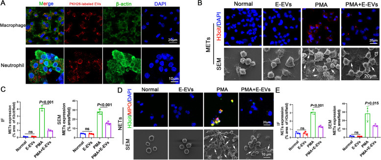
Background: Macrophages and neutrophils are rapidly recruited around Schistosome eggs to form granulomas. Extracellular traps (ETs) of macrophages and neutrophils are part of the pathogen clearance armamentarium of leukocytes. Schistosome eggs possess the ability to resist attack by the host's immune cells and survive by employing various immune evasion mechanisms, including the release of extracellular vesicles (EVs). However, the specific mechanisms by which Schistosome egg-derived EVs (E-EVs) evade the immune response and resist attack from macrophage and neutrophil ETs remain poorly understood. In this study, we aimed to investigate the association between E-EVs and macrophage/neutrophil ETs.

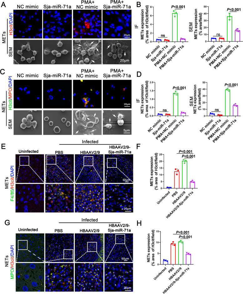
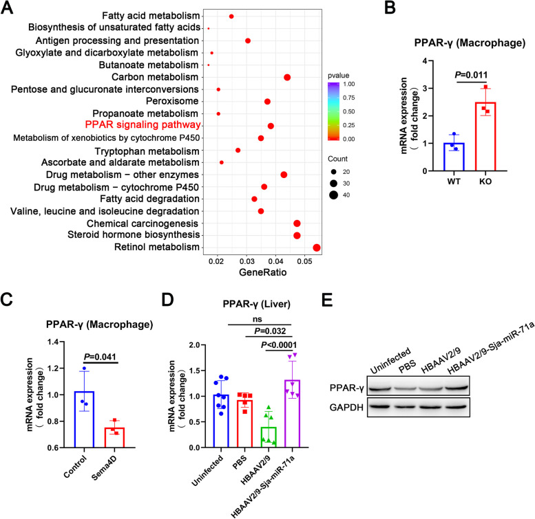
Methods: EVs were isolated from the culture supernatant of S. japonicum eggs and treated macrophages and neutrophils with E-EVs and Sja-miR-71a. The formation of ETs was then observed. Additionally, we infected mice with S. japonicum, administered HBAAV2/9-Sja-miR-71a, and the formation of macrophage ETs (METs) and neutrophil ETs (NETs) in the livers was measured. Sema4D-knockout mice, RNA sequencing, and trans-well assay were used to clarify Sja-miR-71a in E-EVs inhibits METs and NETs formation via the Sema4D/ PPAR-γ/ IL-10 axis.

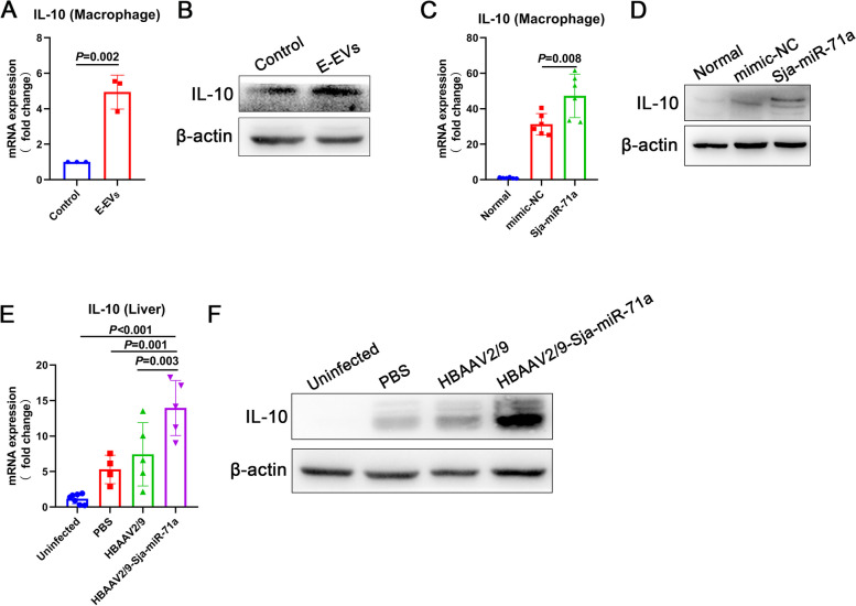
Results: Our findings revealed that E-EVs were internalized by macrophages and neutrophils, leading to the inhibition of METs and NETs formation. The highly expressed Sja-miR-71a in E-EVs targeted Sema4D, resulting in the up-regulation of IL-10 and subsequent inhibition of METs and NETs formation. Sema4D knockout up-regulated IL-10 expression and inhibited the formation of METs and NETs. Furthermore, we further demonstrated that Sja-miR-71a inhibits METs and NETs formation via the Sema4D/ PPAR-γ/ IL-10 axis.

Conclusions: In summary, our findings provide new insights into the immune evasion abilities of Schistosome eggs by demonstrating their ability to inhibit the formation of METs and NETs through the secretion of EVs. This study enhances our understanding of the host-pathogen interaction and may have implications for the development of novel therapeutic approaches. Video Abstract.

Keywords: Extracellular traps; Extracellular vesicles; Schistosoma japonicum eggs; Sema4D; Sja-miR-71a.


Conflict of interest statement

The authors declare no competing interests.

Figures

Fig. 1
Fig. 1
S. japonicum egg-derived extracellular vesicles (E-EVs) were internalized by macrophages and neutrophils and inhibited METs and NETs formation. A Macrophages and neutrophils were incubated with PKH26-labeled E-EVs, and E-EVs internalization was examined using laser scanning confocal microscopy. B-E Macrophages and neutrophils were treated with E-EVs (10 μg/mL, 24 h), PMA (500 nM, 5 h), or PMA (500 nM, 5 h) + E-EVs (10 μg/mL, 24 h). Macrophage extracellular traps (METs) and neutrophil extracellular traps (NETs) were observed using immunofluorescence (IF) and scanning electron microscopy (SEM) (B and D) and were quantified (C and E). METs were detected on IF based on H3cit. NETs were detected based on H3cit and MPO co-localization, Arrows: METs or NETs. IF was quantified based on area the of H3cit, and SEM was quantified based on the area of METs and NETs. e, g, representative of n = 3 independent experiments
Fig. 2
Fig. 2
Sja-miR-71a in E-EVs inhibits METs and NETs formation. A-D Macrophages and neutrophils were treated with normal control (NC) mimic (50 nM, 24 h), Sja-miR-71a (50 nM, 24 h), PMA (500 nM, 5 h) + NC mimic (50 nM, 24 h), or PMA (500 nM, 5 h) + Sja-miR-71a (50 nM, 24 h). METs and NETs were observed using IF and SEM (A and C), and quantified (B and D). (E-H) METs (E) and NETs (G) in liver sections were detected and quantified (F and H). Arrows: METs or NETs. B, D, F, H representative of n = 3 independent experiments
Fig. 3
Fig. 3
E-EVs and Sja-miR-71a up-regulate IL-10. A and B Macrophages were treated with E-EVs, and expression levels of IL-10 were analyzed with qRT-PCR and western blot. C and D Macrophages were treated with normal control (NC) mimic (50 nM, 24 h) and Sja-miR-71a (50 nM, 24 h). IL-10 expression was analyzed with qRT-PCR and western blot. E and F HBAAV2/9-Sja-miR-71a recombinant adeno-associated virus (rAAV) was constructed. Mice infected with S. japonicum were administered HBAAV2/9-Sja-miR-71a. Liver IL-10 expression was analyzed with qRT-PCR and western blot. A, representative of n = 3 independent experiments; C, representative of n = 6 independent experiments; E, n = 4-7 per group
Fig. 4
Fig. 4
Sja-miR-71a in E-EVs targets Sema4D to up-regulate IL-10, thereby inhibiting METs and NETs formation. A Macrophages and neutrophils were isolated and treated with E-EVs, Sema4D expression was analyzed using qRT-PCR. B The expression of Sema4D in macrophages and neutrophils of the liver was detected by IF. C Macrophages and neutrophils were treated with Sja-miR-71a, Sema4D expression was analyzed using qRT-PCR. D and E IL-10 expression in Sema4D deletion (KO) and WT macrophages was analyzed using qRT-PCR and western blot. F and G IL-10 expression in the livers of Sema4D knockout (Sema4D-KO) and WT mice infected with S. japonicum were analyzed using qRT-PCR and western blot. H Macrophages were treated with recombinant Sema4D (10 μg/ml, 24 h), and the level of IL-10 was measured using western blot. I and G Sema4D-KO and WT macrophages and neutrophils were treated with PMA (500 nM, 5 h) or had no treatment at all. METs and NETs were observed using IF and SEM and quantified. K and L Sema4D-KO and WT macrophages and neutrophils were treated with recombinant Sema4D (10 μg/ml, 24 h), PMA (500 nM, 5 h), or PMA (500 nM, 5 h) + Sema4D (10 μg/ml, 24 h), METs and NETs were observed using IF and SEM and quantified. M Macrophages and neutrophils were treated with PMA (500 nM, 5 h) + NC mimic (50 nM, 24 h), PMA (500 nM, 5 h) + Sja-miR-71a (50 nM, 24 h), or PMA (500 nM, 5 h) + Sja-miR-71a (50 nM, 24 h) + IL-10 antibodies (15 μg/ml, 24 h), METs and NETs were observed using IF. Arrows: METs or NETs. A and C, n = 4-6 per group; D, n = 5 per group; F, n = 6 per group; I-L representative of n = 3 independent experiments
Fig. 5
Fig. 5
Sja-miR-71a increases the expression of PPAR-γ. A KEGG was used to analyze the up-regulated signaling pathway in S. japonicum-infected Sema4D-KO compared with S. japonicum-infected WT mice. B Expression levels of PPAR-γ in Sema4D-KO and WT macrophages were analyzed using qRT-PCR. C The PPAR-γ expression of macrophages treated with recombinant Sema4D (10 μg/ml, 24 h) was determined using qRT-PCR. D and E PPAR-γ expression in the livers of mice infected with S. japonicum was analyzed using qRT-PCR and western blot. D, n = 5-8 per group; B, C, representative of n = 3 independent experiments
Fig. 6
Fig. 6
Sja-miR-71a inhibits METs and NETs formation via the Sema4D/ PPAR-γ/ IL-10 axis. A-H Macrophages and neutrophils were treated with a PPAR-γ agonist (30μΜ, 24 h), PPAR-γ antagonist (20μΜ, 24 h), PMA (500 nM, 5 h), PMA (500 nM, 5 h) + PPAR-γ agonist (30μΜ, 24 h), or a PMA (500 nM, 5 h) + PPAR-γ antagonist (20μΜ, 24 h). METs and NETs were observed (A, C, E and G), and quantified using IF and SEM (B, D, F, and H). I and J Macrophages and neutrophils were treated with a PPAR-γ agonist (30μΜ, 24 h) AND PPAR-γ antagonist (20μΜ, 24 h) (DMSO as control), then co-cultured for 24 h via trans-well, NETs were observed using IF and SEM (I) and quantified (J). K and L Macrophages and neutrophils were treated with a PPAR-γ agonist (30μΜ, 24 h) AND PPAR-γ antagonist (20μΜ, 24 h) (DMSO as control), then co-cultured for 24 h via trans-well. Neutrophils were treated with PMA (500 nM) for the last 5 hours, NETs were observed using IF and SEM (K) and quantified (L). M Macrophages and neutrophils were treated with PMA, PPAR-γ antagonist, and Sja-miR-71a simultaneously, METs and NETs were observed using IF. Arrows: METs or NETs. Representative of n = 3 independent experiments

References

    1. Lo NC, Bezerra FSM, Colley DG, et al. Review of 2022 WHO guidelines on the control and elimination of schistosomiasis. Lancet Infect Dis. 2022;22(11):e327–ee35. doi: 10.1016/S1473-3099(22)00221-3. - DOI - PubMed
    1. Global, regional, and national disability-adjusted life-years (DALYs) for 359 diseases and injuries and healthy life expectancy (HALE) for 195 countries and territories, 1990-2017: a systematic analysis for the global burden of disease study 2017. Lancet. 2018;392(10159):1859–922. - PMC - PubMed
    1. Wiedemann M, Voehringer D. Immunomodulation and immune escape strategies of gastrointestinal helminths and Schistosomes. Front Immunol. 2020;11:572865. doi: 10.3389/fimmu.2020.572865. - DOI - PMC - PubMed
    1. Smith P, Fallon RE, Mangan NE, et al. Schistosoma mansoni secretes a chemokine binding protein with antiinflammatory activity. J Exp Med. 2005;202(10):1319–1325. doi: 10.1084/jem.20050955. - DOI - PMC - PubMed
    1. Everts B, Perona-Wright G, Smits HH, et al. Omega-1, a glycoprotein secreted by Schistosoma mansoni eggs, drives Th2 responses. J Exp Med. 2009;206(8):1673–1680. doi: 10.1084/jem.20082460. - DOI - PMC - PubMed

Publication types

LinkOut - more resources