. 2023 Dec 21;20(1):308.
doi: 10.1186/s12974-023-02998-1.

Succinate-induced macrophage polarization and RBP4 secretion promote vascular sprouting in ocular neovascularization

Affiliations

Succinate-induced macrophage polarization and RBP4 secretion promote vascular sprouting in ocular neovascularization

Tianyi Shen et al. J Neuroinflammation. .

Abstract

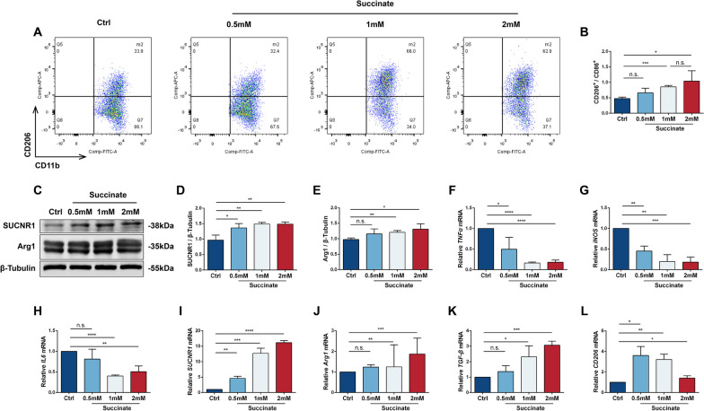
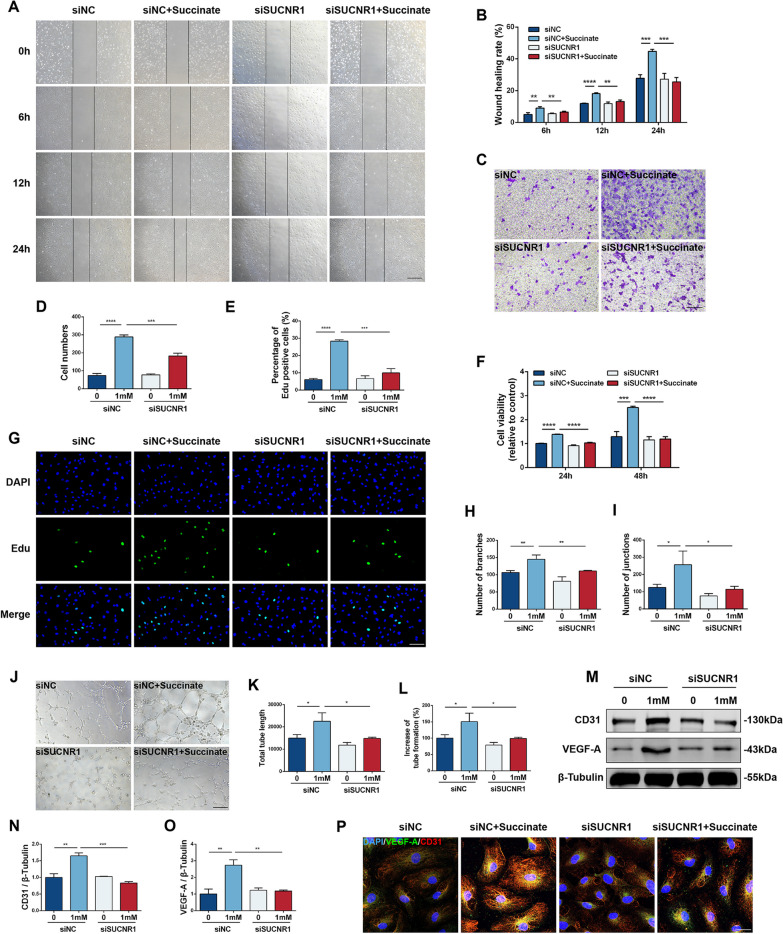
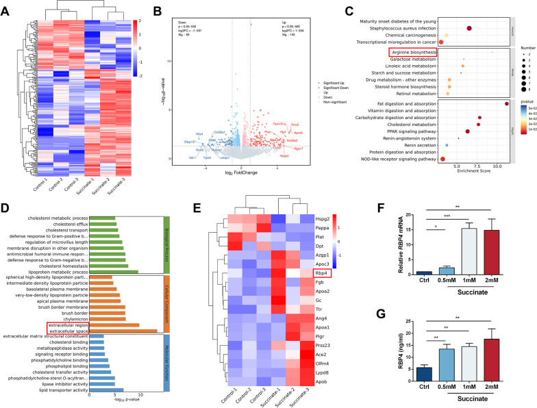
Pathological neovascularization is a pivotal biological process in wet age-related macular degeneration (AMD), retinopathy of prematurity (ROP) and proliferative diabetic retinopathy (PDR), in which macrophages (Mφs) play a key role. Tip cell specialization is critical in angiogenesis; however, its interconnection with the surrounding immune environment remains unclear. Succinate is an intermediate in the tricarboxylic acid (TCA) cycle and was significantly elevated in patients with wet AMD by metabolomics. Advanced experiments revealed that SUCNR1 expression in Mφ and M2 polarization was detected in abnormal vessels of choroidal neovascularization (CNV) and oxygen-induced retinopathy (OIR) models. Succinate-induced M2 polarization via SUCNR1, which facilitated vascular endothelial cell (EC) migration, invasion, and tubulation, thus promoting angiogenesis in pathological neovascularization. Furthermore, evidence indicated that succinate triggered the release of RBP4 from Mφs into the surroundings to regulate endothelial sprouting and pathological angiogenesis via VEGFR2, a marker of tip cell formation. In conclusion, our results suggest that succinate represents a novel class of vasculature-inducing factors that modulate Mφ polarization and the RBP4/VEGFR2 pathway to induce pathological angiogenic signaling through tip cell specialization.

Keywords: Choroidal neovascularization; Macrophage; Oxygen-induced retinopathy; Succinate; Tip cells.


Conflict of interest statement

The authors declared no competing or financial interests.

Figures

Fig. 1
Fig. 1
Metabolic analysis between the CAT group and wAMD group. A Heatmap analysis between the two groups. B Metabonomic pathway analysis. C Different metabolites in the TCA cycle according to the KEGG database. D Display of meaningful metabolites in the TCA cycle. Red represents elevated expression, and blue represents decreased expression. EL Specific concentrations of pyruvate, citrate, cis-aconitate, isocitrate, α-ketoglutarate, succinate, l-malate and fumarate (n = 5). *P < 0.05, **P < 0.01, ***P < 0.001
Fig. 2
Fig. 2
Relative protein expression in animal CNV and OIR models. A, B Choroidal IB4 staining and areas of CNV in control and on days 3, 7 and 14 after Matrigel induction. Scale bar: 200 μm (n = 3). C, E Relative expression of SUCNR1 and Arg1 in control and CNV animals (n = 3). F Immunofluorescence staining of DAPI (blue), Arg1 (green) and SUCNR1 (red) in control and CNV eyes. Scale bar: 100 μm. G, H Immunofluorescence staining of IB4, SUCNR1 and Arg1 in the retinas of control and OIR models. Scale bar: 200 μm. *P < 0.05, **P < 0.01, ***P < 0.001
Fig. 3
Fig. 3
Mφ phenotypes after induction with different concentrations of succinate (0, 0.5 mM, 1 mM and 2 mM for 48 h). A Flow cytometry analysis of Mφs after treatment with different concentrations of succinate. B The ratio of CD206+/CD86+ cells in Mφs in the four groups (n = 3). CE Relative protein levels of SUCNR1 and Arg1 in the four groups (n = 3). FL Relative RNA expression of TNFα, iNOS, IL6, SUCNR1, Arg1, TGF-β and CD206 (n = 3). *P < 0.05, **P < 0.01, ***P < 0.001, ****P < 0.0001
Fig. 4
Fig. 4
Effect of SUCNR1 inhibition on the Mφ phenotype. Mφs were treated with siRNAs and 1 mM succinate for 48 h and divided into four groups: siNC, siNC + succinate, siSUCNR1 and siSUCNR1 + succinate. A Relative RNA expression of iNOS, TNFα, IL6, TGF-β, CD206 and Arg1 in the four groups (n = 3). BG Relative protein levels of SUCNR1, TGF-β, Arg1, IL6 and TNFα in the four groups (n = 3). H Immunofluorescence staining of Mφs with DAPI (blue), TGF-β (green) and Arg1 (red). Scale bar: 50 μm. I, J Relative fluorescence intensity of TGF-β and Arg1 (n = 3). K Flow cytometry analysis of Mφs in the four groups. L The ratio of CD206+/CD86+ cells in Mφs (n = 3). *P < 0.05, **P < 0.01, ***P < 0.001, ****P < 0.0001
Fig. 5
Fig. 5
Migration, proliferation and angiogenesis ability of HUVECs co-cultured with four groups of Mφs (siNC, siNC + succinate, siSUCNR1 and siSUCNR1 + succinate). A, B Images were measured at 0 h, 6 h, 12 h and 24 h in the scratch wound healing test. The cell migration rate was used to indicate migratory ability as described in the article. Scale bar: 200 μm (n = 3). C, D In the Transwell assay, ImageJ software was used to calculate the stained cells in the four groups. Scale bar: 200 μm (n = 3). E, G EdU assays revealed the proliferation of HUVECs, and the percentage of EdU-positive cells in the four groups was calculated. Scale bar: 200 μm (n = 3). F Cell viability was tested by CCK8 assay, and the OD at 450 nm was recorded to calculate cell viability at 24 h and 48 h (n = 3). J Tubular formation of four groups was recorded after being cultured on Matrigel for 6 h. Scale bar: 200 μm. The number of branches (H), junctions (I), total tube length (K) and increase of tube formation (%) (L) were qualified by ImageJ software (n = 3). MO Relative protein levels of CD31 and VEGF-A in the four groups (n = 3). P Immunofluorescence staining for DAPI (blue), VEGF-A (green) and CD31 (red) in HUVECs. Scale bar: 50 μm. *P < 0.05, **P < 0.01, ***P < 0.001, ****P < 0.0001
Fig. 6
Fig. 6
Knockdown of SUCNR1 reduced the severity of succinate-induced CNV and RNV in animals. C57 mice were randomly divided into four groups: control, CNV + shNC, CNV + shNC + succinate, and CNV + shSUCNR1 + succinate. (A, top row) B Choroidal IB4 staining and areas of CNV in four groups on day 7 after Matrigel induction. Scale bar: 200 μm (n = 3). (A, second row) H&E-stained sections of eyeballs. Scale bar: 100 μm. (A, third and fourth row) FA images of four groups. C, D Waveform and amplitude of ERGs in each group (n = 3). EJ Protein levels of SUCNR1, TGF-β, Arg1, IL6 and TNFα in animals (n = 3). K The Arg1/IL6 protein level ratio (n = 3). LN Retinal IB4 staining and size of neovascular area as well as avascular area in OIR models. Scale bar: 400 μm (n = 3). *P < 0.05, **P < 0.01, ***P < 0.001, ****P < 0.0001
Fig. 7
Fig. 7
Analysis of gene expression profiles between control and succinate (1 mM for 48 h) groups in Mφs. A Heatmap of different genes in the two groups. B Log2-fold change for succinate treatment compared to the control. C KEGG pathway enrichment analysis of the top 20 differentially expressed pathways. D GO analysis of the top 10 differentially expressed pathways in the BP, CC and MF categories. E Heatmap of significantly different secreted genes. F Relative mRNA expression of RBP4 in succinate-treated Mφs (n = 3). G RBP4 concentration in Mφ supernatant after succinate intervention (n = 3). *P < 0.05, **P < 0.01, ***P < 0.001
Fig. 8
Fig. 8
Effect of RBP4 on sprouting in vivo and in vitro. HUVECs were treated with 10 ng/ml RBP4 for 48 h, with or without 1 μM Ki8751 pretreatment for 12 h. A Relative mRNA expression of ANGPT2, TIE1, HEY1 and DLL4 in RBP4-treated HUVECs (n = 3). B Relative mRNA expression of ANGPT2, TIE1, HEY1 and DLL4 in the control, RBP4 and RBP4 + Ki8751 groups (n = 3). C, D Protein levels of VEGFR2 in the three groups (n = 3). E Immunofluorescence staining of DAPI (blue) and VEGFR2 (red) in HUVECs. Scale bar: 50 μm. F Co-IP examination of VEGFR2-RBP4 interaction. H, J, K Retinal IB4 staining of P14 in OIR and relative number of tip cells as well as filopodia. Scale bar: 200 μm (n = 3). G, I Choroidal sprouting analysis in three groups (n = 3). Scale bar: 500 μm. *P < 0.05, **P < 0.01, ***P < 0.001, ****P < 0.0001

References

    1. Liu Z-L, Chen H-H, Zheng L-L, Sun L-P, Shi L. Angiogenic signaling pathways and anti-angiogenic therapy for cancer. Signal Transduct Target Ther. 2023;8(1):198. doi: 10.1038/s41392-023-01460-1. - DOI - PMC - PubMed
    1. Xia W, Li C, Chen Q, Huang J, Zhao Z, Liu P, et al. Intravenous route to choroidal neovascularization by macrophage-disguised nanocarriers for mTOR modulation. Acta Pharm Sin B. 2022;12(5):2506–2521. doi: 10.1016/j.apsb.2021.10.022. - DOI - PMC - PubMed
    1. Tsutsumi C, Sonoda K-H, Egashira K, Qiao H, Hisatomi T, Nakao S, et al. The critical role of ocular-infiltrating macrophages in the development of choroidal neovascularization. J Leukoc Biol. 2003;74(1):25–32. doi: 10.1189/jlb.0902436. - DOI - PubMed
    1. Cherepanoff S, McMenamin P, Gillies MC, Kettle E, Sarks SH. Bruch's membrane and choroidal macrophages in early and advanced age-related macular degeneration. Br J Ophthalmol. 2010;94(7):918–925. doi: 10.1136/bjo.2009.165563. - DOI - PubMed
    1. Zhou Y, Yoshida S, Nakao S, Yoshimura T, Kobayashi Y, Nakama T, et al. M2 macrophages enhance pathological neovascularization in the mouse model of oxygen-induced retinopathy. Invest Ophthalmol Vis Sci. 2015;56(8):4767–4777. doi: 10.1167/iovs.14-16012. - DOI - PubMed

LinkOut - more resources