

[Research on Runx2 gene induced differentiation of human amniotic mesenchymal stem cells into ligament fibroblasts in vitro and promotion of tendon-bone healing in rabbits]
- PMID: 38130197
- PMCID: PMC10739672
- DOI: 10.7507/1002-1892.202306010
[Research on Runx2 gene induced differentiation of human amniotic mesenchymal stem cells into ligament fibroblasts in vitro and promotion of tendon-bone healing in rabbits]
Abstract
Objective: To investigate whether the Runx2 gene can induce the differentiation of human amniotic mesenchymal stem cells (hAMSCs) to ligament fibroblasts in vitro and promote the tendon-bone healing in rabbits.
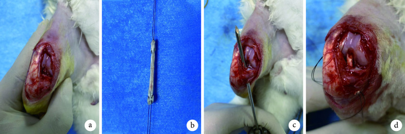
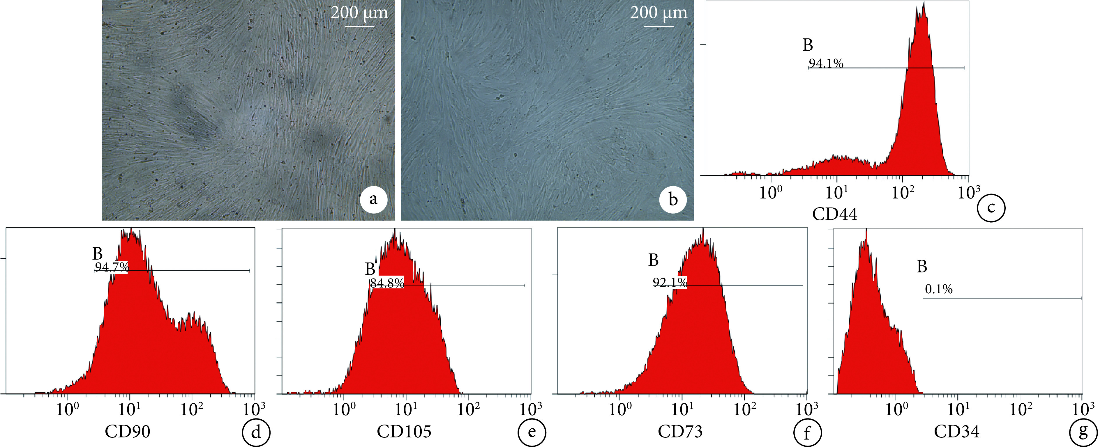
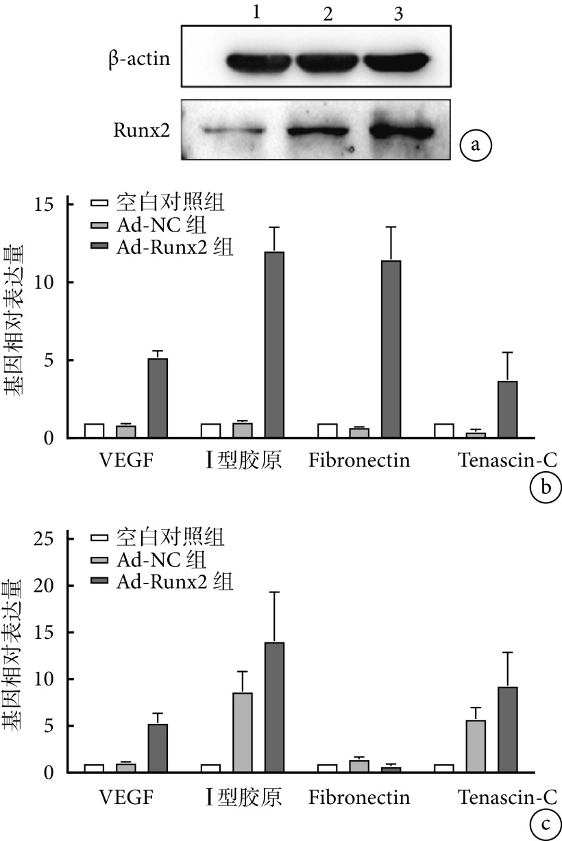
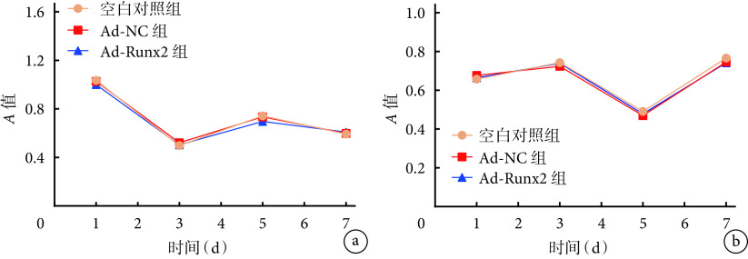
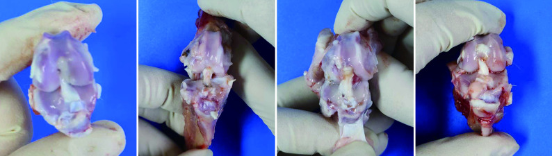
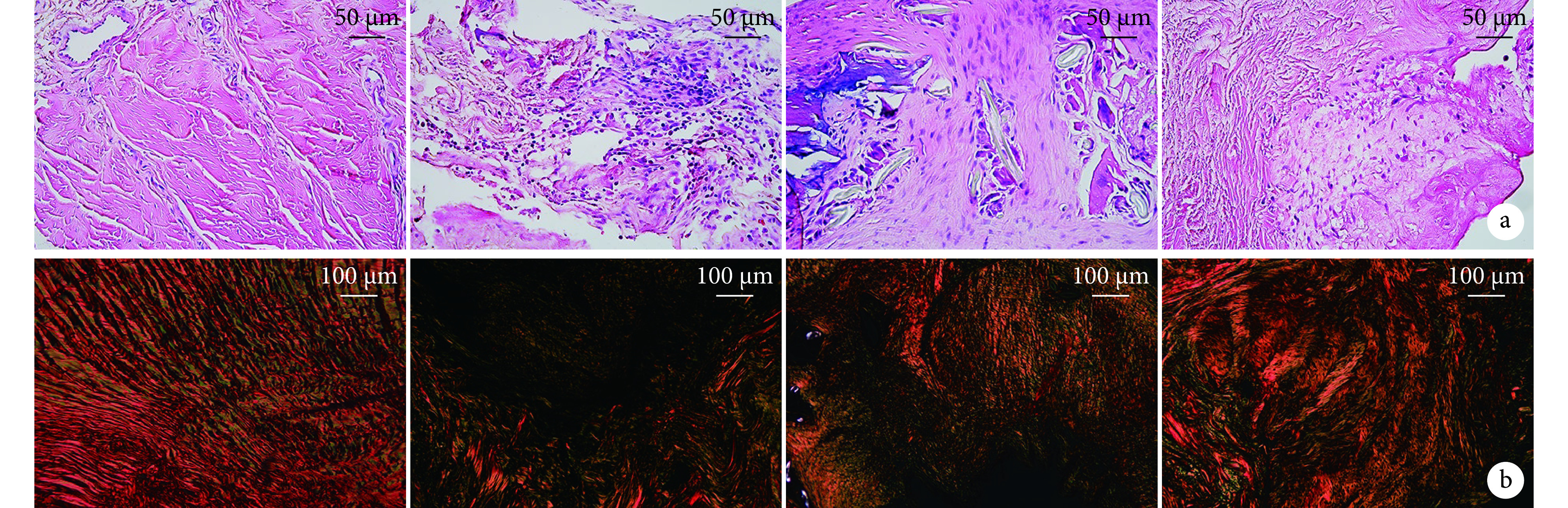
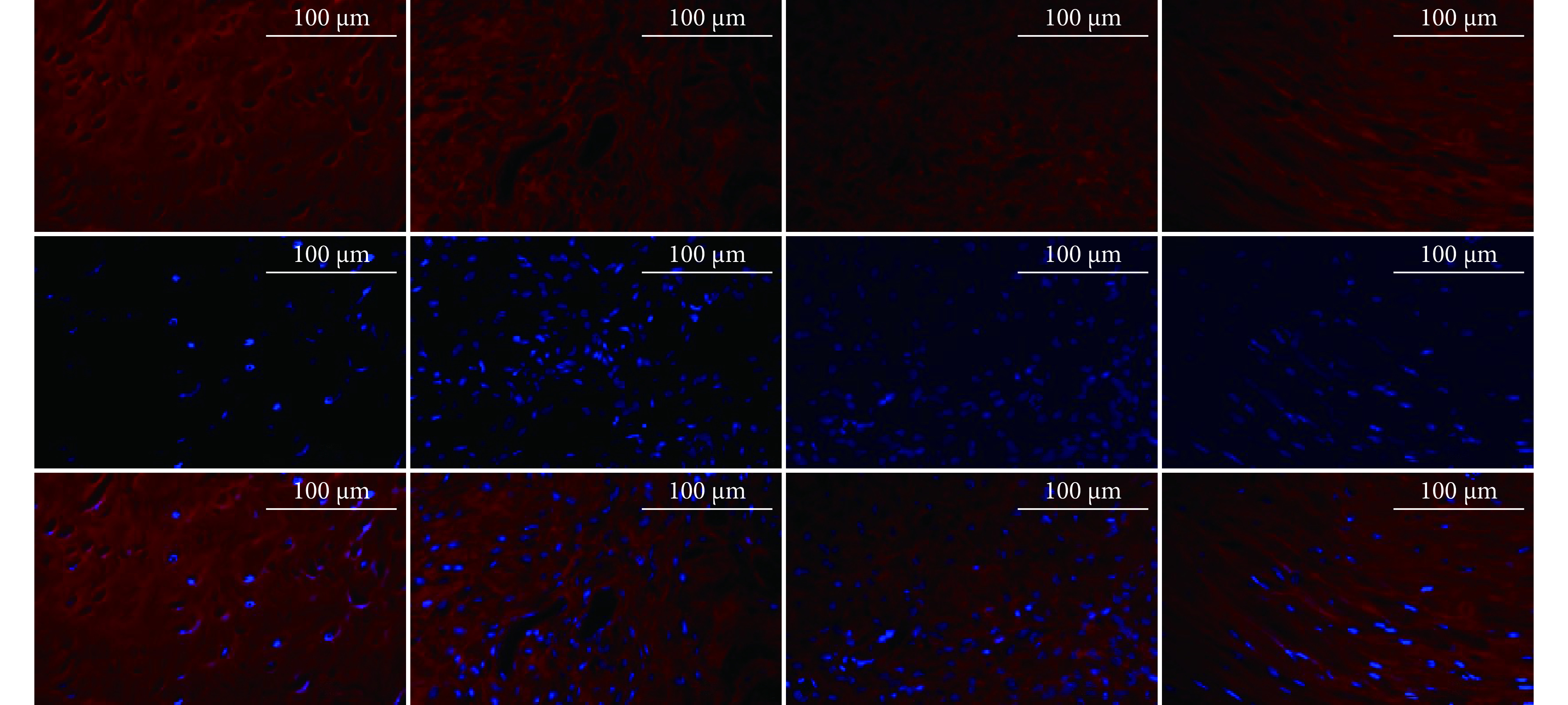
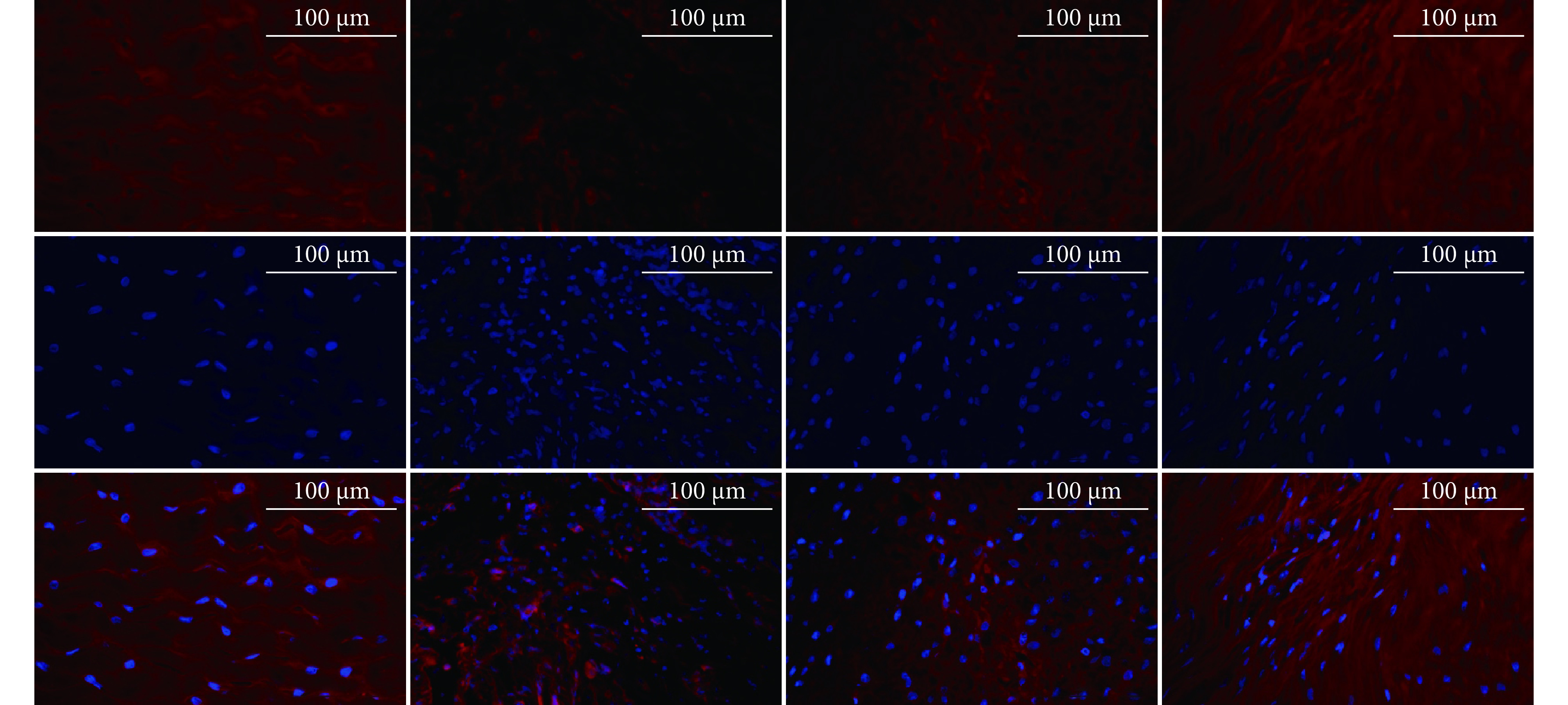
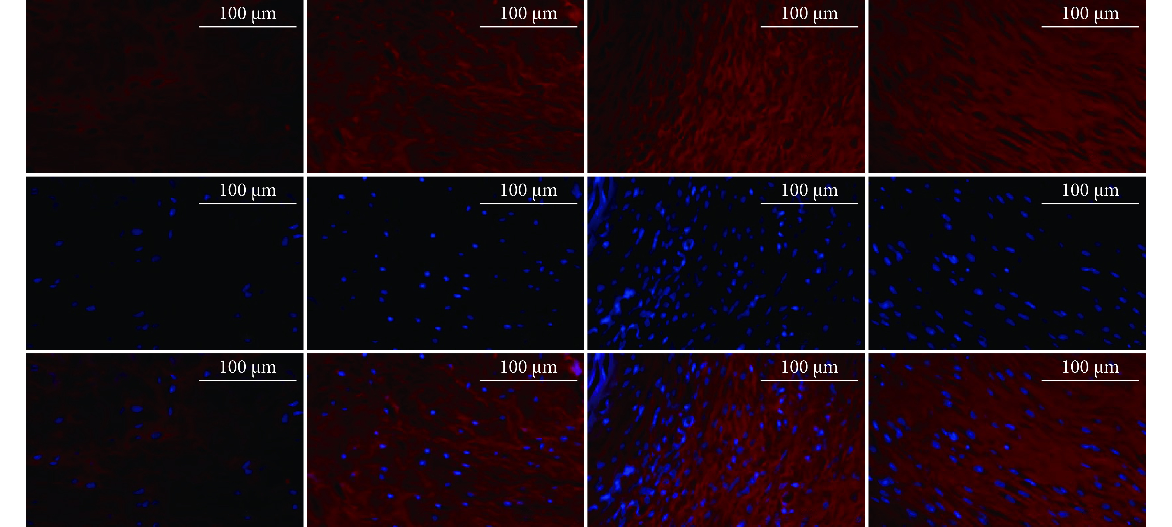
Methods: hAMSCs were isolated from the placentas voluntarily donated from healthy parturients and passaged, and then identified by flow cytometric identification. Adenoviral vectors carrying Runx2 gene (Ad-Runx2) and empty vector adenovirus (Ad-NC) were constructed and viral titer assay; then, the 3rd generation hAMSCs were transfected with Ad-Runx2 (Ad-Runx2 group) or Ad-NC (Ad-NC group). The real-time fluorescence quantitative PCR and Western blot were used to detect Runx2 gene and protein expression to verify the effectiveness of Ad-Runx2 transfection of hAMSCs; and at 3 and 7 days after transfection, real-time fluorescence quantitative PCR was further used to detect the expressions of ligament fibroblast-related genes [vascular endothelial growth factor (VEGF), collagen type Ⅰ, Fibronectin, and Tenascin-C]. The hAMSCs were used as a blank control group. The hAMSCs, hAMSCs transfected with Ad-NC, and hAMSCs were mixed with Matrigel according to the ratio of 1 : 1 and 1 : 2 to construct the cell-scaffold compound. Cell proliferation was detected by cell counting kit 8 (CCK-8) assay, and the corresponding cell-scaffold compound with better proliferation were taken for subsequent animal experiments. Twelve New Zealand white rabbits were randomly divided into 4 groups of sham operation group (Sham group), anterior cruciate ligament reconstruction group (ACLR group), anterior cruciate ligament reconstruction+hAMSCs transfected with Ad-NC-scaffold compound group (Ad-NC group), and anterior cruciate ligament reconstruction+hAMSCs transfected with Ad-Runx2-scaffold compound group (Ad-Runx2 group), with 3 rabbits in each group. After preparing the ACL reconstruction model, the Ad-NC group and the Ad-Runx2 group injected the optimal hAMSCs-Matrigel compunds into the bone channel correspondingly. The samples were taken for gross, histological (HE staining and sirius red staining), and immunofluorescence staining observation at 1 month after operation to evaluate the inflammatory cell infiltration as well as collagen and Tenascin-C content in the ligament tissues.
Results: Flow cytometric identification of the isolated cells conformed to the phenotypic characteristics of MSCs. The Runx2 gene was successfully transfected into hAMSCs. Compared with the Ad-NC group, the relative expressions of VEGF and collagen type Ⅰ genes in the Ad-Runx2 group significantly increased at 3 and 7 days after transfection ( P<0.05), Fibronectin significantly increased at 3 days ( P<0.05), and Tenascin-C significantly increased at 3 days and decreased at 7 days ( P<0.05). CCK-8 detection showed that there was no significant difference ( P>0.05) in the cell proliferation between groups and between different time points after mixed culture of two ratios. So the cell-scaffold compound constructed in the ratio of 1∶1 was selected for subsequent experiments. Animal experiments showed that at 1 month after operation, the continuity of the grafted tendon was complete in all groups; HE staining showed that the tissue repair in the Ad-Runx2 group was better and there were fewer inflammatory cells when compared with the ACLR group and the Ad-NC group; sirius red staining and immunofluorescence staining showed that the Ad-Runx2 group had more collagen typeⅠ and Ⅲ fibers, tending to form a normal ACL structure. However, the fluorescence intensity of Tenascin-C protein was weakening when compared to the ACLR and Ad-NC groups.
Conclusion: Runx2 gene transfection of hAMSCs induces directed differentiation to ligament fibroblasts and promotes tendon-bone healing in reconstructed anterior cruciate ligament in rabbits.
目的: 探讨Runx2基因是否具有体外诱导人羊膜MSCs(human amniotic MSCs,hAMSCs)向韧带成纤维细胞分化以及在体内能否促进兔前交叉韧带重建后腱-骨愈合。.
方法: 取健康产妇自愿捐赠胎盘分离培养hAMSCs并传代后行流式细胞鉴定。构建携带Runx2基因的腺病毒载体(Ad-Runx2)以及空质粒载体腺病毒(Ad-NC),经病毒滴度测定后分别转染第3代hAMSCs(Ad-Runx2组、Ad-NC组),实时荧光定量PCR及Western blot检测Runx2基因及蛋白表达,验证Ad-Runx2基因转染hAMSCs有效性;然后于转染后培养3、7 d时,进一步采用实时荧光定量PCR检测韧带成纤维细胞相关基因 [VEGF、Ⅰ型胶原、纤连蛋白(Fibronectin)和肌腱蛋白C(Tenascin-C)] 表达;以单纯hAMSCs作为空白对照组。将单纯hAMSCs以及转染Ad-NC、Ad-Runx2的hAMSCs分别与基质胶(Matrigel)按照体积比1∶1和1∶2混合构建复合物,采用细胞计数试剂盒8(cell counting kit 8,CCK-8)检测细胞增殖,取增殖较好的对应复合物进行后续动物实验。将12只新西兰白兔随机分为假手术组(Sham组)、前交叉韧带重建组(ACLR组)、前交叉韧带重建+Ad-NC组(Ad-NC组)、前交叉韧带重建+Ad-Runx2组(Ad-Runx2组)4组,每组3只。制备前交叉韧带重建模型后,Ad-NC组、Ad-Runx2组于骨道内对应注射最佳hAMSCs-Matrigel复合物。术后1个月取材行大体、组织学(HE染色及天狼猩红染色)、免疫荧光染色观察,评价韧带组织中炎症细胞浸润以及Ⅰ/Ⅲ型胶原、Tenascin-C含量。.
结果: 流式细胞鉴定分离培养细胞符合MSCs表型特征。Runx2基因成功转染至hAMSCs;与Ad-NC组相比,Ad-Runx2组转染后VEGF及Ⅰ型胶原基因相对表达量于培养3、7 d时均增高( P<0.05),Fibronectin仅3 d时增高( P<0.05),而Tenascin-C于3 d时增高、7 d时降低( P<0.05)。CCK-8检测示两种比例混合培养后,细胞增殖组间以及组内各时间点间差异均无统计学意义( P>0.05),选择1∶1比例构建复合物进行后续实验。动物实验显示,术后1个月时,大体观察各组移植肌腱连续性完整;HE染色示Ad-Runx2组组织修复情况优于ACLR组和Ad-NC组、炎症细胞更少;天狼猩红染色及免疫荧光染色均示Ad-Runx2组韧带组织的Ⅰ、Ⅲ型胶原纤维增多,趋于形成正常前交叉韧带结构,但Tenascin-C蛋白荧光强度与ACLR组和Ad-NC组相比减弱。.
结论: Runx2基因转染hAMSCs后可诱导其向韧带成纤维细胞定向分化,并促进兔前交叉韧带损伤重建的腱-骨愈合。.
Keywords: Runx2 gene; human amniotic mesenchymal stem cells; ligament fibroblasts; ligament tissue engineering; tenbon-bone healing.
Conflict of interest statement
利益冲突 在课题研究和文章撰写过程中不存在利益冲突;经费支持没有影响文章观点和对研究数据客观结果的统计分析及其报道
Figures
References
-
- 熊波涵, 余洋, 卢晓君, 等 促进前交叉韧带重建腱骨愈合的研究与进展. 中国组织工程研究. 2023;27(5):779–786.
Publication types
MeSH terms
Substances
LinkOut - more resources
Full Text Sources