. 2023 Dec 6:17:1252850.
doi: 10.3389/fnins.2023.1252850. eCollection 2023.

Characterization of dynamic patterns of human fetal to neonatal brain asymmetry with deformation-based morphometry

Affiliations

Characterization of dynamic patterns of human fetal to neonatal brain asymmetry with deformation-based morphometry

Céline Steger et al. Front Neurosci. .

Abstract

Introduction: Despite established knowledge on the morphological and functional asymmetries in the human brain, the understanding of how brain asymmetry patterns change during late fetal to neonatal life remains incomplete. The goal of this study was to characterize the dynamic patterns of inter-hemispheric brain asymmetry over this critically important developmental stage using longitudinally acquired MRI scans.

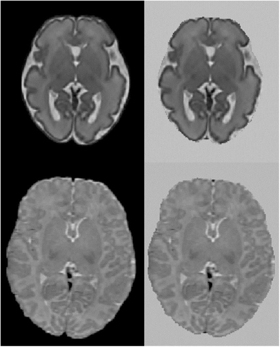
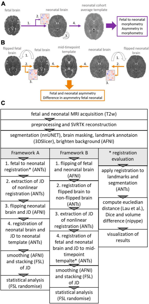
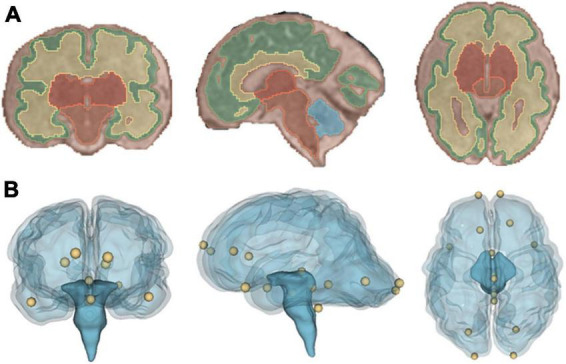
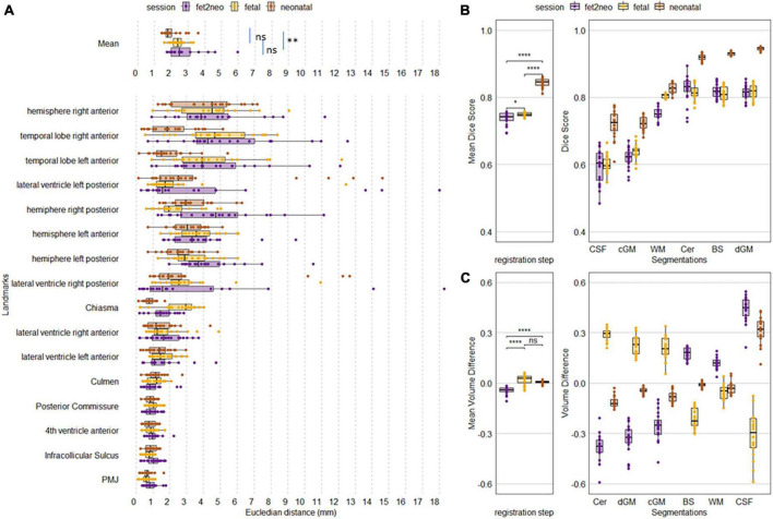
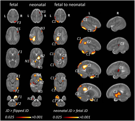
Methods: Super-resolution reconstructed T2-weighted MRI of 20 neurotypically developing participants were used, and for each participant fetal and neonatal MRI was acquired. To quantify brain morphological changes, deformation-based morphometry (DBM) on the longitudinal MRI scans was utilized. Two registration frameworks were evaluated and used in our study: (A) fetal to neonatal image registration and (B) registration through a mid-time template. Developmental changes of cerebral asymmetry were characterized as (A) the inter-hemispheric differences of the Jacobian determinant (JD) of fetal to neonatal morphometry change and the (B) time-dependent change of the JD capturing left-right differences at fetal or neonatal time points. Left-right and fetal-neonatal differences were statistically tested using multivariate linear models, corrected for participants' age and sex and using threshold-free cluster enhancement.

Results: Fetal to neonatal morphometry changes demonstrated asymmetry in the temporal pole, and left-right asymmetry differences between fetal and neonatal timepoints revealed temporal changes in the temporal pole, likely to go from right dominant in fetal to a bilateral morphology in neonatal timepoint. Furthermore, the analysis revealed right-dominant subcortical gray matter in neonates and three clusters of increased JD values in the left hemisphere from fetal to neonatal timepoints.

Discussion: While these findings provide evidence that morphological asymmetry gradually emerges during development, discrepancies between registration frameworks require careful considerations when using DBM for longitudinal data of early brain development.

Keywords: DBM = deformation-based morphometry; brain asymmetry; fetal brain; longitudinal; magnetic resonance imaging (MRI); neonatal brain.


Conflict of interest statement

The authors declare that the research was conducted in the absence of any commercial or financial relationships that could be construed as a potential conflict of interest.

Figures

FIGURE 1
FIGURE 1
Axial view of fetal and neonatal image of a participant. Top: fetal, bottom: neonatal. Left original image, right image background replaced with bright values.
FIGURE 2
FIGURE 2
Overview of the two frameworks used to reveal asymmetry patterns and their temporal dynamics. (A) Framework A (violet), calculating the inter-hemispheric asymmetry of fetal to neonatal regional brain growth maps. (B) Framework B (orange), calculating fetal and neonatal inter-hemispheric asymmetry at the given timepoint, registering these to a mid-timepoint template and then calculating the differences between the inter-hemispheric asymmetry. (C) Flowchart showing steps required to extract JD maps and perform asymmetry analysis for framework A and B and steps required to extract metrics for the registration evaluation. *Indicates registration step was evaluated (see * registration evaluation).
FIGURE 3
FIGURE 3
Visualization of the registration evaluation metrics used in our study. (A) Coronal, sagittal and axial view of segmentation overlaid on a T2-weighted SR image of a fetus. (B) Coronal, sagittal and axial view of a glass brain rendering showing all sixteen, manually placed landmarks that were used during the registration evaluation.
FIGURE 4
FIGURE 4
Achieved metric accuracy for each participant for each registration step. (A) Landmark accuracy as Euclidean distance in mm. Overall mean landmark distance per registration step (Mean, top row) and individual landmark distance. (B) Overall mean Dice score and Dice per segmentation label. (C) Overall mean volume difference and volume difference per segmentation label. fet2neo = fetal to neonatal registration, fetal = fetal to template registration, neonatal = neonatal to template registration. PMJ, pontomesencephalic junction. ns = p-value > 0.05, * = p-value < 0.05, ** = p-value < 0.01, **** = p-value < 0.0001.
FIGURE 5
FIGURE 5
Inter-hemispheric asymmetry of the regional fetal to neonatal morphometry change maps (framework A). We illustrated the anatomical clusters in which flipped and non-flipped morphometry change maps were significantly different, referring to an inter-hemispheric difference in the regional fetal to neonatal morphometry change (TFCE-corrected p < 0.025). Left panel: population mean, voxel-level JD values in the left and right hemispheres characterizing regional brain growth (flipped to illustrate anatomical match). Right panel: anatomical clusters overlaid on a neonatal T2-weighted template.
FIGURE 6
FIGURE 6
Fetal and neonatal inter-hemispheric asymmetry and dynamic, age-related changes of inter-hemispheric asymmetry (framework B). (Fetal/neonatal) Clusters corresponding to anatomical regions showing (static) inter-hemispheric asymmetry in either the fetal or neonatal time points. (Fetal to neonatal) Clusters corresponding to anatomical regions where inter-hemispheric asymmetry showed differences between the neonatal and fetal timepoints. We only illustrate clusters where neonatal JD was higher than fetal JD, since the opposite statistical test (fetal JD > neonatal) would reveal very similar findings in the homotopic ipsilateral anatomical regions. F1…N: clusters representing regional asymmetry in the fetal images (significant TFCE-corrected clusters where flipped JD > non-flipped JD). N1…N: clusters representing regional asymmetry in the neonatal images (significant TFCE-corrected clusters where flipped JD > non-flipped JD). C1…N: clusters where inter-hemispheric asymmetry changed from fetal to neonatal age.

References

    1. Ahmad S., Wu Z., Li G., Wang L., Lin W., Yap P., et al. (2019). Surface-constrained volumetric registration for the early developing brain. Med. Image Anal. 58:101540. 10.1016/j.media.2019.101540 - DOI - PMC - PubMed
    1. Ami O., Maran J., Musset D., Dubray C., Mage G., Boyer L. (2022). Using magnetic resonance imaging during childbirth to demonstrate fetal head moldability and brain compression: Prospective cohort study. JMIR Form. Res. 6:e27421. 10.2196/27421 - DOI - PMC - PubMed
    1. Avants B., Epstein C., Grossman M., Gee J. (2008). Symmetric diffeomorphic image registration with cross-correlation: Evaluating automated labeling of elderly and neurodegenerative brain. Med. Image Anal. 12 26–41. 10.1016/j.media.2007.06.004 - DOI - PMC - PubMed
    1. Bozek J., Makropoulos A., Schuh A., Fitzgibbon S., Wright R., Glasser M., et al. (2018). Construction of a neonatal cortical surface atlas using multimodal surface matching in the developing human connectome project. Neuroimage 179 11–29. 10.1016/j.neuroimage.2018.06.018 - DOI - PMC - PubMed
    1. Cai S., Zhang G., Zhang H., Wang J. (2020). Normative linear and volumetric biometric measurements of fetal brain development in magnetic resonance imaging. Childs Nerv. Syst. 36 2997–3005. 10.1007/s00381-020-04633-3 - DOI - PubMed

LinkOut - more resources