

Rare case of lupus enteritis presenting as colorectum involvement: A case report and review of literature
- PMID: 38130788
- PMCID: PMC10731182
- DOI: 10.12998/wjcc.v11.i34.8176
Rare case of lupus enteritis presenting as colorectum involvement: A case report and review of literature
Abstract
Background: Systemic lupus erythematosus (SLE) is a multisystem autoimmune disease that can affect the gastrointestinal tract. Most cases of lupus enteritis (LE) involve the small intestine, while the involvement of the whole colon and rectum without the small intestine being affected is extremely rare.
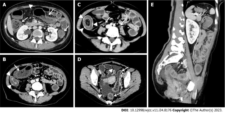
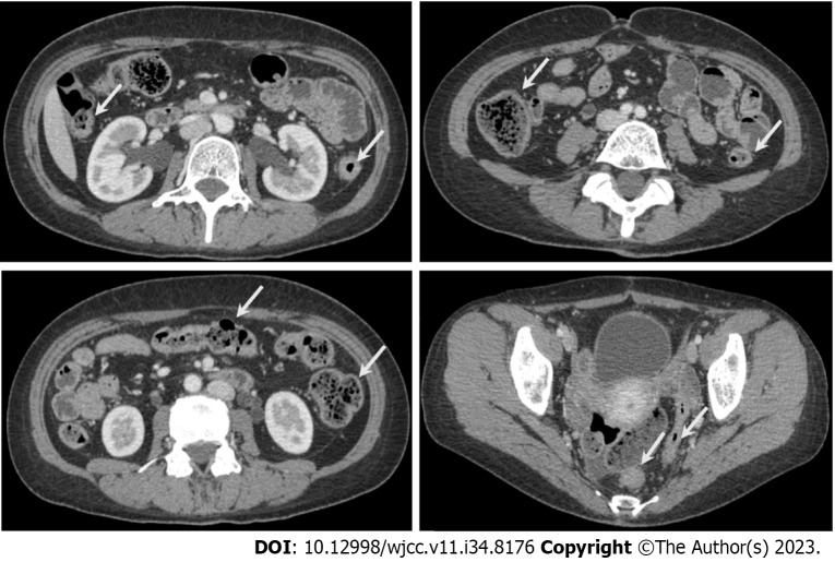
Case summary: A 35-year-old woman was diagnosed with colorectal LE after initially presenting with intermittent abdominal pain and vomiting for two months. She had a regular medication history for five years following the diagnosis of SLE but had been irregular in taking medications, which may have contributed to the onset of LE and led to her current hospital admission. According to the 2019 Classification criteria for SLE of the European League Against Rheumatism/American College of Rheumatology, this case scored 14. Additionally, abdominal computed tomography revealed significant wall edema of the colon and rectum, ischemia and hyperemia of the ascending colon intestinal wall, mesenteric vessel engorgement, increased mesangial fat attenuation, ascites, and bilateral ureter-hydronephrosis, all indicative of colon and rectum LE. Laboratory tests also showed lower levels of complement C3 and C4, with an antinuclear antibody titer of 1:100. Overall, it was clear that this case involved the colon and rectum without affecting the small intestine, representing a rare manifestation of SLE. The patient received treatment with 10 mg of methylprednisolone sodium succinate, 100 mL of 0.9% sodium chloride, hydroxychloroquine (100 mg), and nutrition support. After one week of methylprednisolone and hydroxychloroquine therapy, her SLE symptoms and disease activity improved significantly.
Conclusion: Although colorectal LE without small intestine involvement is very rare, early diagnosis and excellent management with corticosteroids prevented the need for surgical intervention. Physicians should be aware of colorectal LE without small intestine involvement as a manifestation of lupus flare.
Keywords: Case report; Colon and rectum; Comb sign; Lupus enteritis; Methylprednisolone and hydroxychloroquine; Systemic lupus erythematosus; Target sign.
©The Author(s) 2023. Published by Baishideng Publishing Group Inc. All rights reserved.
Conflict of interest statement
Conflict-of-interest statement: All the authors report no relevant conflicts of interest for this article.
Figures

Similar articles
-
Enteritis: a window to the diagnosis of systemic lupus erythematosus in an adolescent girl: case report.Paediatr Int Child Health. 2024 May;44(1):42-47. doi: 10.1080/20469047.2023.2299581. Epub 2024 Jan 7. Paediatr Int Child Health. 2024. PMID: 38184810
-
Lupus enteritis as the only active manifestation of systemic lupus erythematosus: A case report.World J Clin Cases. 2019 Jun 6;7(11):1315-1322. doi: 10.12998/wjcc.v7.i11.1315. World J Clin Cases. 2019. PMID: 31236395 Free PMC article.
-
Acute acalculous cholecystitis as the initial manifestation of systemic lupus erythematous: A case report.Medicine (Baltimore). 2021 Jun 4;100(22):e26238. doi: 10.1097/MD.0000000000026238. Medicine (Baltimore). 2021. PMID: 34087909 Free PMC article. Review.
-
How to diagnose lupus enteritis early? Lessons learned from a multicenter case series.Acta Reumatol Port. 2019 Apr-Jun;44(2):145-150. Acta Reumatol Port. 2019. PMID: 31300632 English.
-
Rare case of systemic lupus with manifestation of FS Jaccoud arthropathy: a case report and literature review.BMC Musculoskelet Disord. 2025 Jun 3;26(1):544. doi: 10.1186/s12891-025-08696-8. BMC Musculoskelet Disord. 2025. PMID: 40461984 Free PMC article. Review.
Cited by
-
A Rare Case of Lupus Nephritis with Predominant Gastrointestinal Manifestation: A Case Study.J Pharm Bioallied Sci. 2024 Dec;16(Suppl 5):S4890-S4892. doi: 10.4103/jpbs.jpbs_1204_24. Epub 2025 Jan 30. J Pharm Bioallied Sci. 2024. PMID: 40061682 Free PMC article.
References
-
- Li YF, Wei MJ. Acute pancreatitis in childhood-onset systemic lupus erythematosus: Case report. Arch Argent Pediatr. 2019;117:e279–e283. - PubMed
-
- Aringer M, Costenbader K, Daikh D, Brinks R, Mosca M, Ramsey-Goldman R, Smolen JS, Wofsy D, Boumpas DT, Kamen DL, Jayne D, Cervera R, Costedoat-Chalumeau N, Diamond B, Gladman DD, Hahn B, Hiepe F, Jacobsen S, Khanna D, Lerstrøm K, Massarotti E, McCune J, Ruiz-Irastorza G, Sanchez-Guerrero J, Schneider M, Urowitz M, Bertsias G, Hoyer BF, Leuchten N, Tani C, Tedeschi SK, Touma Z, Schmajuk G, Anic B, Assan F, Chan TM, Clarke AE, Crow MK, Czirják L, Doria A, Graninger W, Halda-Kiss B, Hasni S, Izmirly PM, Jung M, Kumánovics G, Mariette X, Padjen I, Pego-Reigosa JM, Romero-Diaz J, Rúa-Figueroa Fernández Í, Seror R, Stummvoll GH, Tanaka Y, Tektonidou MG, Vasconcelos C, Vital EM, Wallace DJ, Yavuz S, Meroni PL, Fritzler MJ, Naden R, Dörner T, Johnson SR. 2019 European League Against Rheumatism/American College of Rheumatology Classification Criteria for Systemic Lupus Erythematosus. Arthritis Rheumatol. 2019;71:1400–1412. - PMC - PubMed
Publication types
LinkOut - more resources
Full Text Sources
Miscellaneous
