. 2024 Jan 10;146(1):677-694.
doi: 10.1021/jacs.3c10406. Epub 2023 Dec 22.

Z-Form Adoption of Nucleic Acid is a Multi-Step Process Which Proceeds through a Melted Intermediate

Affiliations

Z-Form Adoption of Nucleic Acid is a Multi-Step Process Which Proceeds through a Melted Intermediate

Parker J Nichols et al. J Am Chem Soc. .

Abstract

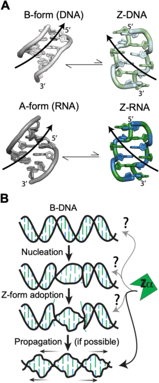
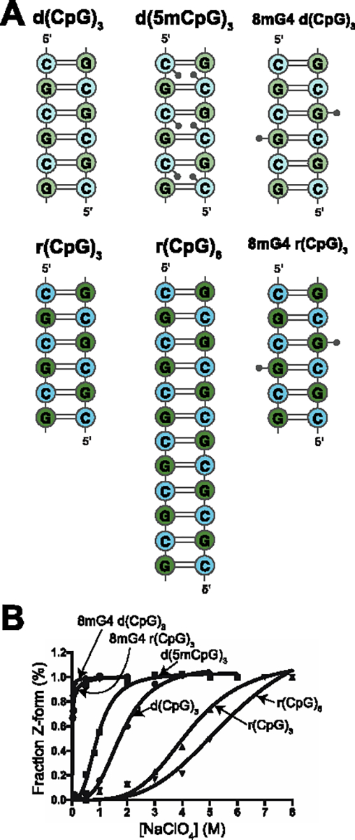
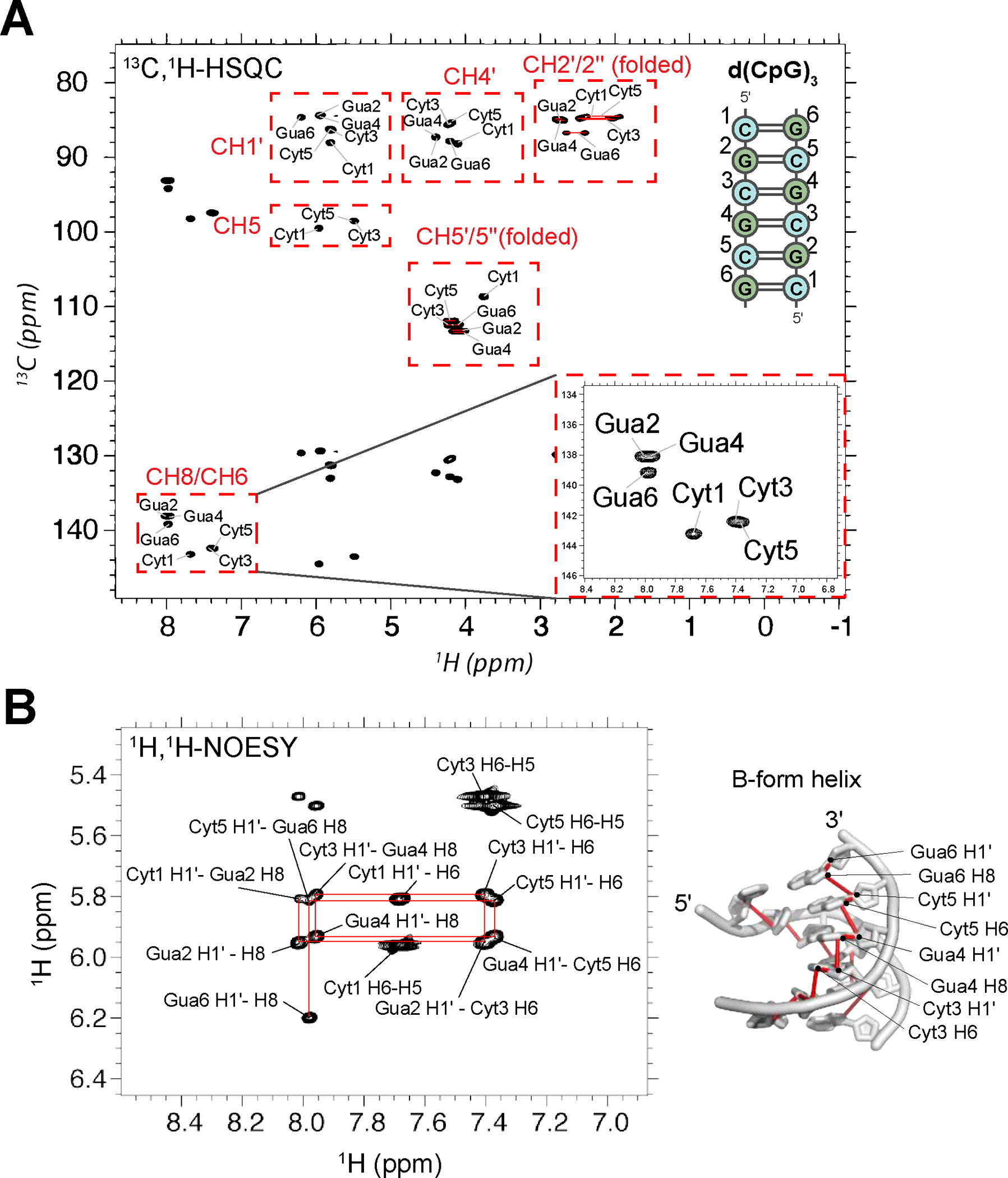
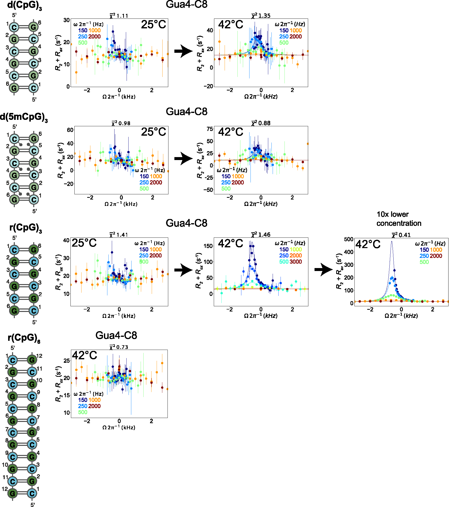
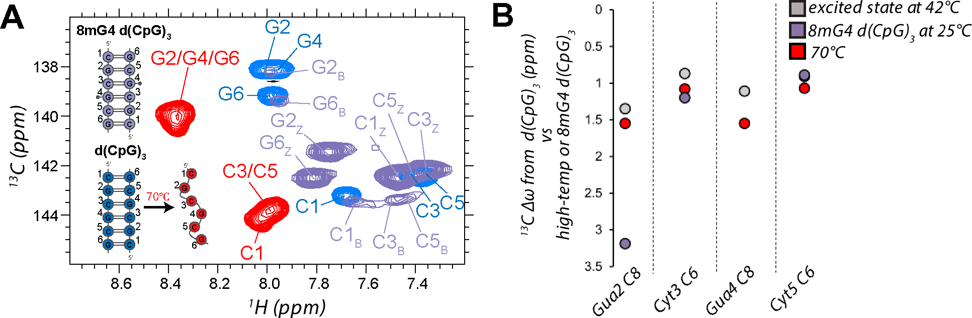
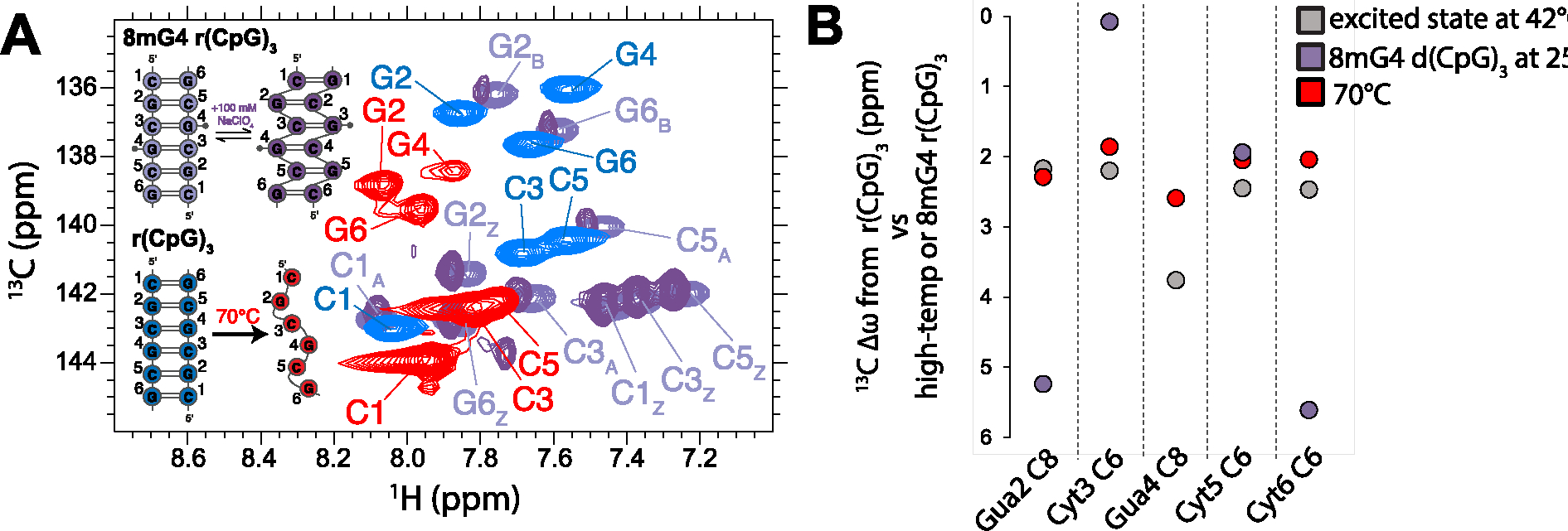
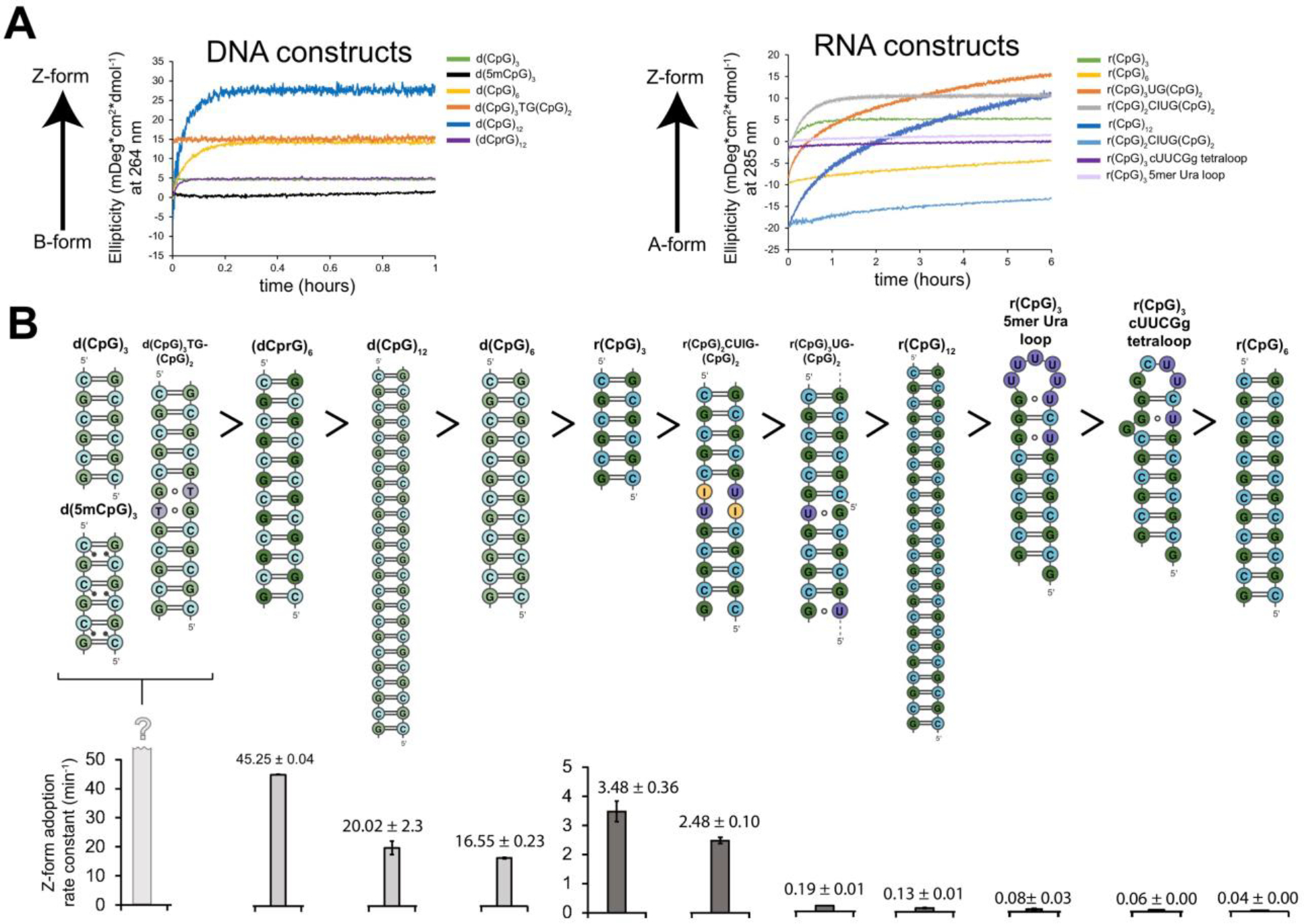
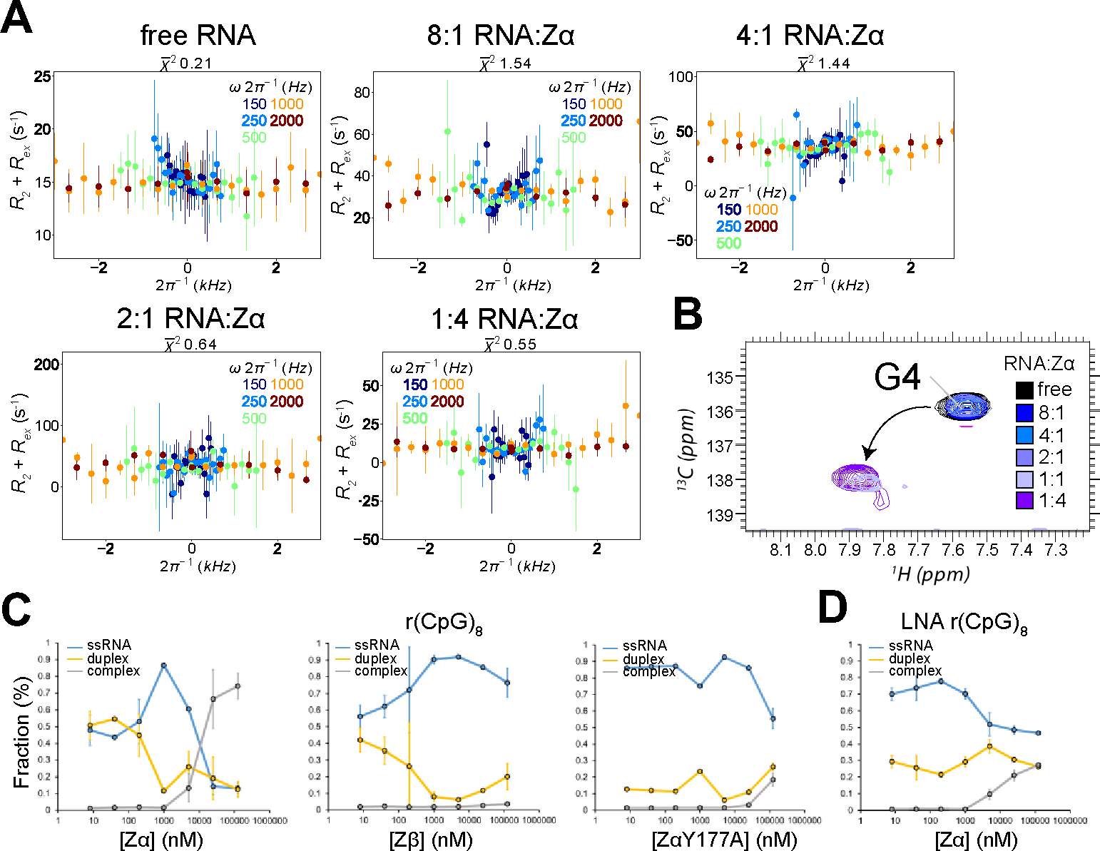
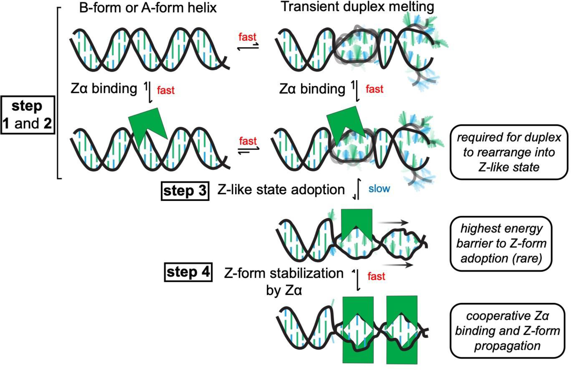
The left-handed Z-conformation of nucleic acids can be adopted by both DNA and RNA when bound by Zα domains found within a variety of innate immune response proteins. Zα domains stabilize this higher-energy conformation by making specific interactions with the unique geometry of Z-DNA/Z-RNA. However, the mechanism by which a right-handed helix contorts to become left-handed in the presence of proteins, including the intermediate steps involved, is poorly understood. Through a combination of nuclear magnetic resonance (NMR) and other biophysical measurements, we have determined that in the absence of Zα, under low salt conditions at room temperature, d(CpG) and r(CpG) constructs show no observable evidence of transient Z-conformations greater than 0.5% on either the intermediate or slow NMR time scales. At higher temperatures, we observed a transient unfolded intermediate. The ease of melting a nucleic acid duplex correlates with Z-form adoption rates in the presence of Zα. The largest contributing factor to the activation energies of Z-form adoption as calculated by Arrhenius plots is the ease of flipping the sugar pucker, as required for Z-DNA and Z-RNA. Together, these data validate the previously proposed "zipper model" for Z-form adoption in the presence of Zα. Overall, Z-conformations are more likely to be adopted by double-stranded DNA and RNA regions flanked by less stable regions and by RNAs experiencing torsional/mechanical stress.


Figures

Figure 1.
Figure 1.
Characteristics of Z-DNA/RNA and Z-conformation adoption. (a) Both dsDNA (top) and dsRNA (bottom), normally in the B- and A-conformations, can adopt the higher-energy left-handed Z-form (right). Z-DNA and Z-RNA are structurally equivalent and will revert to the B/A-form in the absence of stabilizing factors. Pyrimidine (blue) – purine (green) repeats alternate between the C2′-endo and C3′-endo sugar pucker conformations along with the nucleobases between anti and the syn conformations. This leads to a zig-zagged backbone. B-DNA, A-RNA, Z-DNA, and Z-RNA models were made using PDBs 1N1K, 1PBM, 1QBJ, and 2GXB, respectively. (b) Zipper model for the conversion from B-DNA to Z-DNA. First, a high-energy nucleation event allows for helical handedness conversion and a short Z-DNA stretch to be adopted. This can then propagate down the helix in a cooperative manner if the sequence allows it. At what points Zα plays a role in the zipper model is mostly unknown.
Figure 2.
Figure 2.
DNA and RNA constructs selected for NMR measurements. (a) 2D representations of the different d(CpG) and r(CpG) constructs used for NMR measurements in this study. DNA bases are more lightly shaded than RNA ones. Methyl groups for the modified constructs are depicted as small grey circles and indicate their relative position within the duplex. (b) Fits of circular dichroism titrations of NaClO4 into the DNA and RNA constructors shown in (a), with the fraction of the duplex in the Z-conformation on the y-axis and the concentration of NaClO4 (M) on the x-axis. The fraction of Z-DNA and Z-RNA was tracked by following the ellipticity at 266 and 285 nm, respectively, as described in the Methods sections.
Figure 3.
Figure 3.
NMR assignment of the d(CpG)3 construct. (a) Full 13C-1H HSQC spectra assignments for the d(CpG)3 construct are shown (depicted on the right with assignment numbering). The CH2’/2” and CH5’/5” peak positions are folded in from their normal positions around 40 and 66 ppm, respectively. Their proper chemical shift values are indicated in Table S1. CH3’ resonances were not assignable due to water suppression. Inset shows a zoom in of the aromatic assignments. Note that the two strands of the duplex are chemically equivalent and, therefore, have identical chemical shifts. (b) 1H-1H NOESY experiment with a mixing time of 320 ms showing the aromatic H8/H6 to ribose H1′ connectivites. The NOESY “walk” through the B-form helix is indicated with red lines, an example of which is shown on the structure of a B-form helix (PDB: 1N1K1) to the right.
Figure 4.
Figure 4.
Off-Resonance R1ρ relaxation dispersion profiles of the different DNA and RNA constructs at 25°C and 42°C. 2D representations of the d(CpG) and r(CpG) constructs and corresponding Off-Resonance R1ρ relaxation dispersion profiles for the C8 atom of Gua4 carried out at five different spin-lock powers (150, 250, 500, 1000, and 2000 Hz, colored coded according to the legend within each plot) at 25°C and 42°C are shown to the right. The dispersion profile at 10x lower concentration of duplex for the r(CpG)3 construct is also shown. R2 + R2ex (= (RR1cos2θ)/sin2θ, where θ = tan−1(lock power/offset)) values are given as a function of the resonance offset from the major state (Ωoff/2π). Error bars represent experimental uncertainty from a bootstrapping method, as described in the Methods section. The fits (solid lines) were carried out as described in the materials and methods, and fitted parameters are found in Tables 2 and 3.
Figure 5.
Figure 5.
Identification of excited state chemical shift differences extracted from Off-Resonance R1ρ experiments measured on the d(CpG)3. (a) Aromatic 13C-1H HSQC (CH8 of purines and CH6 of pyrimidines) assignments are shown for 8mG4 d(CpG)3 (purple peaks, B-form and Z-form peaks are denoted by subscripts B or Z, respectively) compared to the d(CpG)3 construct at 42°C (folded, blue peaks) and 70°C (melted, red peaks). (b) Chemical shift differences (13C Δω) extracted from Off-Resonance R1ρ experiments measured on the d(CpG)3 construct at 42°C, the difference between the B-form and Z-form peaks in the 13C, 1H HSQC of the 8mG4 d(CpG)3 construct, and the difference between the folded and melted peaks in the 13C, 1H HSQC of the d(CpG)3 construct at 42°C and 70°C. The Z-form chemical shift position for Guanine 4 for the 8mG4 d(CpG)3 construct could not be compared due to the methyl modification.
Figure 6.
Figure 6.
Identification of excited state chemical shift difference extracted from Off-Resonance R1ρ experiments measured on the r(CpG)3 construct. (a) Aromatic 13C-1H HSQC (CH8 of purines and CH6 of pyrimidines) assignments are shown for 8mG4 r(CpG)3 (purple peaks, A-form and Z-form peaks are denoted by subscript A and Z, respectively) compared to the r(CpG)3 construct at 42°C (folded, blue peaks) and 70°C (melted, red peaks). For the 8mG4 r(CpG)3 duplex, the addition of 100 mM NaClO4 promotes the population of Z-RNA while decreasing the population of A-RNA (dark purple peaks). (b) Chemical shift differences (13C Δω) extracted from Off-Resonance R1ρ experiments measured on the r(CpG)3 construct at 42°C, the difference between the A-form and Z-form peaks in the 13C, 1H HSQC of the 8mG4 r(CpG)3 construct, and the difference between the folded and melted peaks in the 13C, 1H HSQC of the r(CpG)3 construct at 42°C and 70°C. The Z-form chemical shift position for Guanine 4 for the 8mG4 r(CpG)3 construct could not be compared due to the methyl modification.
Figure 7.
Figure 7.
Z-form adoption rates in DNA and RNA constructs. (a) Circular dichroism time-course experiments showing the rate of Z-DNA (tracked at 266 nm) and Z-RNA (tracked at 285 nm) adoption after the addition of saturating concentrations of Zα at 25°C. The different constructs are color-coded according to the legend on the right-handed side. (b) 2D depictions of DNA and RNA constructs (above) are shown in descending order according to their Z-form adoption rates at 25°C (below), with their half-times indicated in minutes.
Figure 8.
Figure 8.
Characterization of exchange in the r(CpG)3 construct at increasing concentrations of Zα. (a) Off-Resonance R1ρ relaxation dispersion profiles for the C8 atom of isotopically labeled Gua4 in the r(CpG)3 construct measured with increasing concentrations of Zα. The molar ratio of RNA:Zα can be found at the top of the graphs. Off-resonance R1ρ experiments were carried out at five different spin-lock powers (150, 250, 500, 1000, and 2000 Hz, colored coded according to the legend on the right). R2 + Rex (= R) values are given as a function of the resonance offset from the major state (Ωoff/2π). Error bars represent experimental uncertainty. (b) 1H,13C HSQC spectra showing the C8 atom of isotopically labeled Gua4 in the r(CpG)3 construct is shown at increasing concentrations of Zα, color coded according to the legend on the right. (c) The fraction of ssRNA r(CpG)8, dsRNA r(CpG)8, and r(CpG)8 in complex with Zα, Zβ, and ZαY177A extracted from electrophoretic mobility shift assays (shown in Figure S25). The values shown are an average of two replicates plotted on a log scale. (d) Same as in (c), but with a (CpG)8 LNA construct which cannot adopt the Z-conformation.
Figure 9.
Figure 9.
Model for Z-form adoption by Zα. First, Zα binds to a right-handed B- or A-form helix non-specifically (step 1). Next, the duplex transiently melts, which may be promoted through Zα binding (step 2). The dynamics of these steps occur on a relatively faster time scale (on the micro- to millisecond time regime). From here, the ribose sugar pucker and nucleosides have to rearrange into a high-energy, Z-like state (which occurs slowly taking seconds to hours, step 3). Then, Zα cooperatively binds to and stabilizes the Z-conformation (which occurs fast once a Z-like state is adopted, step 4)).

References

    1. Wolfram S Principles of Nucleic Acid Structure, 1st ed.; Springer; New York: New York, 1984.
    1. Stefl R; Skrisovska L; Allain FHT RNA Sequence- and Shape-Dependent Recognition by Proteins in the Ribonucleoprotein Particle. EMBO Reports. 2005. 10.1038/sj.embor.7400325. - DOI - PMC - PubMed
    1. Battistini F; Hospital A; Buitrago D; Gallego D; Dans PD; Gelpí JL; Orozco M How B-DNA Dynamics Decipher Sequence-Selective Protein Recognition. J. Mol. Biol. 2019. 10.1016/j.jmb.2019.07.021. - DOI - PubMed
    1. Hall K; Cruz P; Tinoco I; Jovin TM; Van De Sande JH “Z-RNA” - A Left-Handed RNA Double Helix. Nature 1984. 10.1038/311584a0. - DOI - PubMed
    1. Schwartz T; Rould MA; Lowenhaupt K; Herbert A; Rich A Crystal Structure of the Zα Domain of the Human Editing Enzyme ADAR1 Bound to Left-Handed Z-DNA. Science (80-. ). 1999. 10.1126/science.284.5421.1841. - DOI - PubMed

Figure references

    1. Monleon D; Esteve V; Celda B. NMR Study of Hexanucleotide d(CCGCGG)2 Containing Two Triplet Repeats of Fragile X Syndrome. Biochem Biophys Res Commun 2003, 303 (1), 81–90. 10.1016/s0006-291x(03)00304-8. - DOI - PubMed
    1. Popenda M; Biala E; Milecki J; Adamiak RW Solution Structure of RNA Duplexes Containing Alternating CG Base Pairs: NMR Study of r(CGCGCG)2 and 2’-O-Me(CGCGCG)2 under Low Salt Conditions. Nucleic Acids Res 1997, 25 (22), 4589–4598. 10.1093/nar/25.22.4589. - DOI - PMC - PubMed
    1. Schwartz T; Rould MA; Lowenhaupt K; Herbert A; Rich A Crystal Structure of the Zalpha Domain of the Human Editing Enzyme ADAR1 Bound to Left-Handed Z-DNA. Science (80-. ). 1999, 11 (284(5421)), 1841–1845. - PubMed
    1. Placido D; Brown BA 2nd; Lowenhaupt K; Rich A; Athanasiadis A. A Left-Handed RNA Double Helix Bound by the Z Alpha Domain of the RNA-Editing Enzyme ADAR1. Structure 2007, 15 (4), 395–404. 10.1016/j.str.2007.03.001. - DOI - PMC - PubMed
    1. Ho PS The Non-B-DNA Structure of d(CA/TG)(n) Does Not Differ from That of Z-DNA. Proc. Natl. Acad. Sci. U. S. A. 1994. 10.1073/pnas.91.20.9549. - DOI - PMC - PubMed

Publication types