. 2023 Dec 18;11(4):184.
doi: 10.3390/diseases11040184.

Protective Effects of Sitagliptin on Streptozotocin-Induced Hepatic Injury in Diabetic Rats: A Possible Mechanisms

Affiliations

Protective Effects of Sitagliptin on Streptozotocin-Induced Hepatic Injury in Diabetic Rats: A Possible Mechanisms

Qamraa H Alqahtani et al. Diseases. .

Abstract

Diabetes is a ubiquitous disease that causes several complications. It is associated with insulin resistance, which affects the metabolism of proteins, carbohydrates, and fats and triggers liver diseases such as fatty liver disease, steatohepatitis, fibrosis, and cirrhosis. Despite the effectiveness of Sitagliptin (ST) as an antidiabetic drug, its role in diabetes-induced liver injury is yet to be fully investigated. Therefore, this study aims to investigate the effect of ST on hepatic oxidative injury, inflammation, apoptosis, and the mTOR/NF-κB/NLRP3 signaling pathway in streptozotocin (STZ)-induced liver injury. Rats were allocated into four groups: two nondiabetic groups, control rats and ST rats (100 mg/kg), and two diabetic groups induced by STZ, and they received either normal saline or ST for 90 days. Diabetic rats showed significant hyperglycemia, hyperlipidemia, and elevation in liver enzymes. After STZ induction, the results revealed remarkable increases in hepatic oxidative stress, inflammation, and hepatocyte degeneration. In addition, STZ upregulated the immunoreactivity of NF-κB/p65, NLRP3, and mTOR but downregulated IKB-α in liver tissue. The use of ST mitigated metabolic and hepatic changes induced by STZ; it also reduced oxidative stress, inflammation, and hepatocyte degeneration. The normal expression of NF-κB/p65, NLRP3, mTOR, and IKB-α were restored with ST treatment. Based on that, our study revealed for the first time the hepatoprotective effect of ST that is mediated by controlling inflammation, oxidative stress, and mTOR/NF-κB/NLRP3 signaling.

Keywords: STZ; diabetic; dipeptidyl peptidase-4 inhibitor; mTOR/NF-κB/NLRP3 signaling; sitagliptin.


Conflict of interest statement

The authors declare no potential conflict of interest related to the research, authorship, and/or publication of this article.

Figures

Figure 1
Figure 1
ST modulates the body weight (A), blood glucose (B), and liver/body weight ratio (C) in STZ-induced hepatic damage in diabetic rats. Diabetic and nondiabetic rats were either treated with physiological saline or ST (100 mg/kg) for 90 days. Body weight, blood glucose levels, and liver weight were measured at the end of the treatment period. ST, Sitagliptin; STZ, Streptozotocin. Data are mean ± SEM (n = 8). *** p < 0.001.
Figure 2
Figure 2
ST reduces the liver function enzymes (AST and ALT) in STZ-induced hepatic damage in diabetic rats. After treatment of animals either with vehicle control or ST (100 mg/kg) for 90 days, the liver enzymes ALT and AST were measured with biochemical kits. AST, aspartate aminotransferase; ALT, alanine aminotransferase; ST, Sitagliptin; STZ, Streptozotocin. Data are mean ± SEM, (n = 8). *** p < 0.001.
Figure 3
Figure 3
ST attenuates hepatic oxidative stress in STZ-induced diabetic rats. Treatment of rats with 100 mg/kg ST for 90 days decreases hepatic MDA (A) and increases SOD (B) and GSH (C) in diabetic model. MDA, malonaldehyde; SOD, superoxide dismutase; GSH, reduce glutathione; ST, Sitagliptin; STZ, Streptozotocin. Data are mean ± SEM, (n = 8). ** p < 0.01, *** p < 0.001.
Figure 4
Figure 4
Photomicrograph of H&E-stained liver section, scale bar = 100 µm. (A,B) represent normal nondiabetic control liver and liver of rat who received ST, respectively, showing normal histological architecture of hepatic lobules and portal tracts. Hepatocytes and the hepatic blood sinusoid are in normal state. (C) represents a liver section from rat receiving STZ as a model of diabetes, showing multiple patches of hepatic degeneration with loss of group of hepatocytes (star). The surrounding tissues show inflammatory cellular infiltrations and hepatocyte cytoplasmic degeneration. Some hepatocytes show nuclear pyknosis. (D) represents liver from diabetic rat who received ST, showing marked decrease in inflammatory cellular infiltrations; most hepatocytes are apparently within normal cytoplasm and nuclei. There is recovery of the hepatic lobular architecture. The degenerated patches decreased in number and size (star). ST, Sitagliptin; STZ, Streptozotocin.
Figure 5
Figure 5
ST Downregulated hepatic inflammatory and apoptotic markers in STZ-diabetic rats. Treatment with ST decreases hepatic IL-6 (A), IL-18 (B), TNF-α (C), and Caspase-1 (D). ST; Sitagliptin, STZ; Streptozotocin. Data are mean ± SEM, (n = 8). * p < 0.05, ** p < 0.01, *** p < 0.001.
Figure 6
Figure 6
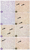
(AD) Photomicrograph of NF-κB/p65 immune-stained liver sections, scale bar = 50 µm. (A,B) represent normal control liver and liver of rat that received ST, respectively, showing normal hepatocytes and absence of nuclear immune signal. (C) represents liver sections from diabetic rats showing many hepatocytes with strongly immune-stained nuclei (arrows), while section from rats treated with ST (D) shows few immune-stained hepatocytes nuclei (arrows). (EH) Photomicrograph of IKB-α immune-stained liver sections, scale bar = 50 µm. (E,F) represent normal control liver and liver of rat received ST, respectively, showing normal positive strong immune reaction of hepatocyte nuclei. (G), liver section from diabetic rat showing few hepatocytes with immune-stained nuclei (arrow), while section from diabetic rat treated with ST (H) shows a marked increase in the number of hepatocytes with positively immune-stained nuclei (arrow). ST, Sitagliptin; STZ, Streptozotocin.
Figure 7
Figure 7
Photomicrograph of NLRP3 immune-stained liver sections, scale bar = 50 µm. (A,B) are of normal control liver and liver of rat who received ST, respectively, showing normal absence of nuclear immune reaction. (C) Liver section from diabetic rat shows many hepatocytes with immune stained nuclei (arrows), while section from ST treated rat (D) shows a marked decrease in the hepatocyte nuclear immune reaction (arrows). ST, Sitagliptin.
Figure 8
Figure 8
Photomicrograph of mTOR immune-stained liver sections, scale bar = 50 µm. (A,B) Photomicrograph of normal control liver and liver of rat who received ST, respectively, showing normal absence of immune reactivity, while (C) represents liver from diabetic rat, shows patches of hepatocytes with strong cytoplasmic immune reactivity (arrowheads); the rest of the hepatocytes show cytoplasm with weak immune reaction (arrows). (D) represents liver from diabetic rat that received ST showing small patches of hepatocytes with weak immune reaction (arrows) while the rest of hepatocytes have no immune reaction. ST, Sitagliptin.

References

    1. American Diabetes Association Diagnosis and Classification of Diabetes Mellitus. Diabetes Care. 2014;37((Suppl. S1)):81–90. doi: 10.2337/dc14-S081. - DOI - PubMed
    1. Saeedi P., Petersohn I., Salpea P., Malanda B., Karuranga S., Unwin N., Colagiuri S., Guariguata L., Motala A.A., Ogurtsova K., et al. Global and regional diabetes prevalence estimates for 2019 and projections for 2030 and 2045: Results from the International Diabetes Federation Diabetes Atlas, 9th edition. Diabetes Res. Clin. Pract. 2019;157:107843. doi: 10.1016/j.diabres.2019.107843. - DOI - PubMed
    1. Jarrar M., Al-Bsheish M., Abusalah M.A.H., Albaker W., Alsyouf A., Al-Mugheed K., Issa M.R., Alumran A. Prevalence of type 2 diabetes mellitus in the general population of Saudi Arabia, 2000–2020: A systematic review and meta-analysis of observational studies. Saudi J. Med. Med. Sci. 2023;11:1. doi: 10.4103/sjmms.sjmms_394_22. - DOI - PMC - PubMed
    1. Galicia-Garcia U., Benito-Vicente A., Jebari S., Larrea-Sebal A., Siddiqi H., Uribe K.B., Ostolaza H., Martín C. Pathophysiology of type 2 diabetes mellitus. Int. J. Mol. Sci. 2020;21:6275. doi: 10.3390/ijms21176275. - DOI - PMC - PubMed
    1. Larter C.Z., Farrell G.C. Insulin resistance, adiponectin, cytokines in NASH: Which is the best target to treat? J. Hepatol. 2006;44:253–261. doi: 10.1016/j.jhep.2005.11.030. - DOI - PubMed

LinkOut - more resources