. 2023 Dec 9;12(24):2807.
doi: 10.3390/cells12242807.

The Identification of Nuclear FMRP Isoform Iso6 Partners

Affiliations

The Identification of Nuclear FMRP Isoform Iso6 Partners

Nassim Ledoux et al. Cells. .

Abstract

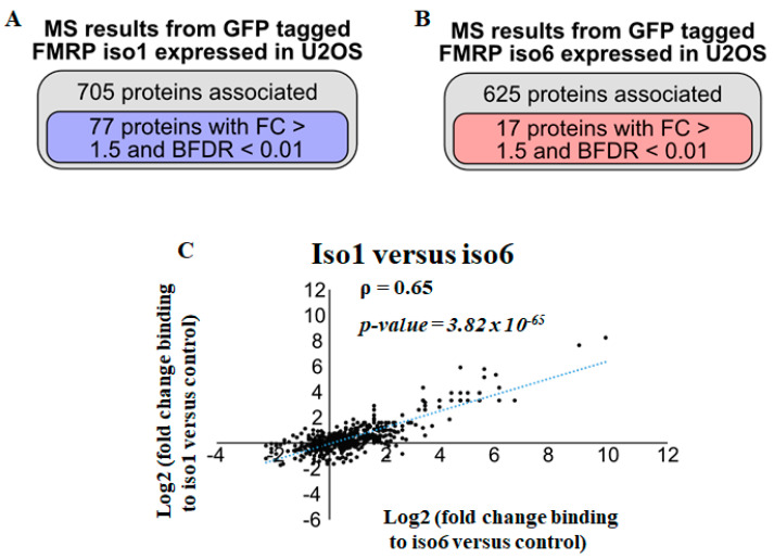
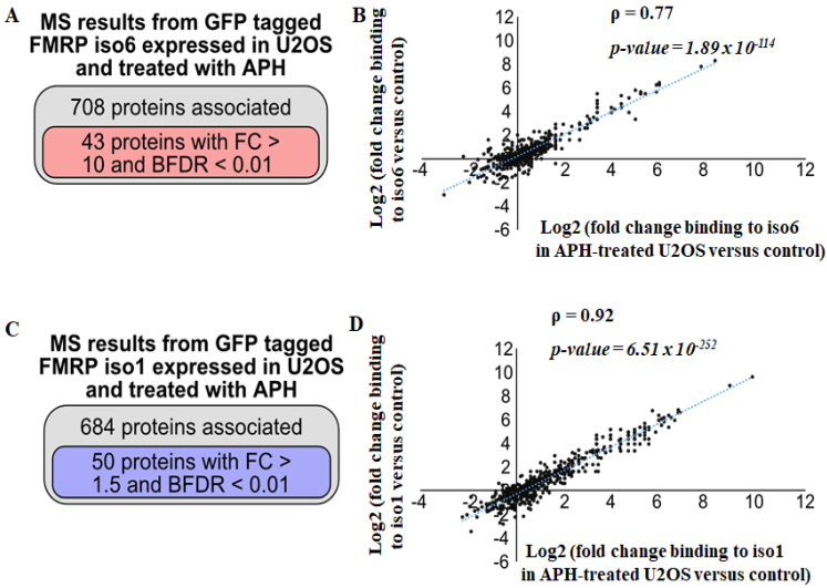
A deficiency of FMRP, a canonical RNA-binding protein, causes the development of Fragile X Syndrome (FXS), which is characterised by multiple phenotypes, including neurodevelopmental disorders, intellectual disability, and autism. Due to the alternative splicing of the encoding FMR1 gene, multiple FMRP isoforms are produced consisting of full-length predominantly cytoplasmic (i.e., iso1) isoforms involved in translation and truncated nuclear (i.e., iso6) isoforms with orphan functions. However, we recently implicated nuclear FMRP isoforms in DNA damage response, showing that they negatively regulate the accumulation of anaphase DNA genomic instability bridges. This finding provided evidence that the cytoplasmic and nuclear functions of FMRP are uncoupled played by respective cytoplasmic and nuclear isoforms, potentially involving specific interactions. While interaction partners of cytoplasmic FMRP have been reported, the identity of nuclear FMRP isoform partners remains to be established. Using affinity purification coupled with mass spectrometry, we mapped the nuclear interactome of the FMRP isoform iso6 in U2OS. In doing so, we found FMRP nuclear interaction partners to be involved in RNA processing, pre-mRNA splicing, ribosome biogenesis, DNA replication and damage response, chromatin remodeling and chromosome segregation. By comparing interactions between nuclear iso6 and cytoplasmic iso1, we report a set of partners that bind specifically to the nuclear isoforms, mainly proteins involved in DNA-associated processes and proteasomal proteins, which is consistent with our finding that proteasome targets the nuclear FMRP iso6. The specific interactions with the nuclear isoform 6 are regulated by replication stress, while those with the cytoplasmic isoform 1 are largely insensitive to such stress, further supporting a specific role of nuclear isoforms in DNA damage response induced by replicative stress, potentially regulated by the proteasome.

Keywords: FMRP; GFP-Trap; RNA-binding proteins; mass spectrometry; proteasome.


Conflict of interest statement

The authors declare no conflict of interest.

Figures

Figure 1
Figure 1
(A,B) U2OS stably expressing either GFP-iso6 or-iso1 was either lysed and its protein extracts analysed for the expression of GFP-iso6 and -iso1 using anti-GFP antibodies or fixed and processed for immunofluorescence to visualise GFP. Blue staining depicts nuclear DNA. Scale bars (10 μm) are shown. Images were acquired using an LSM 900 confocal microscope. For Western blot quantification, the expression level of GFP-iso6 relative to GFP-iso1 was estimated via densitometry quantification of the film signal using Image Studio™ Lite 4.0.21 software upon standardization against total tubulin. Data are represented as mean ± SD of three independent experiments. A two-tailed Student’s t test was used. *** p ≤ 0.001. (C,D) GFP-iso6 interactome. U2OS stably expressing GFP-iso6 or GFP-free U2OS controls were lysed, and their proteins were pulled down in the GFP-trap. Bound proteins from two controls and two experimental GFP-Traps were identified via LC-Ms/Ms. (C) Indicated on the top is a total number of proteins pulled down, showing differentially associated proteins in U2OS-GFP iso6 compared to GFP-free U2OS identified associated with GFP-iso6, and those scored with <1% FDR and a fold change higher than 1.5. On the bottom is a graph representing the fold enrichment of selected proteins that bind to iso6. The values are represented on a log2 scale. p value ≤ 0.05. (D) GST-pull downs. U2OS protein extracts were incubated with GST-iso6 or GST immobilised on glutathione beads. Bound proteins were eluted and analysed via Western blot using antibodies specific to the indicated proteins.
Figure 2
Figure 2
Analysis of GFP-iso6 and GFP-iso1 interactions under stringent conditions. U2OS stably expressing GFP-iso6, -iso1, or GFP-free U2OS controls were lysed, and their proteins pulled down in the GFP-Trap. Bound proteins from 6 controls and 2 experimental experiments were identified via LC-Ms/Ms. (A,B) The total number of proteins pulled down showing differentially associated proteins in either U2OS-GFP-iso1 (A) or -GFP-iso6 is indicated (B) compared to GFP-free U2OS, and those scored with <1% FDR and a fold change higher than 1.5. (C) Correlation of fold change proteins associated with iso1 and iso6. Pearson correlation coefficients (ρ) and linear regression (blue dotted lines) are indicated.
Figure 3
Figure 3
Analysis of GFP-iso6 and GFP-iso1 interactions that occur in APH-treated U2OS under stringent conditions. These experiments were carried out and the data analysed as described in Figure 2, except where U2OS was treated with 0.3 mM APH for 24 h. (A) The total number of proteins pulled down in APH-treated U2OS-GFP-iso6 is indicated, showing differentially associated proteins compared to GFP-free U2OS, and those scored with <1% FDR and a fold change higher than 1.5. (B) Less correlation in fold change is found between proteins associating with GFP-iso6 in APH-treated U2OS versus mock-treated U2OS. (C) The total number of proteins pulled down in APH-treated U2OS-GFP-iso1 is indicated, showing differentially associated proteins compared to GFP-free U2OS, and those scored with <1% FDR and a fold change higher than 1.5. (D) A strong correlation in fold change is found between proteins associating with GFP-iso1 in APH-treated U2OS versus mock-treated U2OS.
Figure 4
Figure 4
(A) Effect of MG132 treatment on the expression of FMRP. U2OS stably expressing either GFP-iso1 or -iso6 was treated with MG132 or DMSO, lysed, and its protein extracts were analysed for the expression of GFP-iso1 (left panels) and -iso6 (right panels) using anti-GFP antibodies. The expression p53 is used to monitor the effect of MG132, while tubulin serves as a loading control for GFP quantification, as shown on the bottom. For these quantifications, the expression level of GFP-iso1 and -iso6 was estimated via densitometry quantification of the film signal using Image Studio™ Lite 4.0.21 Software and standardized against total tubulin. Data are represented as mean ± SD of three independent experiments. A two-tailed Student’s t test was used. ** p ≤ 0.01. * p ≤ 0.05. ns: not significant. (B) The deletion of the C-t domain of iso6 restores the expression of the protein. U2OS stably expressing either GFP-iso1, -iso6, or iso6 (ΔC-t) was lysed, and its protein extracts were analysed for the expression of GFP-iso1 and -iso6 and ΔC-t using anti-GFP (top panels) antibodies. Tubulin (bottom panels) serves as a loading control. The expression level of GFP-ΔC-t relative to -iso6 was estimated via densitometry quantification of the film signal using Image Studio™ Lite Software upon standardization against total tubulin. Data are represented as mean ± SD of three independent experiments. A two-tailed Student’s t test was used. * p ≤ 0.05. (C) The deletion of the C-t domain of iso6 results in a significant cytoplasmic accumulation of the protein. U2OS stably expressing either GFP-iso6 or-ΔC-t were fixed and processed for immunofluorescence to visualise GFP. Blue staining depicts nuclear DNA. Scale bars (5 μm) are shown. Images were acquired by using an LSM 900 confocal microscope. (D) U2OS stably expressing GFP-ΔC-t was treated with MG132 or DMSO, lysed, and its protein extracts were analysed for the expression of GFP-ΔC-t using anti-GFP antibodies. The expression p53 is used to monitor the effect of MG132, while tubulin serves as a loading control for GFP quantification, as shown on the bottom. Data are represented as mean ± SD of three independent experiments. A two-tailed Student’s t test was used. ns: not significant. (E) Treatment with cycloheximide results in a rapid drop of the expression of iso6 but not iso6ΔC-t. U2OS stably expressing either GFP-iso6 or GFP-ΔC-t was treated with cycloheximide for the indicated time before adding puromycin for the last 5 min to monitor ongoing protein synthesis. Cells were then lysed, and their protein extracts were analysed for the expression of GFP-iso6 and ΔC-t using anti-GFP antibodies. Data are represented as mean ± SD of three independent experiments. A two-tailed Student’s t test was used. *** p ≤ 0.001. * p ≤ 0.05. ns: not significant. (F) U2OS was treated with cycloheximide for the indicated time. Cells were then lysed, and their protein extracts analysed to probe puromycylated proteins indicating active translation, while tubulin served as a loading control.

References

    1. Banerjee A., Ifrim M.F., Valdez A.N., Raj N., Bassell G.J. Aberrant RNA translation in fragile X syndrome: From FMRP mechanisms to emerging therapeutic strategies. Brain Res. 2018;1693:24–36. doi: 10.1016/j.brainres.2018.04.008. - DOI - PMC - PubMed
    1. Hagerman R.J., Berry-Kravis E., Hazlett H.C., Bailey D.B., Jr., Moine H., Kooy R.F., Tassone F., Gantois I., Sonenberg N., Mandel J.L., et al. Fragile X syndrome. Nat. Rev. Dis. Primers. 2017;3:17065. doi: 10.1038/nrdp.2017.65. - DOI - PubMed
    1. Dury A.Y., El Fatimy R., Tremblay S., Rose T.M., Cote J., De Koninck P., Khandjian E.W. Nuclear Fragile X Mental Retardation Protein is localized to Cajal bodies. PLoS Genet. 2013;9:e1003890. doi: 10.1371/journal.pgen.1003890. - DOI - PMC - PubMed
    1. Sittler A., Devys D., Weber C., Mandel J.L. Alternative splicing of exon 14 determines nuclear or cytoplasmic localisation of fmr1 protein isoforms. Hum. Mol. Genet. 1996;5:95–102. doi: 10.1093/hmg/5.1.95. - DOI - PubMed
    1. Valverde R., Pozdnyakova I., Kajander T., Venkatraman J., Regan L. Fragile X mental retardation syndrome: Structure of the KH1-KH2 domains of fragile X mental retardation protein. Structure. 2007;15:1090–1098. doi: 10.1016/j.str.2007.06.022. - DOI - PubMed

Publication types

MeSH terms

LinkOut - more resources