Review
. 2023 Dec 12;12(24):2819.
doi: 10.3390/cells12242819.

Age-Related Alternative Splicing: Driver or Passenger in the Aging Process?

Affiliations
Review

Age-Related Alternative Splicing: Driver or Passenger in the Aging Process?

Marco Baralle et al. Cells. .

Abstract

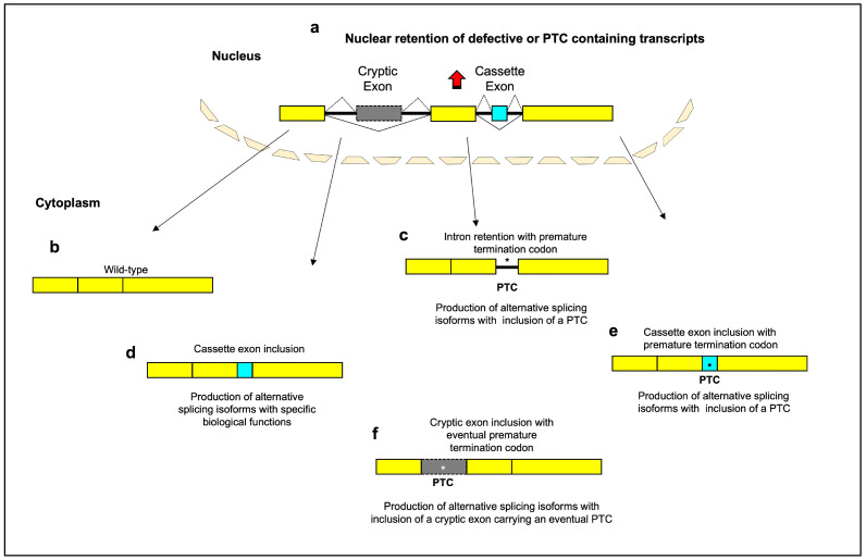
Alternative splicing changes are closely linked to aging, though it remains unclear if they are drivers or effects. As organisms age, splicing patterns change, varying gene isoform levels and functions. These changes may contribute to aging alterations rather than just reflect declining RNA quality control. Three main splicing types-intron retention, cassette exons, and cryptic exons-play key roles in age-related complexity. These events modify protein domains and increase nonsense-mediated decay, shifting protein isoform levels and functions. This may potentially drive aging or serve as a biomarker. Fluctuations in splicing factor expression also occur with aging. Somatic mutations in splicing genes can also promote aging and age-related disease. The interplay between splicing and aging has major implications for aging biology, though differentiating correlation and causation remains challenging. Declaring a splicing factor or event as a driver requires comprehensive evaluation of the associated molecular and physiological changes. A greater understanding of how RNA splicing machinery and downstream targets are impacted by aging is essential to conclusively establish the role of splicing in driving aging, representing a promising area with key implications for understanding aging, developing novel therapeutical options, and ultimately leading to an increase in the healthy human lifespan.

Keywords: age-associated splicing events; age-related diseases; aging; alternative splicing; molecular aging; senescence; splice variants; splicing factors; splicing regulation; transcriptome changes.


Conflict of interest statement

The authors declare no conflict of interest.

Figures

Figure 1
Figure 1
Schematic diagram of the main aging-related alternative splicing events. Most genes are split between exons and introns. Splicing-defective or premature termination codon (PTC)-containing transcripts are retained within nucleus, with inhibition of translation (a). Normal patterns of splice site selection (indicated with solid black lines) join the constitutive exons together to create “wild type” mRNAs (b). Intron-retaining transcripts can include a PTC and undergo degradation by activation of nonsense-mediated decay (c). The inclusion of a cassette exon into mRNA (indicated with dashed black lines) can lead to a gain of functional domains (d) or to the appearance of a PTC (e). The inclusion of a cryptic exon into mRNA can lead to the introduction of frameshifts or PTC and subsequently to the loss of specific domains or to reduction in gene expression (f). A portion of PTC-containing transcripts is recognized and retained in the nucleus before they have a chance to be exported to the cytoplasm for degradation by the NMD system. PTCs in different reading frames can lead to different levels of nuclear retention. This suggests that the mechanism of nuclear retention is not simply due to the presence of a stop codon, but is dependent on the specific sequence context of the PTC [30,31]. The exact mechanisms by which PTC-containing transcripts are retained in the nucleus are still being investigated. One possible mechanism is alternative splicing—the presence of a PTC can affect the splicing of an mRNA, leading to the inclusion of exons that contain nuclear retention signals that prevent export to the cytoplasm. Another possibility is interaction with specific nuclear proteins or RNA binding proteins that bind to the PTC-containing transcripts and retain them in the nucleus, blocking their export. Changes to the mRNA structure induced by the PTC could also prevent transit to the cytoplasm if the folding prevents export. While further research is needed to elucidate the precise retention mechanisms, the nuclear retention itself may serve a protective purpose. By retaining PTC-containing transcripts in the nucleus, truncated proteins are prevented from being translated in the cytoplasm where they could have detrimental effects on the cell [31,32]. Thus, nuclear retention may act more to protect the cell rather than facilitate mRNA repair pathways like splicing to excise the PTC or induce degradation of unrepairable mRNAs. Yellow box: constitutive exon; gray box: cryptic exon; aqua box: cassette exon; asterisk: premature termination codon.
Figure 2
Figure 2
Intron retention: regulation of S-endoglin and of cardiac ankyrin repeat domain 1-i8 (ANKRD1) during senescence. (a) S-endoglin. During endothelial cell senescence, the splicing factor SRSF1 supports generation of the S-endoglin mRNA, by its binding to a cis-element overlapping the branch point, that hampers splicing of intron 13. As a result, SRSF1 stabilizes intron retention and increases S-endoglin mRNA levels. (b) Cardiac ankyrin repeat domain 1 gene (ANKRD1 or CARP). In the ANKRD1-i7,8 splicing isoform retaining both intron 7 and 8, the fourth ankyrin repeat is absent. Higher levels of the ANKRD1-i8 splicing isoform retaining intron 8 were found in infant compared to adult heart tissues. This isoform maintains all conserved ankyrin repeat motifs crucial for its interactions with titin and cardiac calsequestrin. Yellow box: constitutive exon; solid line: intron.
Figure 3
Figure 3
Cassette exon: aging-regulated alternative splicing of SIRT1 and ING1 genes. (a) The human SIRT1 genomic organization and its aging-related splicing isoforms. SIRT1-v1 isoform includes nine exons and no cassette exons (aqua coloured boxes). SIRT1-v2 includes eight exons, and the first exon was named Exon-1′ (E1′). SIRT1-v3 isoform includes seven exons with the Exon-4′ (E4′) in-between exon 4 and 5. (b) The human IGF1 genomic organization (IGF1) and its aging-related splicing isoforms (Ea, Eb, and Ec/MGF). The IGF1-Ea isoform shows skipping of the whole exon 5. The IGF1-Eb isoform shown includes the whole exon 5. The Ec/MGF isoform contains the N-terminal part of exon 5 (consisting of 49 nucleotides, shown as a gray box) and exon 6. Exon 5 inclusion causes the occurrence of an altered C-terminal amino acid sequence as a result of a shift in the reading frame. Yellow box: constitutive exon; solid line: intron; dotted lines: alternative splicing patterns; upwards red arrow: upregulated expression; downwards blue arrow: downregulated expression.
Figure 4
Figure 4
Aging-related cryptic exons. Schematic representation of cryptic exons activated by loss of TDP-43. For UNC13A gene, two alternative acceptor splice sites can be activated, causing the insertion of 128 bp or 178 bp cryptic exons. The exons immediately upstream and downstream of the cryptic exons are shown with the corresponding number. Yellow box: constitutive exon; solid line: intron; dotted lines: alternative splicing patterns; gray box: cryptic exon (CE); red point: the position of the putative TDP-43 binding site within the in-between intron.

References

    1. Johnson F.B., Sinclair D.A., Guarente L. Molecular Biology of Aging. Cell. 1999;96:291–302. doi: 10.1016/S0092-8674(00)80567-X. - DOI - PubMed
    1. Liu Y., Gonzàlez-Porta M., Santos S., Brazma A., Marioni J.C., Aebersold R., Venkitaraman A.R., Wickramasinghe V.O. Impact of Alternative Splicing on the Human Proteome. Cell Rep. 2017;20:1229–1241. doi: 10.1016/j.celrep.2017.07.025. - DOI - PMC - PubMed
    1. Kim E., Magen A., Ast G. Different Levels of Alternative Splicing among Eukaryotes. Nucleic Acids Res. 2007;35:125–131. doi: 10.1093/nar/gkl924. - DOI - PMC - PubMed
    1. Lee Y., Rio D.C. Mechanisms and Regulation of Alternative Pre-mRNA Splicing. Annu. Rev. Biochem. 2015;84:291–323. doi: 10.1146/annurev-biochem-060614-034316. - DOI - PMC - PubMed
    1. Somel M., Guo S., Fu N., Yan Z., Hu H.Y., Xu Y., Yuan Y., Ning Z., Hu Y., Menzel C., et al. MicroRNA, mRNA, and Protein Expression Link Development and Aging in Human and Macaque Brain. Genome Res. 2010;20:1207–1218. doi: 10.1101/gr.106849.110. - DOI - PMC - PubMed