Review
. 2023 Dec 14;12(24):2837.
doi: 10.3390/cells12242837.

Therapeutic Monoclonal Antibodies against Cancer: Present and Future

Affiliations
Review

Therapeutic Monoclonal Antibodies against Cancer: Present and Future

Marisa Delgado et al. Cells. .

Abstract

A series of monoclonal antibodies with therapeutic potential against cancer have been generated and developed. Ninety-one are currently used in the clinics, either alone or in combination with chemotherapeutic agents or other antibodies, including immune checkpoint antibodies. These advances helped to coin the term personalized medicine or precision medicine. However, it seems evident that in addition to the current work on the analysis of mechanisms to overcome drug resistance, the use of different classes of antibodies (IgA, IgE, or IgM) instead of IgG, the engineering of the Ig molecules to increase their half-life, the acquisition of additional effector functions, or the advantages associated with the use of agonistic antibodies, to allow a broad prospective usage of precision medicine successfully, a strategy change is required. Here, we discuss our view on how these strategic changes should be implemented and consider their pros and cons using therapeutic antibodies against cancer as a model. The same strategy can be applied to therapeutic antibodies against other diseases, such as infectious or autoimmune diseases.

Keywords: bioinformatics; cancer database analyses; cancer treatment; therapeutic antibodies; transmembrane proteins.


Conflict of interest statement

The authors declare no conflict of interest.

Figures
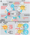
Figure 1
Figure 1
Schematic representation illustrating the mechanisms of action of therapeutic antibodies. (A) Some antibodies block the interaction between ligands and receptors by binding to either the ligand (ligand blocking) or the receptor (receptor blocking), preventing the signaling promoting tumor growth. Other antibodies bind to tumor antigens and then are recognized by natural killer cells (NK), triggering their cytotoxic activity, known as ADCC (antibody-dependent cell cytotoxicity). Another possibility is that when the antibody binds to the tumor antigen, it opsonizes the cell and activates phagocytic cells, thereby triggering antibody-dependent cell phagocytosis (ADCP). Additionally, the antibody can fix complement and trigger complement-dependent cytotoxicity (CDC) after binding to the tumor cell. Furthermore, some antibodies can trigger direct apoptosis after binding to an antigen on the tumor’s cell surface. (B) Other antibodies have agonistic effects. They identify antigens in the antigen-presenting cells and trigger an activating response of these cells similar to the ligand binding. (C) The last group corresponds to antibodies recognizing immune checkpoint antigens, such as CTLA-4, PD-1, or PD-L1. Here, the antibody acts by inhibiting the binding of the ligand and prevents the negative signaling through these receptors. Examples of antibodies functioning through these mechanisms are in red. Stars of different colors indicate antibodies that work through several mechanisms of action.
Figure 2
Figure 2
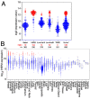
Expression levels of HER2 in breast cancer and other tumor types. Expression levels of HER2 mRNA in (A) different breast cancer subtypes (TNBC = triple-negative breast cancer; None* = breast tumors with no data available on subtype). (B) Tumors expressing high levels of HER2 are in red, whereas tumors expressing low/medium levels of HER2 are in blue. Medians, the first and third quartiles (boxes), and the 10th and 90th percentiles (whiskers) are indicated for each type of tumor. The number of samples for each tumor type (n) is shown in Supplemental Table S2. Expression data were obtained from the GENT2 public database [109].
Figure 3
Figure 3
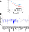
Effects of HER2 on survival and expression in normal tissues. (A) Kaplan–Meier survival curves depending on either high or low/intermediate HER2 expression levels, indicating the mean survival ± SEM as well as the statistical significance of the differences in survival curves determined using the Chi-square test. Data from all breast cancer tumor patients from Figure 2A, for which survival data were available, were analyzed. (B) HER2 expression levels in normal tissue samples. Samples expressing high levels of HER2 are in red, whereas samples expressing low/medium levels of HER2 are in blue. Medians, the first and third quartiles (boxes), and the 10th and 90th percentiles (whiskers) are shown for each normal tissue. The number of samples for each normal tissue (n) is shown in Supplemental Table S2. Expression data were obtained from the GENT2 public database [109].
Figure 4
Figure 4
Cancer incidence and cancer mortality worldwide. (A) cancer incidence [117] and (B) cancer mortality [117] data obtained from GLOBOCAN2020 from the International Agency for Research on Cancer from the World Health Organization [118].
Figure 5
Figure 5
Cancer mortality-to-incidence ratio (MIR). The mortality-to-incidence ratio was calculated from the GLOBOCAN2020 data (presented in Figure 4) by calculating 100 times the mortality-to-incidence ratio, expressed as a percentage [121]. The data were obtained from the International Agency for Research on Cancer from the World Health Organization [118].
Figure 6
Figure 6
Future of personalized medicine using therapeutic antibodies. (A) The idea is to select a series of genes from the human genome, coding for plasma membrane proteins, which are over-expressed in tumors (≥2-fold higher expression levels). On each tumor sample, the tumor/normal expression ratio for each gene allows it to be ranked. Afterward, the scores for the different tumors are analyzed together to make a rank for the genes on that set of tumors. According to the biological features of these genes, the top-ranking ones are used to select mAbs. Then, they are tested for the ability to inhibit or delay tumor growth. This panel of antibodies (in this hypothetical example, represented by 96 different antibodies) will have therapeutic potential for one or more types of tumors. (B) The tumor samples for each patient (tumors 1–5) are screened for the expression of the antigens recognized by the antibodies, allowing to select a series of antibodies, positive for each particular tumor (4 to 6 in the hypothetical example, with some antibodies not recognizing any tumor samples and others recognizing one, two, or even three tumor samples). Thus, a combination containing 2–3 different antibodies could be used for the initial treatment of each patient, allowing the killing of the maximum number of tumor cells from the beginning of the treatment.

References

    1. Köhler G., Milstein C. Continuous cultures of fused cells secreting antibody of predefined specificity. Nature. 1975;256:495–497. doi: 10.1038/256495a0. - DOI - PubMed
    1. Mach J.P., Carrel S., Forni M., Ritschard J., Donath A., Alberto P. Tumor localization of radiolabeled antibodies against carcinoembryonic antigen in patients with carcinoma: A critical evaluation. N. Engl. J. Med. 1980;303:5–10. doi: 10.1056/NEJM198007033030102. - DOI - PubMed
    1. Buchegger F., Vacca A., Carrel S., Schreyer M., Mach J.P. Radioimmunotherapy of human colon carcinoma by 131I-labelled monoclonal anti-CEA antibodies in a nude mouse model. Int. J. Cancer. 1988;41:127–134. doi: 10.1002/ijc.2910410123. - DOI - PubMed
    1. Hanack K., Messerschmidt K., Listek M. Antibodies and Selection of Monoclonal Antibodies. Adv. Exp. Med. Biol. 2016;917:11–22. doi: 10.1007/978-3-319-32805-8_2. - DOI - PubMed
    1. Corraliza-Gorjón I., Somovilla-Crespo B., Santamaria S., Garcia-Sanz J.A., Kremer L. New Strategies Using Antibody Combinations to Increase Cancer Treatment Effectiveness. Front. Immunol. 2017;8:1804. doi: 10.3389/fimmu.2017.01804. - DOI - PMC - PubMed

Publication types

Substances