. 2023 Dec 14;12(24):2833.
doi: 10.3390/cells12242833.

Separable Roles for Neur and Ubiquitin in Delta Signalling in the Drosophila CNS Lineages

Affiliations

Separable Roles for Neur and Ubiquitin in Delta Signalling in the Drosophila CNS Lineages

Konstantina Kalodimou et al. Cells. .

Abstract

The execution of a Notch signal at the plasma membrane relies on the mechanical force exerted onto Notch by its ligand. It has been appreciated that the DSL ligands need to collaborate with a ubiquitin (Ub) ligase, either Neuralized or Mindbomb1, in order to exert this pulling force, but the role of ubiquitylation per se is uncertain. Regarding the Delta-Neur pair, it is documented that neither the Neur catalytic domain nor the Delta intracellular lysines (putative Ub acceptors) are needed for activity. Here, we present a dissection of the Delta activity using the Delta-Notch-dependent expression of Hey in newborn Drosophila neurons as a sensitive in vivo assay. We show that the Delta-Neur interaction per se, rather than ubiquitylation, is needed for activity, pointing to the existence of a Delta-Neur signaling complex. The Neur catalytic domain, although not strictly needed, greatly improves Delta-Neur complex functionality when the Delta lysines are mutated, suggesting that the ubiquitylation of some component of the complex, other than Delta, can enhance signaling. Since Hey expression is sensitive to the perturbation of endocytosis, we propose that the Delta-Neur complex triggers a force-generating endocytosis event that activates Notch in the adjacent cell.

Keywords: Delta; Drosophila; Hey; Neuralized; Notch; asymmetric cell division; endocytosis; neurodevelopment; ubiquitylation.


Conflict of interest statement

The authors declare no conflict of interest. The funders had no role in the design of the study; in the collection, analyses, or interpretation of data; in the writing of the manuscript; or in the decision to publish the results.

Figures
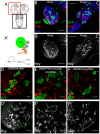
Figure 1
Figure 1
The Ganglion Mother Cells’ Asymmetric Cell Division (GMC ACD) assay (A) Graphic of the larval CNS; the red box depicts a brain hemisphere and the black box the ventral nerve cord. (A’) Graphic of a neuroblast (NB) lineage: the NB undergoes asymmetric divisions to self-renew and generate ganglion mother cells (GMCs). Each GMC divides asymmetrically to give rise to neuron type a (Hey-positive Elav-positive, Notch-ON) and neuron type b (Hey-negative Elav-positive Notch OFF); red arrows show Notch signaling events. (B’,C’) Overview of control mosaic CNS (FRT 82B) with no mutations. (B) Brain hemisphere. (C) Ventral nerve cord. Green: GFP clonal marker; red: Hey, marks only young Notch-ON neurons; Blue: Elav, marks all neurons, but not NBs or GMCs. Scale bar 60 μm. (DF) Examples of genotypes that produce strong (D), moderate (E), or no (F) rescue of Hey expression. Single sections from enlarged areas of the central brain containing GFP-marked clones expressing a Dl variant in various mutant backgrounds, as indicated (for a description of Dl variants, see Figure 3). Red: Hey. (D’F’) Hey pattern alone; the borders of the clones are drawn in white. Green, yellow, and orange arrows point to positive, weakly positive, and negative lineages respectively; the same color code is used in the bar charts of Figure 4, Figure 5, Figure 6 and Figure 7. Scale bar: 20 μm.
Figure 2
Figure 2
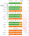
Clonal phenotype classification and quantification for Dl-HA, Dl-K2R-HA, and Dli2ala-HA. Three phenotypic categories are presented as a percentage of the total number (N) of lineages scored: strong (>2 Hey positive cells) is shown in green, weak (1–2 Hey positive cells) is shown in yellow, and negative (no Hey-positive cells) in dark orange. Genotypes (from top to bottom): control (neutral clones); clones expressing Dlwt-HA in DlSer, mibDlSer, or neurDlSer background; clones expressing DlK2R-HA in DlSer, mibDlSer, or neurDlSer background; clones expressing Dli2ala-HA in DlSer, mibDlSer, or neurDlSer background; clones mutant for DlSer, mibDlSer, or neurDlSer without any transgene expression. Selected pairwise comparisons are shown (chi-square): black lines indicate non-significant differences (ns, p > 0.05) and red lines significant differences (****): p < 0.0001. For more comparisons, see Table S1. Dl transgenes’ cartoons as in Figure 3.
Figure 3
Figure 3
Schematic view of the intracellular domain of all Delta variants used in this study (on the left). Only the intracellular domain is shown, as the extracellular and transmembrane domains are intact in all variants used in this study. icd1 stands for intracellular domain 1 and corresponds to residues 630–641, icd2 to residues 682–793, icd3 to residues 742–747. icd1–3 are three strongly conserved motifs across insects [9]. Dli1 and Dli1/2 are precise deletions of icd1 or both icd1 and icd2, respectively. On the right, the amino acid changes present in the remaining variants are shown, highlighted in red. Hatched icd boxes denote changes in part of the motif to alanines, as shown on the right. Red stars denote changes of lysines to arginines. In addition to the nine lysines shown here, there are three more lysine residues directly following the transmembrane domain (at the N-terminus of the segment of Delta shown here—the putative stop-transfer sequence); these, too, have been converted to arginines in all variants bearing the K2R label.
Figure 4
Figure 4
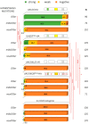
Behavior of Dli1ala variants in GMC asymmetric cell divisions. Graph bar for the Hey phenotype quantification classified in three categories as in Figure 2. Genotypes (from top to bottom): Dl-HA expression in DlSer, mibDlSer, or neurDlSer clones (same data as Figure 2 for comparison); Dli1ala-HA expression in DlSer, mibDlSer, or neurDlSer clones; Dli1/2-V5 expression in DlSer clones; DlK2Ri1ala-HA expression in DlSer, mibDlSer, or neurDlSer clones; mutant clones for DlSer, mibDlSer, or neurDlSer (same data as Figure 2 for comparison). Selected pairwise comparisons are shown (chi-square): red lines for significant differences (*): 0.01 < p < 0.05, (***): 0.0001 < p < 0.001, (****): p < 0.0001. For more comparisons, see Table S1. Dl transgenes’ cartoons as in Figure 3.
Figure 5
Figure 5
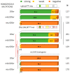
Dli2ala and DlK2Ri2ala variants rescue Hey expression similarly. Graph bar for the Hey phenotype quantification classified in three categories as in Figure 2. Genotypes (from top to bottom): Dli2ala-HA expression in DlSer, mibDlSer, or neurDlSer clones (same data as Figure 2 for comparison); DlK2Ri2ala-HA expression in DlSer, mibDlSer, or neurDlSer clones; mutant clones for DlSer, mibDlSer, or neurDlSer with no transgene expression (same data as Figure 2 for comparison). Selected pairwise comparisons are shown (chi-square): black lines are used for non-significant differences (ns): p > 0.05 and red lines for significant differences, (****): p < 0.0001. For more comparisons, see Table S1. Dl transgenes’ cartoons as in Figure 3.
Figure 6
Figure 6
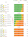
Combination of Delta variants with NeurΔRING. Bar chart for the Hey phenotype quantification classified into three categories as in Figure 2. Genotypes (from top to bottom): DlSer clones expressing Dl-HA, DlK2R-HA, Dli2ala, DlK2Ri2ala (data from Figure 2 and Figure 5 for comparison); neurDlSer clones expressing Dl-HA + EGFP-Neur, Dl-HA + NeurΔRING-EGFP, DlK2R-HA + EGFP-Neur, DlK2RHA + NeurΔRING-EGFP, Dli2ala-HA + NeurΔRING-EGFP, DlK2Ri2ala-HA + NeurΔRING-EGFP, Dl-HA alone, DlK2R-HA alone, Dli2ala-HA alone, DlK2Ri2ala-HA alone (last four bars from Figure 2 and Figure 5 for comparison). Selected pairwise comparisons are shown (chi-square): black lines are used for non-significant differences (ns): p > 0.05 and red lines for significant differences (****): p < 0.0001. For more comparisons, see Table S1. Dl transgenes’ cartoons as in Figure 3.
Figure 7
Figure 7
The role of epsin, dynamin, and DlLDL. (AC) Single sections of enlarged areas of the central brain showing characteristic examples of 3-day mutant clones for epsin (lqf) (A) or DlSer expressing DlLDL (B). Green: GFP, red: Hey. (A’) One Hey-positive lineage is highlighted. (B’) Two neighboring lineages are shown, with their NB highlighted by white arrows, both of which are Hey-negative. (C’) Dl Ser control lineages, all negative. (D) Graph bar for the Hey phenotype quantification for the genotypes shown in (A,B). The results from equivalently aged clones from Dl Ser or Dl Ser + UAS-Dl-HA (from Figure 2) are included for comparison. (EG) 40 h clones of the indicated genotypes; animals were kept at 29 °C after clone induction. Green: GFP, red: Hey. (E’G’) Hey pattern alone; the borders of the lineages are drawn in white. Green, yellow, and orange arrows point to positive, weakly positive, and negative lineages, respectively. (E) DlSer mutation abolishes Hey expression 40 h after clone induction, with few escapers. (F) Flip-out clones overexpressing a dominant negative allele for shi. (G) DlHA; DlSer restores Hey expression 40 h after clone induction. (H) Graph bar for the Hey phenotype quantification for the genotypes shown in (DF). Scale bar: 20 μm. In D and H, selected pairwise comparisons are shown (chi-square): black lines are used for non-significant differences: p > 0.05 and red lines for significant differences (****): p < 0.0001. See also Table S1.
Figure 8
Figure 8
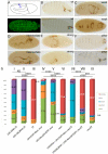
Rescue of the embryo neurogenic phenotype by Delta and Neur variants. (A) Top: graphic of a stage 10 embryo indicating individualized NBs (blue dots) in the left side and clustered NBs in the right side, characteristic of the absence of lateral inhibition in Notch pathway mutations. Bottom: da.G32-Gal4; UAS-neurΔRING-EGFP: uniform expression of the transgene (GFP in green) is shown in this superficial ectodermal section of a stage 10 embryo. (BF) Cuticle preparations of dead embryos that show increasing lack of epidermis. Wild-type embryos (two examples shown in (B)) have normal cephalic structures and denticle belts on their ventral side. Denticle belts are fewer and disorganized in the two examples shown in (C) and small holes are present at the ventral side, which arise from a lack of epidermal cells due to mis-specification to neuroblasts. In (D), the gaps have fused to each other and the entire ventral epidermis is absent. Finally, in (E), almost the entire embryonic ectoderm has transformed to neural tissue, leaving only a tiny fragment of cuticle. We observe also defective embryos with no epidermal holes, like the two presented in (F), which show a twisted body plan. We classify these as “other”. In (AF), all embryos are shown anterior to the right. (G) Percentages of each phenotypic category per genotype. Abbreviations: Dl = DeltaRevF10 mutation, neur = neur1 mutation, neur Dl = neur1 DeltaRevF10 mutations. All UAS transgenes are expressed under the control of da.G32-Gal4. Chi-square comparisons showed significant differences (p < 0.01) for all experimental samples compared to the relevant control (I, II with III; IV, V with VI; and VII, VIII with IX). Other comparisons are shown above the histogram. NS: no significance; ***: 0.0001 < p < 0.001; ****: p < 0.0001 Absolute numbers of embryos scored per category, percentages, and all comparisons with their respective p-values and χ2 are presented analytically in Table S2.
Figure 9
Figure 9
Distinct modes of Delta activation by Neur vs. Mib1. Summary of our working hypothesis of how Neur and Mib1 promote Delta endocytosis and activity. Black arrows indicate interactions, strong (solid) or weak (dashed). Blue arrows indicate ubiquitylations. i1, 2, 3 (red squares) indicate the icd1, 2, 3 motifs of the intracellular domain of Delta. The much larger extracellular domain is not drawn to scale. All lysines of the icd are shown with their residue numbers; all of them are putative Ub acceptors. KRKRKR is the sequence of residues 620–625, which constitute the stop-transfer sequence and could also serve as Ub acceptors. Mib1 interacts with icd2 and 3 [10,18] and can ubiquitylate many Delta lysines (blue arrows labeled 1, exact target lysines have not been mapped). Moreover, this ubiquitylation is needed to promote signaling. Putative endocytic adaptors (EA) are shown that recognize Ub-modified Delta to promote its inclusion in endocytic pits. Neur interacts strongly with icd1 [9] and weakly with icd2 (this work) and ubiquitylates Delta preferentially on K742 (blue arrows labeled 2; [9]), but can also ubiquitylate another substrate (blue arrows labeled 3; this work). It is also an endocytic adaptor itself, shown in this schematic by its direct contact both with the Delta cargo and with other EAs. The presence of Ub molecules, although not absolutely necessary, enhances the activity of Delta, perhaps by strengthening the formation of such adaptor–cargo complexes and promoting the efficient endocytosis of Delta.

References

    1. Artavanis-Tsakonas S., Matthew D., Rand R.J.L. Notch Signaling: Cell Fate Control and Signal Integration in Development. Science. 1999;284:770–776. doi: 10.1126/science.284.5415.770. - DOI - PubMed
    1. Kovall R.A., Gebelein B., Sprinzak D., Kopan R. The Canonical Notch Signaling Pathway: Structural and Biochemical Insights into Shape, Sugar, and Force. Dev. Cell. 2017;41:228–241. doi: 10.1016/j.devcel.2017.04.001. - DOI - PMC - PubMed
    1. Bray S.J. Notch signalling: A simple pathway becomes complex. Nat. Rev. Mol. Cell Biol. 2006;7:678–689. doi: 10.1038/nrm2009. - DOI - PubMed
    1. Gordon W.R., Roy M., Vardar-Ulu D., Garfinkel M., Mansour M.R., Aster J.C., Blacklow S.C. Structure of the Notch1-negative regulatory region: Implications for normal activation and pathogenic signaling in T-ALL. Blood. 2009;113:4381–4390. doi: 10.1182/blood-2008-08-174748. - DOI - PMC - PubMed
    1. Gordon W.R., Vardar-Ulu D., Histen G., Sanchez-Irizarry C., Aster J.C., Blacklow S.C. Structural basis for autoinhibition of Notch. Nat. Struct. Mol. Biol. 2007;14:295–300. doi: 10.1038/nsmb1227. - DOI - PubMed

Publication types

MeSH terms

LinkOut - more resources