Review
. 2023 Dec 8;13(24):3628.
doi: 10.3390/diagnostics13243628.

Acute Colonic Diverticulitis: CT Findings, Classifications, and a Proposal of a Structured Reporting Template

Affiliations
Review

Acute Colonic Diverticulitis: CT Findings, Classifications, and a Proposal of a Structured Reporting Template

Francesco Tiralongo et al. Diagnostics (Basel). .

Abstract

Acute colonic diverticulitis (ACD) is the most common complication of diverticular disease and represents an abdominal emergency. It includes a variety of conditions, extending from localized diverticular inflammation to fecal peritonitis, hence the importance of an accurate diagnosis. Contrast-enhanced computed tomography (CE-CT) plays a pivotal role in the diagnosis due to its high sensitivity, specificity, accuracy, and interobserver agreement. In fact, CE-CT allows alternative diagnoses to be excluded, the inflamed diverticulum to be localized, and complications to be identified. Imaging findings have been reviewed, dividing them into bowel and extra-intestinal wall findings. Moreover, CE-CT allows staging of the disease; the most used classifications of ACD severity are Hinchey's modified and WSES classifications. Differential diagnoses include colon carcinoma, epiploic appendagitis, ischemic colitis, appendicitis, infectious enterocolitis, and inflammatory bowel disease. We propose a structured reporting template to standardize the terminology and improve communication between specialists involved in patient care.

Keywords: abscess; colonic; computed tomography; diverticulitis; fistula; perforation; peritonitis; structured report.


Conflict of interest statement

The authors declare no conflict of interest.

Figures

Figure 1
Figure 1
Axial contrast-enhanced CT images show acute left colonic diverticulitis and associated findings of perivisceritis (a,b). Once the longitudinal axis of the colon is identified (green straight line in (a)), bowel wall thickness is measured perpendicular to the centerline (9.6 mm, red segment in (a)), showing an increase in the maximum distance from the serosal-to-mucosal surface. Additional mesenteric findings (b), such as increased density of pericolic fat (arrow), thickening of latero-conal fascia (“comma sign”) (arrowhead), and abdominal free fluid (asterisk), are found.
Figure 2
Figure 2
Axial contrast-enhanced CT images show wall thickening of the left colon (thick arrow) and the presence of perivisceritis, including the increased density of pericolic fat (fat stranding) (dotted arrow) and thickening of the left lateroconal fascia (so-called comma sign) (arrowhead).
Figure 3
Figure 3
Axial unenhanced (a), axial and coronal contrast-enhanced CT images (b,c) show wall thickening of the left colon and inflammatory mass, without walls, located near the inflamed colonic tract, oval in shape, without an enhancing wall, that represents a peridiverticular phlegmon (arrowheads).
Figure 4
Figure 4
Axial contrast-enhanced arterial (a), portal (b), and delayed (c) CT phases show the presence of arterial active contrast extravasation within a diverticulum (arrowheads), more appreciable in the later phases of the study (b,c); this is a typical finding of active bleeding. Following DSA (d,e) reveals active extravasation of iodinated contrast medium (arrow), allowing identification of the source of the bleeding, which was then treated with coil embolization (dotted arrow in (e)).
Figure 5
Figure 5
Coronal contrast-enhanced CT images show wall thickening of the left colon with signs of perivisceritis (asterisk) and localized air bubbles adjacent to the inflamed colon (arrow).
Figure 6
Figure 6
Axial and coronal contrast-enhanced CT images (a,b) show an abscess, a fluid-containing mass with air and an enhancing wall (white arrow), near the inflamed colonic tract (dotted arrow) and signs of perforation, with distant air bubbles below the left hemidiaphragm (arrowhead in (a)); lung window axial CT image (c) shows distant free air anteriorly to the liver (arrowhead).
Figure 7
Figure 7
Axial (a) and coronal (b) contrast-enhanced CT images show an abscess near the sigmoid colon (arrowhead), located close to the uterine fundus (arrow). An axial unenhanced CT image (c) shows a 10 F pigtail catheter in the abscess collection.
Figure 8
Figure 8
A sagittal contrast-enhanced CT image shows a fistulous tract (arrowhead) between the thickened wall of the sigmoid colon and the posterior bladder wall that appears thickened (dotted arrow) and the presence of free intravesical air (arrow). These findings are suggestive of a colovescical fistula.
Figure 9
Figure 9
Axial contrast-enhanced CT images (a,b) show the presence of pericolic air bubbles and air collections (arrows) and diffuse peritoneal free fluid (asterisks); lung window axial CT (c) shows distant free air (arrowheads) below the abdominal wall.
Figure 10
Figure 10
Axial (a), coronal (b), and sagittal (c) contrast-enhanced CT show stenosing annular colon adenocarcinoma (thick arrow in (a)), with its typical “apple core” appearance and associated malignant lymphadenopathy (thin arrow in (b)). There is also the presence of a colocolic fistula (arrowheads).
Figure 11
Figure 11
Axial contrast-enhanced CT shows an oval mass with adipose tissue density located on the antimesenteric side (circle), which presents fat inflammation, demonstrated by peripheral hyperdensity (arrowhead in detail) and central thrombosed vein (arrow in detail). These findings are consistent with epiploic appendagitis.
Figure 12
Figure 12
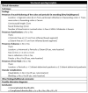
Structured reporting template.

Similar articles

Cited by

References

    1. Hong M.K., Skandarajah A.R., Hayes I.P. Diverticulosis, diverticular disease and diverticulitis—Definitions and differences. J. Stomal Ther. Aust. 2015;35:8–10.
    1. Naves A.D.A., D’Ippolito G., Souza L.R.M.F., Borges S.P., Fernandes G.M. What radiologists should know about tomographic evaluation of acute diverticulitis of the colon. Radiol. Bras. 2017;50:126–131. doi: 10.1590/0100-3984.2015.0227. - DOI - PMC - PubMed
    1. Sugi M.D., Sun D.C., Menias C.O., Prabhu V., Choi H.H. Acute diverticulitis: Key features for guiding clinical management. Eur. J. Radiol. 2020;128:109026. doi: 10.1016/j.ejrad.2020.109026. - DOI - PubMed
    1. Stollman N., Raskin J.B. Diverticular disease of the colon. Lancet. 2004;363:631–639. doi: 10.1016/S0140-6736(04)15597-9. - DOI - PubMed
    1. Onur M.R., Akpinar E., Karaosmanoglu A.D., Isayev C., Karcaaltincaba M. Diverticulitis: A comprehensive review with usual and unusual complications. Insights Imaging. 2017;8:19–27. doi: 10.1007/s13244-016-0532-3. - DOI - PMC - PubMed

LinkOut - more resources