Review
. 2023 Dec 11;13(24):3638.
doi: 10.3390/diagnostics13243638.

The Role of Multimodality Imaging in Patients with Congenital Heart Disease and Infective Endocarditis

Affiliations
Review

The Role of Multimodality Imaging in Patients with Congenital Heart Disease and Infective Endocarditis

Sara Moscatelli et al. Diagnostics (Basel). .

Abstract

Infective endocarditis (IE) represents an important medical challenge, particularly in patients with congenital heart diseases (CHD). Its early and accurate diagnosis is crucial for effective management to improve patient outcomes. Multimodality imaging is emerging as a powerful tool in the diagnosis and management of IE in CHD patients, offering a comprehensive and integrated approach that enhances diagnostic accuracy and guides therapeutic strategies. This review illustrates the utilities of each single multimodality imaging, including transthoracic and transoesophageal echocardiography, cardiac computed tomography (CCT), cardiovascular magnetic resonance imaging (CMR), and nuclear imaging modalities, in the diagnosis of IE in CHD patients. These imaging techniques provide crucial information about valvular and intracardiac structures, vegetation size and location, abscess formation, and associated complications, helping clinicians make timely and informed decisions. However, each one does have limitations that influence its applicability.

Keywords: cardiac computed tomography; cardiac magnetic resonance imaging; congenital heart disease; echocardiography; infective endocarditis; multimodality imaging; nuclear imaging.


Conflict of interest statement

The authors declare no conflict of interest.

Figures
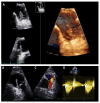
Figure 1
Figure 1
Transthoracic echocardiography of an adult patient with previous Ross procedure for bicuspid aortic valve using a pulmonary homograft. There are large mobile vegetations (max ~18 × 7 mm, (A,B), black arrows) on the pulmonary homograft. Severe pulmonary regurgitation (regurgitant jet originating in the main pulmonary artery, vena contracta 8.5 mm, (C)); no significant stenosis (mean gradient 13 mmHg, peak gradient 26 mmHg, (D)). The findings are consistent with infective endocarditis. Images courtesy of Ms Joane Daradar—Echocardiography Department, Royal Brompton and Harefield Hospitals, London.
Figure 2
Figure 2
Right ventricle outflow tract bSSFP CMR image with endocarditis vegetation of a thirty-year-old male with Tetralogy of Fallot and a bio pulmonic conduit in the pulmonary position, with positive blood cultures for S. Mitis following dental extraction.
Figure 3
Figure 3
Cardiac computed tomography infective endocarditis visualisation. The images show right-ventricular pulmonary artery (RV-PA) conduit involvement (panels (A,B)) and systemic to pulmonary shunt involvement with a calcified lesion (panel (C,D)). The arrows indicate the vegetations.
Figure 4
Figure 4
A case of pulmonary valve/valve duct endocarditis of a 38-year-old female patient (surgically treated for Fallot tetralogy in 1988 and 2012), evaluated with 18F-fluorodeoxyglucose (18F-FDG) positron emission tomography/computed tomography (PET/CT): axial CT, PET/CT, and PET images (AC) and maximum intensity projection (MIP) image (D) show hypermetabolism in the pulmonary valve prosthesis and in the valve duct (orange arrows), with extension to aortic wall (demonstrating a disease involvement in the aorta as well). Moreover, the images depict multiple reactive hylo-mediastinal lymph-nodes with high uptake of the radiopharmaceutical (blue arrows) and hypermetabolism in an area of consolidation in the right lung (manifestation of pneumonia, green arrows). In addition, image D shows high uptake of 18F-FDG in spleen and bone marrow, as a sign of systemic inflammatory response.

References

    1. Moore B., Cao J., Kotchetkova I., Celermajer D.S. Incidence, predictors and outcomes of infective endocarditis in a contemporary adult congenital heart disease population. Int. J. Cardiol. 2017;249:161–165. doi: 10.1016/j.ijcard.2017.08.035. - DOI - PubMed
    1. Montanaro C., Dimopoulos K., Shore D.F. Infective endocarditis in patients with congenital heart disease: When, where and how. Int. J. Cardiol. 2017;249:171–172. doi: 10.1016/j.ijcard.2017.09.186. - DOI - PubMed
    1. Habib G., Erba P.A., Iung B., Donal E., Cosyns B., Laroche C., Popescu B.A., Prendergast B., Tornos P., Sadeghpour A., et al. Clinical presentation, aetiology and outcome of infective endocarditis. Results of the ESC-EORP EURO-ENDO (European infective endocarditis) registry: A prospective cohort study. Eur. Heart J. 2019;40:3222–3232. doi: 10.1093/eurheartj/ehz620. Erratum in Eur. Heart J. 2020, 41, 2091. - DOI - PubMed
    1. Baddour L.M., Wilson W.R., Bayer A.S., Fowler V.G., Jr., Tleyjeh I.M., Rybak M.J., Barsic B., Lockhart P.B., Gewitz M.H., Levison M.E., et al. Infective Endocarditis in Adults: Diagnosis, Antimicrobial Therapy, and Management of Complications: A Scientific Statement for Healthcare Professionals from the American Heart Association. Circulation. 2015;132:1435–1486. doi: 10.1161/CIR.0000000000000296. Erratum in Circulation 2015, 132, e215. Erratum in Circulation 2016, 134, e113. Erratum in Circulation 2018, 138, e78–e79. - DOI - PubMed
    1. Reynolds H.R., Jagen M.A., Tunick P.A., Kronzon I. Sensitivity of transthoracic versus transesophageal echocardiography for the detection of native valve vegetations in the modern era. J. Am. Soc. Echocardiogr. 2003;16:67–70. doi: 10.1067/mje.2003.43. - DOI - PubMed

LinkOut - more resources