. 2023 Dec 14;13(24):3663.
doi: 10.3390/diagnostics13243663.

Rare Pancreatic/Peripancreatic Cystic Lesions Can Be Accurately Characterized by EUS with Through-the-Needle Biopsy-A Unique Pictorial Essay with Clinical and Histopathological Correlations

Affiliations

Rare Pancreatic/Peripancreatic Cystic Lesions Can Be Accurately Characterized by EUS with Through-the-Needle Biopsy-A Unique Pictorial Essay with Clinical and Histopathological Correlations

Maria Cristina Conti Bellocchi et al. Diagnostics (Basel). .

Abstract

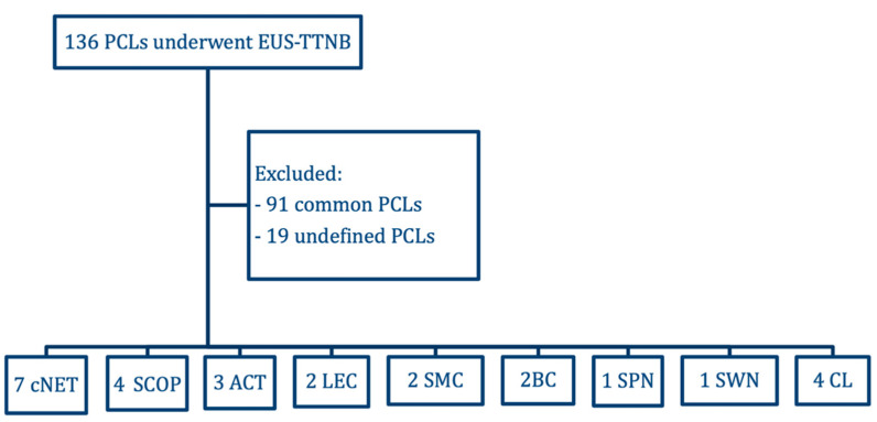
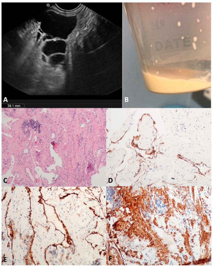
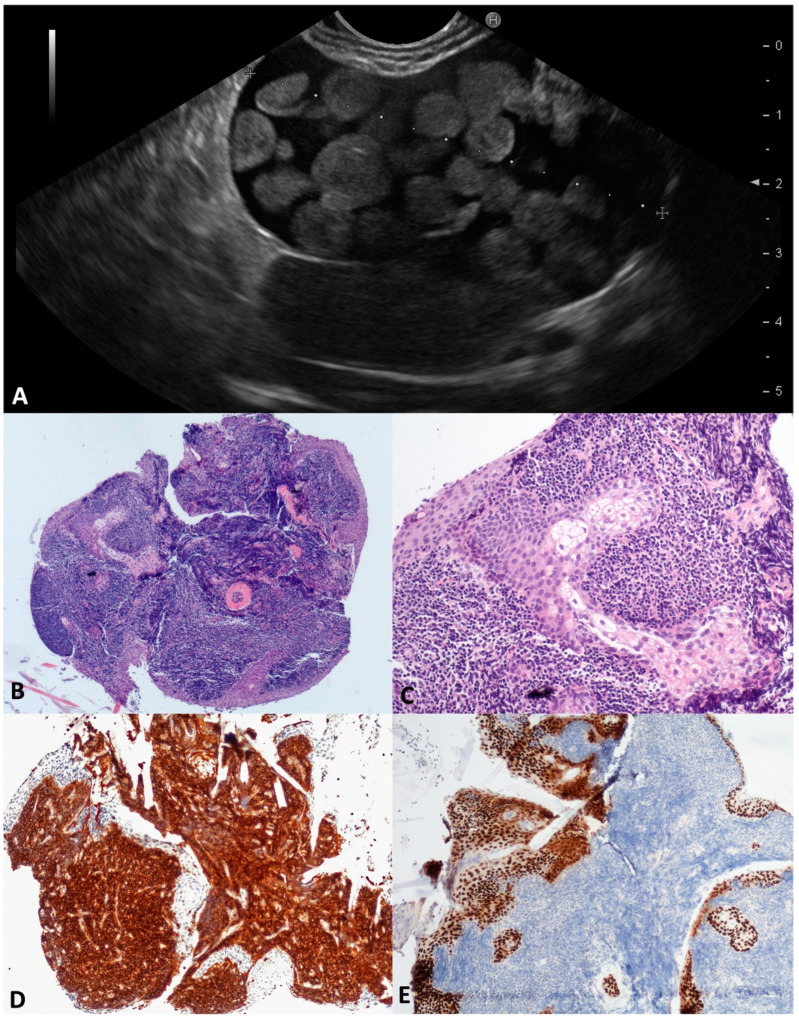
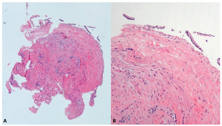
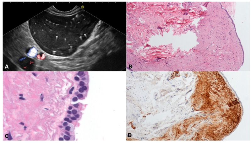
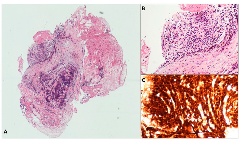
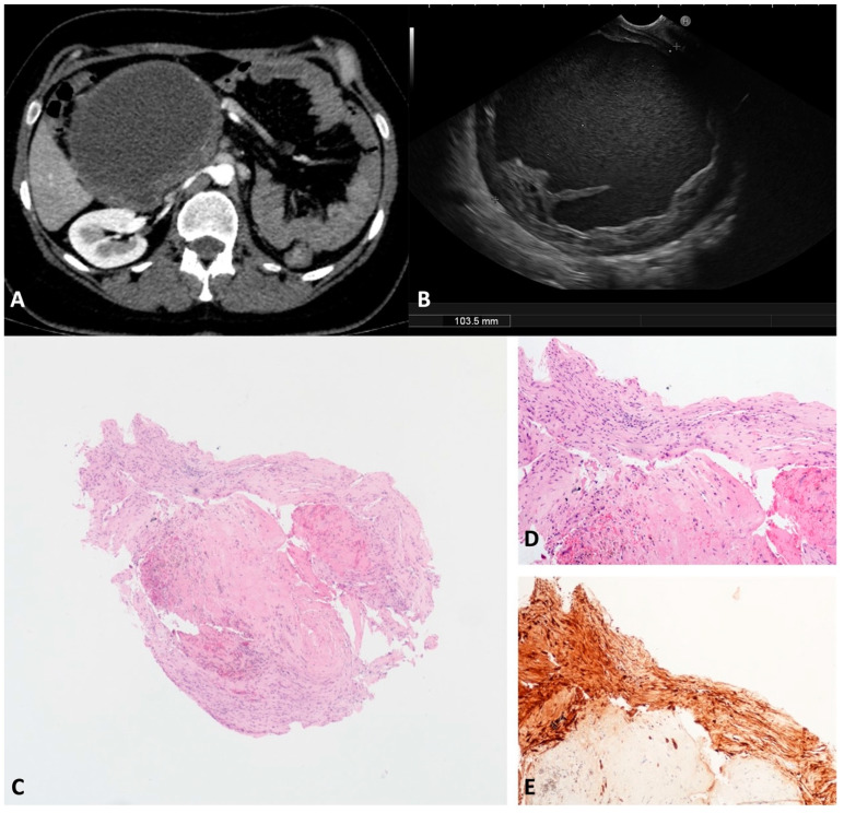
Due to their aspecific macroscopic appearance, uncommon pancreatic cystic lesions (PCLs) are often misdiagnosed as mucinous lesions and improperly resected. We aimed to evaluate the endoscopic ultrasound (EUS)-guided through-the-needle biopsy (TTNB) capacity of the preoperative diagnosis of uncommon PCLs. Overall, 136 patients with PCLs who underwent EUS-TTNB between 2016 and 2022 were retrospectively identified. Common histotypes (e.g., IPMN, serous cystadenoma, and mucinous cystadenoma) were excluded and 26 (19.1%) patients (15 female, mean age 52.9 ± 10.4) were analyzed. The EUS findings, adverse events (AEs), and TTNB outcomes in uncommon PCLs were evaluated. The cysts histotype was accurately diagnosed by TTNB in 24/26 (92.3%) cases (seven cystic neuroendocrine tumors, four squamoid cysts, three acinar cells cystadenomas, two lymphoepithelial cysts, two mucinous non-neoplastic cysts, two bronchogenic cysts, two cystic lymphangiomas, one solid-pseudopapillary neoplasm, and one schwannoma). In the remaining two cases, lymphangioma was eventually diagnosed after resection. Surgery was performed in 15/26 (57.7%) patients. The mean follow-up of non-surgical patients was 32.5 months. One severe acute case of pancreatitis (3.8%) that required surgery occurred after EUS-TTNB. Uncommon pancreatic/peripancreatic lesions represent the 19.1% of PCLs in our series, with mainly benign histotypes. TTNB demonstrated a high diagnostic performance with a low rate of AEs in this setting, representing a reliable tool with which to avoid useless surgery.

Keywords: endoscopic ultrasound; fine-needle aspiration; pancreatic cancer; pancreatic cyst; pancreatic surgery; through-the-needle biopsy.


Conflict of interest statement

The authors declare no conflict of interest.

Figures

Figure 1
Figure 1
Flowchart of the study. EUS-TTNB, endoscopic ultrasound-guided through-the-needle biopsy; PCLs, pancreatic cystic lesions; cNET, cystic neuroendocrine tumors; SCOP, squamoid cyst of pancreatic duct; ACT, acinar cystic transformation; LEC, lymphoepithelial cyst; SMC, simple mucinous cyst; BC, bronchogenic cyst; SPN, solid pseudopapillary neoplasm; SWN, schwannoma; CL, cystic lymphangioma.
Figure 2
Figure 2
Cystic neuroendocrine neoplasm. Contrast-harmonic endoscopic ultrasound showing hyper-enhanced walls of an unilocular cyst (A). Endoscopic ultrasound-guided through-the-needle biopsy targeting the thickened walls (B). The cyst wall is almost completely composed of small, tightly packed epithelial cells (C) that stain intensely for the neuroendocrine marker Synaptophysin (D). Hematoxylin–eosin original magnification ×100 (C). Synaptophysin original magnification ×100 (D).
Figure 3
Figure 3
Squamoid cyst of pancreatic ducts. T2-weighted magnetic resonance imaging of a pancreatic cyst located in the head, with a nodule inside (A). The same cyst on an endoscopic ultrasound scan containing round vegetation that resulted in avascular at contrast-harmonic evaluation (B). The fibrous thin wall of the cyst is lined by stratified epithelium (C) without atypia and keratinization (D). Hematoxylin–eosin original magnification ×100 (C), ×200 (D).
Figure 4
Figure 4
Acinar Cystic Transformation. The whole mounted section of the multilocular cyst with fibrous wall (A). The lining epithelium is composed of cuboidal cells with abundant eosinophilic and granular cytoplasm, organized both in monolayer and acinar aggregates (B). The acinar markers BCL10 (C) and trypsin (D) are detected in epithelial cells. Hematoxylin–eosin original magnification ×40 (A), ×100 (B); BCL10 original magnification ×100 (C); Trypsin original magnification ×100 (D).
Figure 5
Figure 5
Cystic lymphangioma. Endoscopic ultrasound revealed an irregularly shaped multilocular cyst close to the pancreatic parenchyma, with a thin wall and septa (A). The aspirated fluid appeared as thick “milky” white-yellowish fluid (B). Numerous narrow lymphatic vessels lined by a cuboidal epithelium were separated by bundles of smooth muscle with few aggregates of lymphocytes (C). Immunolabelling for endothelial markers D2-40 (D) and CD31 (E). Bundles of smooth muscle actin (SMA) positive cells (F). Hematoxylin–eosin original magnification ×100 (C); D2-40 original magnification ×200 (D); CD31 original magnification ×200 (E); SMA original magnification ×200 (F).
Figure 6
Figure 6
Lymphoepithelial cyst. During the endoscopic ultrasound, a large cystic lesion with a regular wall and central thickened septum containing multiple round, echoic, and avascular floating balls after contrast injection was documented (A). The whole mounted section of the cyst wall forceps biopsy (B). Large aggregates of lymphoid cells are separated by squamous epithelial cells with sebaceous glands (C). Lymphocytes are highlighted by CD45 (D) and squamous cells by P63 (E) immunostaining, respectively. Hematoxylin–eosin original magnification ×40 (B), ×200 (C); CD45 original magnification ×100 (D); P63 original magnification ×100 (E).
Figure 7
Figure 7
Simple mucinous cyst. The whole fully mounted section of the cyst wall forceps biopsy (A). At higher magnification, the thick fibrous cist wall is lined with a monolayer of mucinous epithelial cells, without atypia (B). Hematoxylin–eosin original magnification ×40 (A), ×200 (B).
Figure 8
Figure 8
Bronchogenic cyst. Typical appearance of bronchogenic cyst on endoscopic ultrasound. The content of the cyst is similar to the liver parenchyma, with multiple white spots with a comet-tail artifact (A). The cyst wall biopsy shows a thick layer of eosinophilic, dense, hypocellular tissue beneath the pseudostratified epithelium (B). The epithelium lining the cyst wall is composed of ciliated and goblet cells (C). The subepithelial connective tissue is rich in smooth muscle fibers, which are Calponin-positive (D). Hematoxylin–eosin original magnification ×100 (B), ×400 (C); Calponin original magnification ×100 (D).
Figure 9
Figure 9
Solid pseudopapillary tumor. The whole mounted section of the cyst wall tissue fragment shows an altered histological architecture with an area of crash artefact caused by the forceps sampling procedure (A). In an area with better preserved morphology, the epithelial cells are round, small and tightly packed (B). Immunolabelling for β-catenin with aberrant nuclear positivity is conclusive for the diagnosis (C). Hematoxylin–eosin original magnification ×40 (A), ×200 (B); β-catenin original magnification ×200 (C).
Figure 10
Figure 10
Schwannoma. Computed tomography appearance of a large cystic lesion located between the second/third portion of the duodenum and the pancreatic head that appeared compressed and dislodged (A). On the endoscopic ultrasound, the lesion appeared as a round unilocular cyst with smooth borders and an irregular thickened wall (B). The whole mounted section of the cyst wall biopsy shows no epithelium lining the cyst (C). A well visible fascicle of spindle cells depicts the peripheral profile of the biopsy (D). Diffuse and intense immunolabelling for S100 in spindle cells (E). Hematoxylin–eosin original magnification ×40 (C), ×100 (D). S100 original magnification ×100 (E).

References

    1. Mizuno S., Nakai Y., Yoshikawa T., Ishigaki K., Matsubara S., Yamamoto N., Ijichi H., Tateishi K., Tada M., Hayashi N., et al. Prevalence of pancreatic cystic lesions is associated with diabetes mellitus and obesity: An analysis of 5296 individuals who underwent a preventive medical examination. Pancreas. 2017;46:801–805. - PubMed
    1. Tanaka M., Fernández-Del Castillo C., Kamisawa T., Jang J.Y., Levy P., Ohtsuka T., Salvia R., Shimizu Y., Tada M., Wolfgang C.L. Revisions of international consensus Fukuoka guidelines for the management of IPMN of the pancreas. Pancreatology. 2017;17:738–753. doi: 10.1016/j.pan.2017.07.007. - DOI - PubMed
    1. Salvia R., Marchegiani G., Pennacchio S., Paiella S., Paini M., Pea A., Butturini G., Pederzoli P., Bassi C. Pancreatic resections for cystic neoplasms: From the surgeon’s presumption to the pathologist’s reality. Surgery. 2012;152:S135–S142. doi: 10.1016/j.surg.2012.05.019. - DOI - PubMed
    1. de Pretis N., Aryal-Khanal A., Bi Y., Takahashi N., Chari S. Pancreatic cysts: Diagnostic accuracy and risk of inappropriate resections. Pancreatology. 2017;17:267–272. doi: 10.1016/j.pan.2017.01.002. - DOI - PubMed
    1. Canakis A. State-of-the-Art Update of Pancreatic Cysts. Dig. Dis. Sci. 2022;67:1573–1587. doi: 10.1007/s10620-021-07084-1. - DOI - PMC - PubMed

LinkOut - more resources