Case Reports
. 2023 Nov 30;11(12):278.
doi: 10.3390/dj11120278.

Biomechanically and Periodontally-Based Orthodontic Treatment of a Patient with Upper Canine Affected by External Cervical Resorption (ECR): A Case Report

Affiliations
Case Reports

Biomechanically and Periodontally-Based Orthodontic Treatment of a Patient with Upper Canine Affected by External Cervical Resorption (ECR): A Case Report

Marino Musilli et al. Dent J (Basel). .

Abstract

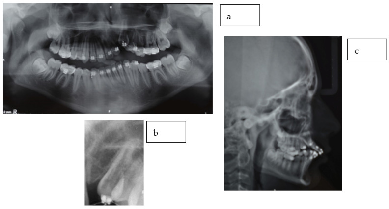
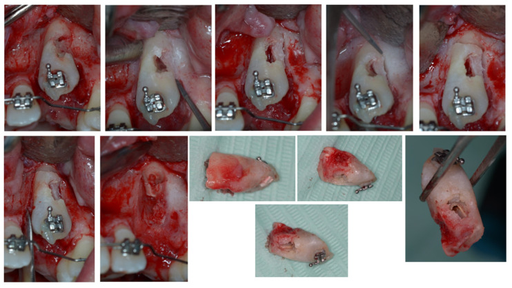
(1) Background: Orthodontic treatment may be a potential predisposing factor for ECR. The affected tooth goes to ankylosis, which could lead to a malocclusion. Although teeth severely affected by ECR (class IV Heithersay) are usually extracted, this case report aims to present the use of an ECR class IV upper canine, both as ankylosed to solve the malocclusion and the occlusal plane canting, as well as not ankylosed to correct its ridge defect with orthodontic extrusion. (2) Methods: A 14-year-old male, complaining of an ugly smile and a failed orthodontic attempt to recover an impacted canine, was referred to the orthodontic clinic. He was diagnosed with class II right subdivision, midline deviation, both upper and lower occlusal plane canting, and an upper left canine, previously impacted, showing ECR class IV. The treatment first included canting resolution with a cantilever and a spring, exploiting the anchorage offered by the ankylosed ECR canine. Then, a coronectomy, endodontic treatment, and orthodontic extrusion of that canine were performed to obtain the implant site development. (3) Results: Clinical and radiographic outcomes showed normocclusion and better bony conditions for safer implant placement in the aesthetic zone. (4) Conclusions: The high aesthetics and the periodontal and bony conditions obtained are probably not achievable by other therapeutic alternatives.

Keywords: ankylosed upper canine; case report; external cervical resorption; occlusal plane canting; orthodontic extrusion.


Conflict of interest statement

The authors declare no conflict of interest.

Figures

Figure A1
Figure A1
CARE Guidelines Completed Checklist.
Figure 1
Figure 1
Intra-oral patient baseline presentation (with multibrackets fixed appliance elsewhere used to recover the impacted canine).
Figure 2
Figure 2
Extra-oral patient baseline presentation (with multibracket fixed appliance elsewhere used to recover the impacted canine).
Figure 3
Figure 3
X-rays: orthopantomography (a), intraoral X-ray (b), teleradiography (c). (a,b) showed a radio transparency on the distal surface of the upper left canine. Cantings were also evident in (a,c). No other anomalies could be detected.
Figure 4
Figure 4
The end of the canting resolution phase is shown intra-orally. Here, the ECR upper left canine was located apical to its ideal position and far from the occlusal plane.
Figure 5
Figure 5
The end of the canting resolution phase extra-orally is shown. The ECR upper left canine was located apical to its ideal position and far from the occlusal plane.
Figure 6
Figure 6
Surgical canine exposure to remove the damaged ankylosed distal surface with coronectomy.
Figure 7
Figure 7
Endodontic treatment of 2.3 element residual root.
Figure 8
Figure 8
Cantilever bonded to the residual root.
Figure 9
Figure 9
The extrusion system with the Elgiloy cantilever was inserted in two palatal mini-screws and connected to the canine residual root.
Figure 10
Figure 10
A 0.9 mm cantilever in stainless steel replaced the first one to increase the extrusion at the orthopaedic force of 500 g.
Figure 11
Figure 11
As soon as the residual root erupted, the canine was orthodontically splinted with a small composite veneer for aesthetic reasons.
Figure 12
Figure 12
Malocclusion of class II left subdivision to correct with class II elastics.
Figure 13
Figure 13
Final normo-occlusion.
Figure 14
Figure 14
Good aesthetic facial and profile.
Figure 15
Figure 15
Normal panoramic X-ray features.

References

    1. Ezoddini A.F., Sheikhha M.H., Ahmadi H. Prevalence of dental developmental anomalies: A radiographic study. Community Dent. Health. 2007;24:140–144. - PubMed
    1. Becker A., Chaushu G., Chaushu S. Analysis of failure in the treatment of impacted maxillary canines. Am. J. Orthod. Dentofac. Orthop. 2010;137:743–754. doi: 10.1016/j.ajodo.2008.07.022. - DOI - PubMed
    1. Becker A., Abramovitz I., Chaushu S. Failure of treatment of impacted canines associated with invasive cervical root resorption. Angle Orthod. 2013;83:870–876. doi: 10.2319/090812-716.1. - DOI - PMC - PubMed
    1. Tronstad L. Root resorption—Etiology, terminology and clinical manifestations. Dent. Traumatol. 1988;4:241–252. doi: 10.1111/j.1600-9657.1988.tb00642.x. - DOI - PubMed
    1. Patel S., Mavridou A.M., Lambrechts P., Saberi N. External cervical resorption-part 1: Histopathology, distribution and presentation. Int. Endod. J. 2018;51:1205–1223. doi: 10.1111/iej.12942. - DOI - PubMed

Publication types

LinkOut - more resources