Review
. 2023 Dec 12;11(4):45.
doi: 10.3390/jdb11040045.

Phase Separation as a Driver of Stem Cell Organization and Function during Development

Affiliations
Review

Phase Separation as a Driver of Stem Cell Organization and Function during Development

Amalia S Parra et al. J Dev Biol. .

Abstract

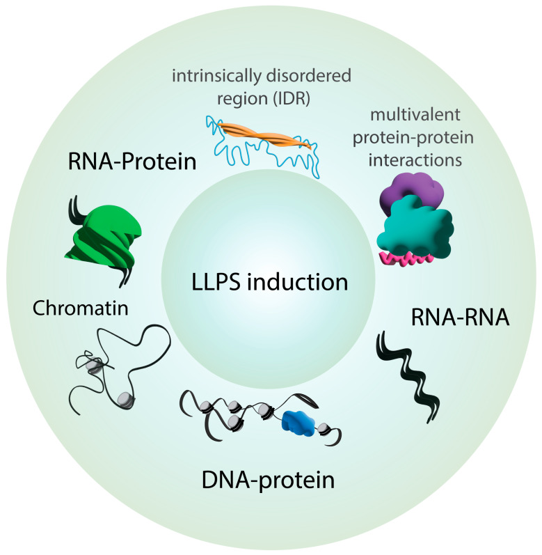
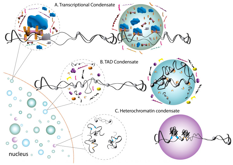
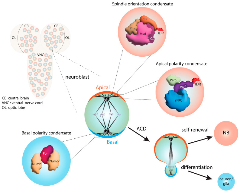
A properly organized subcellular composition is essential to cell function. The canonical organizing principle within eukaryotic cells involves membrane-bound organelles; yet, such structures do not fully explain cellular complexity. Furthermore, discrete non-membrane-bound structures have been known for over a century. Liquid-liquid phase separation (LLPS) has emerged as a ubiquitous mode of cellular organization without the need for formal lipid membranes, with an ever-expanding and diverse list of cellular functions that appear to be regulated by this process. In comparison to traditional organelles, LLPS can occur across wider spatial and temporal scales and involves more distinct protein and RNA complexes. In this review, we discuss the impacts of LLPS on the organization of stem cells and their function during development. Specifically, the roles of LLPS in developmental signaling pathways, chromatin organization, and gene expression will be detailed, as well as its impacts on essential processes of asymmetric cell division. We will also discuss how the dynamic and regulated nature of LLPS may afford stem cells an adaptable mode of organization throughout the developmental time to control cell fate. Finally, we will discuss how aberrant LLPS in these processes may contribute to developmental defects and disease.

Keywords: asymmetric cell division; cell fate; cell polarity; chromatin; phase separation; spindle orientation; stem cell.


Conflict of interest statement

The authors declare no conflict of interest.

Figures

Figure 1
Figure 1
Molecular features commonly found in biomolecular condensate components. Some chemical properties of proteins can drive LLPS induction. Intrinsically disordered regions (IDRs), multivalent protein–protein interactions, and nucleic acids are commonly found in membranelle compartments and are associated with the formation of biomolecular condensates.
Figure 2
Figure 2
Summary of phase separation events in the regulation of gene expression. Changes in gene expression occur in condensates that sequester the transcriptional machinery. (A) Homogenous mixtures (dashed circles) contain accessory proteins (purple, pink, orange) and co-activators (gold, gray) that facilitate transcription (DNA; black, histones; grey) in response to a stimulus or return to the homogenous state in the absence or with the withdrawal of stimuli. Transcriptional condensates often form through IDRs found within the activation domain of specific transcription factors. (B) Topologically associated domains (TADs) are three-dimensional organizations of chromatin regions that span large genomic distances to control gene expression and contain histone proteins and marks (gray and orange). Chromatin loops are often conserved across cell types and are monitored by regulatory elements (yellow, pink). (C) Gene repression occurs via condensate-mediated sequestration of heterochromatin protein 1 (HP1) (blue) into heterochromatin condensates. These condensates form around promoters on chromatin (black) and histones (gray). Histone-associated HP1 (orange) promotes gene silencing and the organization of higher-order chromatin structures.
Figure 3
Figure 3
Summary of the signaling pathways regulated by phase separation during development. (Left): Hippo signaling pathway regulates cell growth, proliferation, and survival, including in many stem cells. Mechanoregulation, polarity complexes, and GPCR signaling are upstream regulatory signals modulating Hippo activity. Hippo (Mst1/2) signaling is a phosphorylation cascade that inactivates STRIPAK. This leads to the phosphorylation of YAP/TAZ by LATS1/2, thereby preventing the nuclear translocation of the transcriptional regulator. Partitioning of Mst1/2 and STRIPAK to distinct condensates prevents YAP/TAZ phosphorylation, leading to the nuclear translocation and transcriptional activation of target genes. (Right): Wnt signaling also plays a critical role in development and occurs through binding to the frizzled receptor and LRP5/6 co-receptor (‘Active’). Wnt pathway activation is facilitated by the formation of β-catenin signalosome condensates consisting of disheveled, GSK/CK1 kinases, APC, and axin. This complex causes the stabilization of β-catenin by preventing its phosphorylation and allowing its nuclear translocation for the activation of target genes. The absence of Wnt (‘Inactive’) promotes the phosphorylation of β-catenin within distinct condensates containing a destruction complex composed of active GSK/CK1 kinases, APC, axin, and β-catenin. Phosphorylated β-catenin is targeted for proteasomal degradation.
Figure 4
Figure 4
Summary of phase separation functions in asymmetric cell division. Drosophila neuroblasts (NBs), like most stem cells, divide via asymmetric cell division (ACD). ACD is established by the coordination of mitotic spindle orientation and cell polarity cues. Distinct condensates containing apical (orange) and basal (blue) complexes have been identified. The apical polarity complex (orange) consists of the conserved proteins atypical protein kinase C (aPKC), Par3, and Par6. The intrinsically disordered region of Par3 facilitates the phase separation of this complex. The apical spindle orientation complex contains Pins, Mud, and Hu li tai shao (Hts). Formation of the spindle orientation condensate is promoted by multivalent protein–protein interactions mediated by the Mud coiled-coil domain and an IDR within the Hts C-terminus. The basal domain (blue) of NBs contains the differentiation factors Numb and partner of numb (Pon). These components form a basal polarity condensate through multivalent interactions of Numb with tandem binding sequences in Pon. The phase-separated complexes are inherited by the daughter cell that will differentiate to form neurons or glial cells, following ACD.

References

    1. Banani S.F., Lee H.O., Hyman A.A., Rosen M.K. Biomolecular condensates: Organizers of cellular biochemistry. Nat. Rev. Mol. Cell Biol. 2017;18:285–298. doi: 10.1038/nrm.2017.7. - DOI - PMC - PubMed
    1. Shin Y., Brangwynne C.P. Liquid phase condensation in cell physiology and disease. Science. 2017;357:eaaf4382. doi: 10.1126/science.aaf4382. - DOI - PubMed
    1. Bracha D., Walls M.T., Brangwynne C.P. Probing and engineering liquid-phase organelles. Nat. Biotechnol. 2019;37:1435–1445. doi: 10.1038/s41587-019-0341-6. - DOI - PubMed
    1. Dignon G.L., Best R.B., Mittal J. Biomolecular Phase Separation: From Molecular Driving Forces to Macroscopic Properties. Annu. Rev. Phys. Chem. 2020;71:53–75. doi: 10.1146/annurev-physchem-071819-113553. - DOI - PMC - PubMed
    1. Rhine K., Vidaurre V., Myong S. RNA Droplets. Annu. Rev. Biophys. 2020;49:247–265. doi: 10.1146/annurev-biophys-052118-115508. - DOI - PMC - PubMed

LinkOut - more resources