Review
. 2023 Dec 11;9(12):1187.
doi: 10.3390/jof9121187.

Exploring the Molecular Underpinnings of Cancer-Causing Oncohistone Mutants Using Yeast as a Model

Affiliations
Review

Exploring the Molecular Underpinnings of Cancer-Causing Oncohistone Mutants Using Yeast as a Model

Xinran Zhang et al. J Fungi (Basel). .

Abstract

Understanding the molecular basis of cancer initiation and progression is critical in developing effective treatment strategies. Recently, mutations in genes encoding histone proteins that drive oncogenesis have been identified, converting these essential proteins into "oncohistones". Understanding how oncohistone mutants, which are commonly single missense mutations, subvert the normal function of histones to drive oncogenesis requires defining the functional consequences of such changes. Histones genes are present in multiple copies in the human genome with 15 genes encoding histone H3 isoforms, the histone for which the majority of oncohistone variants have been analyzed thus far. With so many wildtype histone proteins being expressed simultaneously within the oncohistone, it can be difficult to decipher the precise mechanistic consequences of the mutant protein. In contrast to humans, budding and fission yeast contain only two or three histone H3 genes, respectively. Furthermore, yeast histones share ~90% sequence identity with human H3 protein. Its genetic simplicity and evolutionary conservation make yeast an excellent model for characterizing oncohistones. The power of genetic approaches can also be exploited in yeast models to define cellular signaling pathways that could serve as actionable therapeutic targets. In this review, we focus on the value of yeast models to serve as a discovery tool that can provide mechanistic insights and inform subsequent translational studies in humans.

Keywords: budding yeast; cancer; epigenetics; fission yeast; histone; oncohistone.


Conflict of interest statement

The authors declare no conflict of interest.

Figures

Figure 1
Figure 1
Nucleosome structure and the consequences of an oncohistone mutant. (A) Nucleosomes are comprised of two copies of each of the four core histone proteins: H2A, H2B, H3, and H4. When only wild type histones are present, cells display normal growth. (B) When an oncohistone missense mutation occurs, such as H3K36M (red), various functional consequences may result. Even with many wildtype H3 proteins present, a single mutation in one allele can confer oncogenic growth through a variety of mechanisms, including altered post translational modifications, modification of the genomic landscape, and/or disruption of gene expression.
Figure 2
Figure 2
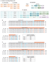
A comparison of histone H3 from S. cerevisiae, S. pombe, and H. sapiens. (A) The number of gene copies and protein variants differs greatly between yeast species (S. cerevisiae and S. pombe), which are termed HHT genes, and humans (H. sapiens), which are termed H3XX, with the exception of the specialized centromeric histone CENPA for histone H3. Nomenclature guidelines for each species were followed. The genes that encode each variant are color coordinated with their respective H3 Variants. * Non-canonical H3 genes and variants. (B) Protein sequence alignments for histone H3 in S. cerevisiae and the three canonical H3 variants in H. sapiens. Blue residues represent conservative changes, where the biochemical properties of the amino acid are maintained, and orange residues represent non conservative changes, where the biochemical properties are altered. (C) Protein sequence alignments for histone H3 in S. pombe and the three canonical H3 variants in H. sapiens. Color coding is the same as for (B).
Figure 3
Figure 3
Mutations that alter histone H3 sequence recur in human cancers. (A) A cross-cancer mutation analysis was performed utilizing 5406 adult patient samples obtained from the COSMIC and cBioPortal databases. Samples from the two databases were cross-referenced and duplicate entries were removed. The data were then visualized using PRISM GraphPad. This summary includes missense mutations found in H3C1, H3C2, H3C3, H3C4, H3C5, H3C6, H3C7, H3C8, H3C10, H3C11, H3C12, H3C13, H3C14, H3C15, H3-3A, H3-3B, and H3-4. The number of patients that have a mutation (missense or premature termination codon as indicated by *) located in the codon corresponding to each specific amino acid residue along the protein is indicated on the Y-axis (# Mutations across Cancers) with the residues that the amino acid is altered to being indicated by the colors or the asterisk (*). Only residues that are conserved in S. cerevisiae H3 are shown along the X-axis as these represent potential oncohistones that could be modeled in budding yeast. (B) The number of events for which a given amino acid is converted into all other amino acids was assessed, regardless of position in the protein sequence. Premature termination codons were excluded to focus on missense mutants. Same dataset as in (A).
Figure 4
Figure 4
The strength of modeling oncohistone mutants in budding yeast. (A) Budding yeast can be easily engineered to maintain wildtype H3 genes (top), express one mutant and one wildtype H3 (middle), or express only mutant H3 with the wildtype knocked out (bottom). The resulting ratio of wildtype to mutant H3 proteins in a pair of nucleosomes for the given genotypes is depicted on the right. (B) In this serial dilution growth assay, budding yeast cultures were diluted to OD = 5 and serially diluted tenfold before plating onto control or drug plates. The analysis compares H3K36 mutant cells that either have one mutant and one wildtype histone protein (hht2-K36M/R HHT1) and cells that contain mutant histone as the sole copy of histone H3 (hht2-K36M/R hht1Δ) to control wildtype (WT) cells. Cells on control plates were grown for two days, cells on caffeine were grown for five days, and cells on phleomycin were grown for three days. The cellular damage caused by the drugs is indicated below the plates, and the ratio of wildtype to mutant H3 in the nucleosomes within each model is depicted to the right.

References

    1. Xu J., Murphy S.L., Kochanek K.D., Arias E. Mortality in the United States. National Center for Health Statistics; Hyattsville, MD, USA: 2022. pp. 1–8. 2021 NCHS Data Brief.
    1. Hartwell L.H. Yeast and cancer. Biosci. Rep. 2004;24:523–544. doi: 10.1007/s10540-005-2743-6. - DOI - PubMed
    1. Smith M.M. Histone structure and function. Curr. Opin. Cell Biol. 1991;3:429–437. doi: 10.1016/0955-0674(91)90070-F. - DOI - PubMed
    1. Millar C.B., Grunstein M. Genome-wide patterns of histone modifications in yeast. Nat. Rev. Mol. Cell Biol. 2006;7:657–666. doi: 10.1038/nrm1986. - DOI - PubMed
    1. Luger K., Mäder A.W., Richmond R.K., Sargent D.F., Richmond T.J. Crystal structure of the nucleosome core particle at 2.8 A resolution. Nature. 1997;389:251–260. doi: 10.1038/38444. - DOI - PubMed

LinkOut - more resources