. 2023 Dec 3;14(12):563.
doi: 10.3390/jfb14120563.

Model-Based Design to Enhance Neotissue Formation in Additively Manufactured Calcium-Phosphate-Based Scaffolds

Affiliations

Model-Based Design to Enhance Neotissue Formation in Additively Manufactured Calcium-Phosphate-Based Scaffolds

Bingbing Liang et al. J Funct Biomater. .

Abstract

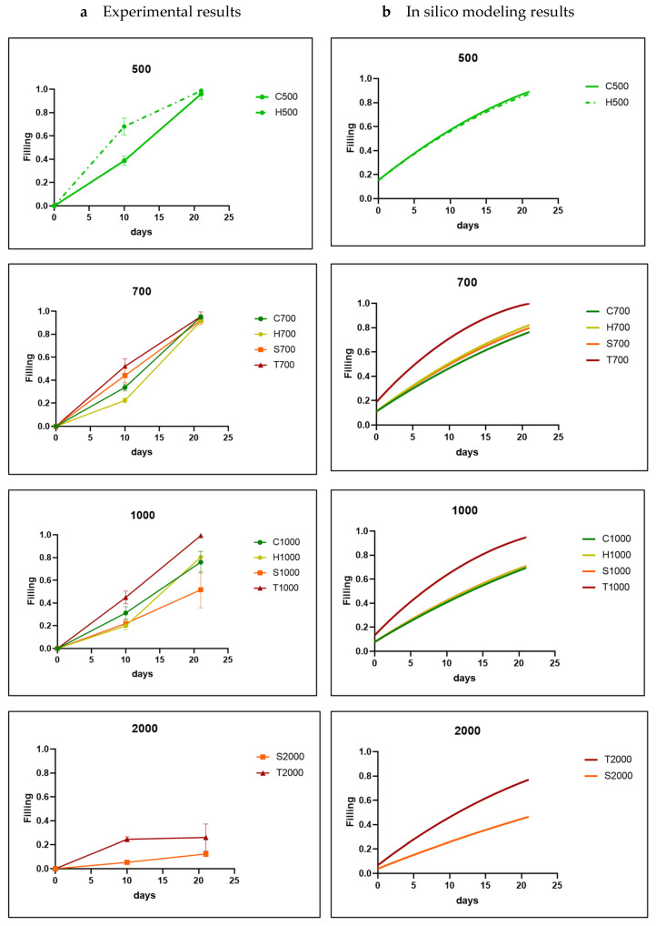
In biomaterial-based bone tissue engineering, optimizing scaffold structure and composition remains an active field of research. Additive manufacturing has enabled the production of custom designs in a variety of materials. This study aims to improve the design of calcium-phosphate-based additively manufactured scaffolds, the material of choice in oral bone regeneration, by using a combination of in silico and in vitro tools. Computer models are increasingly used to assist in design optimization by providing a rational way of merging different requirements into a single design. The starting point for this study was an in-house developed in silico model describing the in vitro formation of neotissue, i.e., cells and the extracellular matrix they produced. The level set method was applied to simulate the interface between the neotissue and the void space inside the scaffold pores. In order to calibrate the model, a custom disk-shaped scaffold was produced with prismatic canals of different geometries (circle, hexagon, square, triangle) and inner diameters (0.5 mm, 0.7 mm, 1 mm, 2 mm). The disks were produced with three biomaterials (hydroxyapatite, tricalcium phosphate, and a blend of both). After seeding with skeletal progenitor cells and a cell culture for up to 21 days, the extent of neotissue growth in the disks' canals was analyzed using fluorescence microscopy. The results clearly demonstrated that in the presence of calcium-phosphate-based materials, the curvature-based growth principle was maintained. Bayesian optimization was used to determine the model parameters for the different biomaterials used. Subsequently, the calibrated model was used to predict neotissue growth in a 3D gyroid structure. The predicted results were in line with the experimentally obtained ones, demonstrating the potential of the calibrated model to be used as a tool in the design and optimization of 3D-printed calcium-phosphate-based biomaterials for bone regeneration.

Keywords: 3D printing; biomaterials; bone tissue engineering; computer modeling and simulation; dental bone regeneration; in silico medicine; optimal design; porosity; porous scaffold.


Conflict of interest statement

The authors declare no conflict of interest. Justine Pirson is an employee of Wishone SA. The paper reflects the views of the scientists, and not the company. The funders had no role in the design of the study; in the collection, analyses, or interpretation of data; in the writing of the manuscript; or in the decision to publish the results.

Figures

Figure A1
Figure A1
Comparison between experimental results (a) and in silico results (b) for all channel shapes for TCP disks. The parameter A was fixed at 0.01 during Bayesian optimization. The shapes are labelled by a letter (T: triangle; S: square; H: hexagon; C: circle) and a number indicating the channel diameter in micrometers. The experimental data are shown as the mean ± SD.
Figure A2
Figure A2
Comparison between experimental results (a) and in silico results (b) for all channel sizes for TCP disks. The parameter A was fixed at 0.01 during Bayesian optimization. The shapes are labelled by a letter (T: triangle; S: square; H: hexagon; C: circle) and a number indicating the channel diameter in micrometers. The experimental data are shown as the mean ± SD.
Figure A3
Figure A3
Comparison between experimental results (a) and in silico results (b) for all channel shapes for BCP disks. The parameter A was fixed at 0.001 during Bayesian optimization. The shapes are labelled by a letter (T: triangle; S: square, H: hexagon; C: circle) and a number indicating the channel diameter in micrometers. The experimental data are shown as the mean ± SD.
Figure A4
Figure A4
Comparison between experimental results (a) and in silico results (b) for all channel sizes for BCP disks. The parameter A was fixed at 0.001 during Bayesian optimization. The shapes are labelled by a letter (T: triangle; S: square; H: hexagon; C: circle) and a number indicating the channel diameter in micrometers. The experimental data are shown as the mean ± SD.
Figure 1
Figure 1
In silico–in vitro experimental design element. (a) Schematic representation of the different domains of the level set method showing the curvature-based growth velocity (in blue) as well as the interface (in yellow) between the neotissue (in green, φ > 0) and void space (in white, φ < 0). (b) Individual channel geometries and sizes (indicated by d). (c) Additively manufactured disks shown in the wells (diameter 14 mm) of a 24-well plate submerged in culture medium. (d) 3D scaffold with gyroid design.
Figure 2
Figure 2
Neotissue growth results in the different channels for HAp disks (representative images) for the different channel shapes and diameters over time. Looking vertically, it is evident that for every shape and size, curvature-driven neotissue formation is taking place over time. Scale bar (0.5 mm) is the same for all panels.
Figure 3
Figure 3
Quantification of experimental results. (a) Percentage of channel cross-section filled with neotissue after 10 and 21 days for the different channel shapes, shown as the mean. The labels in the legend refer to the material used (HAp, TCP, BCP), the shape (C: circle, H: hexagon, S: square, T: triangle), and the channel diameter in micrometers. (b) Percentage of channels with a diameter of 0.7 mm filled with neotissue after 10 and 21 days comparing different CaP biomaterials, shown as the mean of various shapes ± SD, and (c) percentage of channels with a diameter of 0.7 mm filled with neotissue after 10 and 21 days comparing different shapes, shown as the mean of various biomaterials ± SD. Statistical significance is calculated by two-way ANOVA test; * p < 0.05.
Figure 4
Figure 4
Comparison between experimental results (a) and in silico results (b) for each channel size (parameter A was fixed at 0.3 during Bayesian optimization) for HAp disks. The shapes are labelled by a letter (T: triangle; S: square; H: hexagon; C: circle) and a number indicating the channel diameter in micrometers. The experimental data are shown as mean ± SD.
Figure 5
Figure 5
Comparison between experimental results (a) and in silico results (b) for each channel shape (parameter A was fixed at 0.3 during Bayesian optimization) for HAp disks. The shapes are labeled by a letter (T: triangle; S: square; H: hexagon; C: circle) and a number indicating the channel diameter in micrometer. The experimental data are shown as mean ± SD.
Figure 6
Figure 6
Comparison between in vitro experiment and simulations for 3D HAp gyroid structure. (a) Quantification of the neotissue formation (% of filling as a function of time (days)) in the experiment (points) and simulations (full line). L1 = initial thickness of neotissue layer, 10 µm; L2 = initial thickness of neotissue layer, 1 µm. (b) Quantitative view of simulation results on day 10 and day 21. (c) Contrast-enhanced nanoCT images of in vitro experiments on day 10 and day 21, with neotissue in green pseudo-color.

References

    1. Baldwin P., Li D.J., Auston D.A., Mir H.S., Yoon R.S., Koval K.J. Autograft, Allograft, and Bone Graft Substitutes: Clinical Evidence and Indications for Use in the Setting of Orthopaedic Trauma Surgery. J. Orthop. Trauma. 2019;33:203–213. doi: 10.1097/BOT.0000000000001420. - DOI - PubMed
    1. Schmidt A.H. Autologous Bone Graft: Is It Still the Gold Standard? Injury. 2021;52:S18–S22. doi: 10.1016/j.injury.2021.01.043. - DOI - PubMed
    1. Bone|Definition, Anatomy, & Composition|Britannica. [(accessed on 30 October 2023)]. Available online: https://www.britannica.com/science/bone-anatomy.
    1. Peacock M. Calcium Metabolism in Health and Disease. J. Am. Soc. Nephr. 2010;5:S23–S30. doi: 10.2215/CJN.05910809. - DOI - PubMed
    1. Foreman M.A., Gu Y., Howl J., Jones S., Publicover S.J. Group III Metabotropic Glutamate Receptor Activation Inhibits Ca2+ Influx and Nitric Oxide Synthase Activity in Bone Marrow Stromal Cells. J. Cell. Physiol. 2005;204:704–713. doi: 10.1002/jcp.20353. - DOI - PubMed

LinkOut - more resources