. 2023 Dec 6;21(12):629.
doi: 10.3390/md21120629.

Polar Lipids of Marine Microalgae Nannochloropsis oceanica and Chlorococcum amblystomatis Mitigate the LPS-Induced Pro-Inflammatory Response in Macrophages

Affiliations

Polar Lipids of Marine Microalgae Nannochloropsis oceanica and Chlorococcum amblystomatis Mitigate the LPS-Induced Pro-Inflammatory Response in Macrophages

Tiago Conde et al. Mar Drugs. .

Abstract

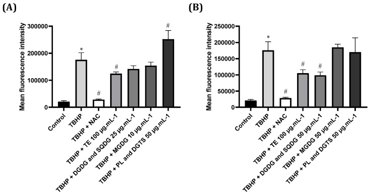
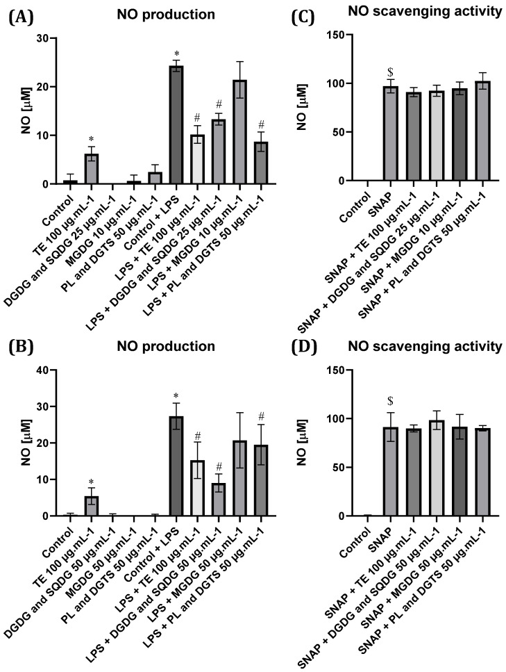
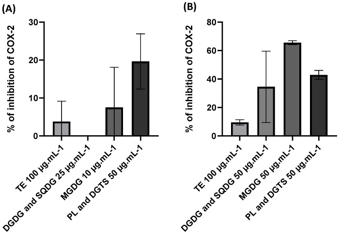
Microalgae are recognized as a relevant source of bioactive compounds. Among these bioactive products, lipids, mainly glycolipids, have been shown to present immunomodulatory properties with the potential to mitigate chronic inflammation. This study aimed to evaluate the anti-inflammatory effect of polar lipids isolated from Nannochloropsis oceanica and Chlorococcum amblystomatis. Three fractions enriched in (1) digalactosyldiacylglycerol (DGDG) and sulfoquinovosyldiacylglycerol (SQDG), (2) monogalactosyldiacylglycerol (MGDG), and (3) diacylglyceryl-trimethylhomoserine (DGTS) and phospholipids (PL) were obtained from the total lipid extracts (TE) of N. oceanica and C. amblystomatis, and their anti-inflammatory effect was assessed by analyzing their capacity to counteract nitric oxide (NO) production and transcription of pro-inflammatory genes Nos2, Ptgs2, Tnfa, and Il1b in lipopolysaccharide (LPS)-activated macrophages. For both microalgae, TE and Fractions 1 and 3 strongly inhibited NO production, although to different extents. A strong reduction in the LPS-induced transcription of Nos2, Ptgs2, Tnfa, and Il1b was observed for N. oceanica and C. amblystomatis lipids. The most active fractions were the DGTS-and-PL-enriched fraction from N. oceanica and the DGDG-and-SQDG-enriched fraction from C. amblystomatis. Our results reveal that microalgae lipids have strong anti-inflammatory capacity and may be explored as functional ingredients or nutraceuticals, offering a natural solution to tackle chronic inflammation-associated diseases.

Keywords: Chlorococcum amblystomatis; Nannochloropsis oceanica; anti-inflammatory activity; betaine lipids; glycolipids; lipidomics; lipids; microalgae; phospholipids.


Conflict of interest statement

The authors declare that author from Allmicroalgae—Natural Products S.A. did not have any role in designing and conducting of the study and analyzing the data.

Figures

Figure 1
Figure 1
Composition of fractions 1–3 obtained after solid-phase extraction of total extracts (TE) of the microalgae Nannochloropsis oceanica (A) and Chlorococcum amblystomatis (B). Fraction 1 was mainly composed of digalactosyldiacylglycerol (DGDG) and sulfoquinovosyldiacylglycerol (SQDG) species, fraction 2 was constituted mainly by monogalactosyldiacylglycerol (MGDG) species, and fraction 3 was majorly composed of diacylglyceryltrimethylhomoserine (DGTS), monoacylglyceryltrimethylhomoserine (MGTS), and phosphatidylglycerol (PG) species.
Figure 2
Figure 2
Effect of (A) Nannochloropsis oceanica and (B) Chlorococcum amblystomatis total lipid extracts and three fractions enriched in lipids from the DGDG and SQDG, MGDG, and PL and DGTS classes, respectively, on the murine cell line Raw 264.7 viability. Cells were treated with total lipid extracts and fractions at concentrations of 10, 25, 50, 100, and 200 µg·mL−1 from (A) N. oceanica or (B) C. amblystomatis for 24 h. Cell viability is expressed as a percentage of resazurin reduction in comparison to control cells (100% viability). Each value represents the mean ± standard deviation of three independent experiments performed in duplicate. Statistical differences between groups were calculated using a One-way ANOVA followed by Dunnet’s post hoc test (* p < 0.05).
Figure 3
Figure 3
Antioxidant potential of (A) Nannochloropsis oceanica and (B) Chlorococcum amblystomatis lipid extracts and fractions against THBP-induced production of reactive oxygen species (ROS) in Jurkat cells. N-acetyl-cysteine (NAC) was used as a positive control against THBP-induced production of ROS. Values represent the mean ± standard deviation of three independent experiments performed in duplicate. Statistical differences between control and THBP groups (*) and treatment conditions and THBP (#) were evaluated using One-way ANOVA followed by Dunnet’s post hoc test (p < 0.05).
Figure 4
Figure 4
Nitrite (NO) production in Raw264.7 cells treated with (A) Nannochloropsis oceanica and (B) Chlorococcum amblystomatis lipid extracts and fractions. Effect of (C) N. oceanica and (D) C. amblystomatis lipid extracts and fractions on NO scavenging. Each value represents the mean ± standard deviation of three experiments performed in duplicate. Statistical differences between control and LPS or treatment groups (*), treatment condition and LPS (#), and control and SNAP ($) were analyzed using One-way ANOVA followed by Dunnet’s post hoc test (p < 0.05).
Figure 5
Figure 5
Inhibition of cyclooxygenase-2 (COX-2) activity in chemico using Nannochloropsis oceanica (A) and Chlorococcum amblystomatis (B) total extracts (TE) and fractions enriched in DGDG and SQDG, MGDG, and PL and DGTS. Each value represents the mean ± standard deviation of three independent experiments.
Figure 6
Figure 6
Modulation of LPS-induced transcription of pro-inflammatory genes (Nos2, Ptgs2, Tnfa, and Il1b) using Nannochloropsis oceanica (AD) and Chlorococcum amblystomatis (EH) lipid extracts (TE) and fractions (DGDG and SQDG, MGDG, and PL and DGTS) in Raw 264.7 cells. The mRNA levels were assessed with quantitative Real-Time RT-PCR. Results are presented as fold change relative to control and normalized with Hprt1 as a housekeeping gene. Each value represents the mean ± standard deviation from three independent biological experiments. Statistical differences between control and LPS-stimulated cells (*) and treatment condition and LPS (#) were evaluated using One-way ANOVA followed by Dunnet’s post hoc test (p < 0.05).
Figure 7
Figure 7
Schematic representation of solid-phase extraction to separate different lipid classes from microalgae extracts. Lipid extracts of Nannochloropsis oceanica and Chlorococcum amblystomatis were loaded into the column, and elution occurred as follows: (1) dichloromethane was used to separate neutral lipids; (2) diethylether/acetic acid (98:2 v/v) was used to separate pigments; (3) a (1:1 v/v) mixture of diethylether/acetic acid (98:2 v/v) and acetone/methanol (9:1 v/v) was used to separate MGDG; (4) acetone/methanol (9:1 v/v) was used to separate DGDG and SQDG; and (5) methanol was used to separate betaine lipids and phospholipids.

References

    1. Malhotra S., Singh A.P. Algae, Traditional Medicine, and Pharmacological Advances. Int. J. Algae. 2008;10:299–308. doi: 10.1615/InterJAlgae.v10.i3.80. - DOI
    1. Pradhan B., Nayak R., Patra S., Jit B.P., Ragusa A., Jena M. Bioactive Metabolites from Marine Algae as Potent Pharmacophores against Oxidative Stress-Associated Human Diseases: A Comprehensive Review. Molecules. 2020;26:37. doi: 10.3390/molecules26010037. - DOI - PMC - PubMed
    1. Lomartire S., Gonçalves A.M.M. An Overview of Potential Seaweed-Derived Bioactive Compounds for Pharmaceutical Applications. Mar. Drugs. 2022;20:141. doi: 10.3390/md20020141. - DOI - PMC - PubMed
    1. Silva M., Kamberovic F., Uota S.T., Kovan I.-M., Viegas C.S.B., Simes D.C., Gangadhar K.N., Varela J., Barreira L. Microalgae as Potential Sources of Bioactive Compounds for Functional Foods and Pharmaceuticals. Appl. Sci. 2022;12:5877. doi: 10.3390/app12125877. - DOI
    1. Conde T.A., Zabetakis I., Tsoupras A., Medina I., Costa M., Silva J., Neves B., Domingues P., Domingues M.R. Microalgal Lipid Extracts Have Potential to Modulate the Inflammatory Response: A Critical Review. Int. J. Mol. Sci. 2021;22:9825. doi: 10.3390/ijms22189825. - DOI - PMC - PubMed