. 2023 Nov 30;30(4):567-585.
doi: 10.3390/pathophysiology30040041.

Combination Therapy with Enalapril and Paricalcitol Ameliorates Streptozotocin Diabetes-Induced Testicular Dysfunction in Rats via Mitigation of Inflammation, Apoptosis, and Oxidative Stress

Affiliations

Combination Therapy with Enalapril and Paricalcitol Ameliorates Streptozotocin Diabetes-Induced Testicular Dysfunction in Rats via Mitigation of Inflammation, Apoptosis, and Oxidative Stress

Magdy Y Elsaeed et al. Pathophysiology. .

Abstract

Background: As the impacts of diabetes-induced reproductive damage are now evident in young people, we are now in urgent need to devise new ways to protect and enhance the reproductive health of diabetic people. The present study aimed to evaluate the protective effects of enalapril (an ACE inhibitor) and paricalcitol (a vitamin D analog), individually or in combination, on streptozotocin (STZ)-diabetes-induced testicular dysfunction in rats and to identify the possible mechanisms for this protection.

Material and methods: This study was carried out on 50 male Sprague-Dawley rats; 10 normal rats were allocated as a non-diabetic control group. A total of 40 rats developed diabetes after receiving a single dose of STZ; then, the diabetic rats were divided into four groups of equivalent numbers assigned as diabetic control, enalapril-treated, paricalcitol-treated, and combined enalapril-and-paricalcitol-treated groups. The effects of mono and combined therapy with paricalcitol and enalapril on testicular functions, sperm activity, glycemic state oxidative stress, and inflammatory parameters, as well as histopathological examinations, were assessed in comparison with the normal and diabetic control rats.

Results: As a result of diabetes induction, epididymal sperm count, sperm motility, serum levels of testosterone, follicle-stimulating hormone (FSH) as well as luteinizing hormone (LH), and the antioxidant enzyme activities, were significantly decreased, while abnormal sperm (%), insulin resistance, nitric oxide (NO), malondialdehyde (MDA), interleukin-6 (IL-6), and tumor necrosis factor-α (TNF-α) were significantly increased, along with severe distortion of the testicular structure. Interestingly, treatment with paricalcitol and enalapril, either alone or in combination, significantly improved the sperm parameters, increased antioxidant enzyme activities in addition to serum levels of testosterone, FSH, and LH, reduced insulin resistance, IL-6, and TNF-α levels, and finally ameliorated the diabetes-induced testicular oxidative stress and histopathological damage, with somewhat superior effect for paricalcitol monotherapy and combined therapy with both drugs compared to monotherapy with enalapril alone.

Conclusions: Monotherapy with paricalcitol and its combination therapy with enalapril has a somewhat superior effect in improving diabetes-induced testicular dysfunction (most probably as a result of their hypoglycemic, antioxidant, anti-inflammatory, and anti-apoptotic properties) compared with monotherapy with enalapril alone in male rats, recommending a synergistic impact of both drugs.

Keywords: diabetes; enalapril; oxidative stress; paricalcitol; testicular dysfunction.


Conflict of interest statement

The authors declare no conflict of interest.

Figures

Figure 1
Figure 1
Impact of treatment with enalapril and paricalcitol on body weight (A), and testicular weight (B). a Significantly different from the non-diabetic control group; b significantly different from the diabetic vehicle-treated group.
Figure 2
Figure 2
Impact of treatment with enalapril and paricalcitol on sperm motility (A), sperm abnormalities (B), and sperm count (C). a Significantly different from the non-diabetic control group; b significantly different from the diabetic vehicle-treated group.
Figure 3
Figure 3
Effects of treatment with enalapril and paricalcitol on testosterone (A), FSH (B), and LH (C) levels. a Significantly different from the non-diabetic control group; b significantly different from the diabetic vehicle-treated group.
Figure 4
Figure 4
Effects of treatment with enalapril and paricalcitol on FBG, fasting blood glucose (A), PPG, postprandial glucose (B), insulin level (C), and HOMA-IR, the homeostasis model assessment of insulin resistance (D). a Significantly different from the non-diabetic control group; b significantly different from the diabetic vehicle-treated group; c significantly different from the diabetic enalapril-treated group; d significantly different from the diabetic paricalcitol-treated group.
Figure 5
Figure 5
Effects of treatment with paricalcitol and enalapril on TNF-α, tumor necrosis factor-α (A); and IL-6, interleukin-6 (B). a Significantly different from the non-diabetic control group; b significantly different from the diabetic vehicle-treated group.
Figure 6
Figure 6
Effects of treatment with enalapril and paricalcitol on testicular oxidative stress parameters. NO, nitric oxide; MDA, malondialdehyde; GSH, reduced glutathione; GPx, glutathione peroxidase; SOD, superoxide dismutase; CAT, catalase; a significantly different from the non-diabetic control group; b significantly different from the diabetic vehicle-treated group; c significantly different from the diabetic enalapril-treated group; d significantly different from the diabetic paricalcitol-treated group.
Figure 7
Figure 7
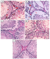
Photomicrographs of testicular sections from non-diabetic control (A), diabetic control rats (B), diabetic enalapril-treated group (C), diabetic paricalcitol-treated animals (D), and diabetic enalapril + paricalcitol-treated group (E). Regarding the control group, the seminiferous tubules were densely packed, and the lumen was filled with spermatogonia (curved arrow), primary spermatocytes (Sp), spermatids (St), as well as sperms (S). Leydig cells (thick arrow) were located in the interstitial space that separates tubules and is bordered by a basement membrane (arrowhead). Diabetic animals’ testes exhibited pronounced vacuolations (V) caused by germinal epithelium detachment from the tubular basement membrane (V) in numerous seminiferous tubules characterized by an irregularly thick basement membrane (arrowhead) as well as an enlargement of the interstitial tissue, including the Leydig cells and dilated blood vessels (thin arrow), while testicular sections in other groups treated with either enalapril or paricalcitol or both showed restoration of the previous changes, with noticeably marked improvement in rats treated with both drugs (H&E ×400). Scale bars, 100 µm.
Figure 8
Figure 8
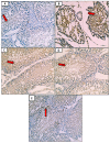
Micrographs of testicular tissue stained with Masson trichrome reveal that collagen fiber deposition (green color) was absent or minimal in the non-diabetic control group (A) but extensive in the diabetes control group. (B): weak deposition of collagen fibers (red arrow) in the diabetic groups treated with either enalapril (C) or paricalcitol (D), or both (E), with noticeably minimal expression in rats treated with both drugs (Masson trichrome stain ×400). Scale bars, 100 µm.
Figure 9
Figure 9
Photomicrographs of immunostained testicular sections for caspase-3 showing no or weakest expression of caspase-3 (red arrow) in control group (A); marked expression of caspase-3 (red arrow) in the diabetic control group (B); weak expression of caspase-3 (red arrow) in the diabetic groups treated with either enalapril (C) or paricalcitol (D), or both (E), with noticeably minimal expression (red arrow) in the group treated with combination of both (anti-caspase-3 immunostaining ×400). Scale bars, 100 µm.
Figure 10
Figure 10
Scheme summarizing how diabetes induces testicular damage, and the mechanisms of improvement after treatment with enalapril and paricalcitol.

References

    1. Saeedi P., Petersohn I., Salpea P., Malanda B., Karuranga S., Unwin N., Colagiuri S., Guariguata L., Motala A.A., Ogurtsova K., et al. Global and regional diabetes prevalence estimates for 2019 and projections for 2030 and 2045: Results from the International Diabetes Federation Diabetes Atlas, 9th edition. Diabetes Res. Clin. Pract. 2019;157:107843. doi: 10.1016/j.diabres.2019.107843. - DOI - PubMed
    1. Aksu I., Baykara B., Kiray M., Gurpinar T., Sisman A.R., Ekerbicer N., Tas A., Gokdemir-Yazar O., Uysal N. Serum IGF-1 levels correlate negatively to liver damage in diabetic rats. Biotech. Histochem. 2013;88:194–201. doi: 10.3109/10520295.2012.758311. - DOI - PubMed
    1. Amaral S., Moreno A.J., Santos M.S., Seiça R., Ramalho-Santos J. Effects of hyperglycemia on sperm and testicular cells of Goto-Kakizaki and streptozotocin-treated rat models for diabetes. Theriogenology. 2006;66:2056–2067. doi: 10.1016/j.theriogenology.2006.06.006. - DOI - PubMed
    1. Palmeira C.M., Santos D.L., Seiça R., Moreno A.J., Santos M.S. Enhanced mitochondrial testicular antioxidant capacity in Goto-Kakizaki diabetic rats: Role of coenzyme Q. Am. J. Physiol. Physiol. 2001;281:C1023–C1028. doi: 10.1152/ajpcell.2001.281.3.C1023. - DOI - PubMed
    1. Shrilatha B., Muralidhara Occurrence of oxidative impairments, response of antioxidant defences and associated biochemical perturbations in male reproductive milieu in the Streptozotocin-diabetic rat. Int. J. Androl. 2007;30:508–518. doi: 10.1111/j.1365-2605.2007.00748.x. - DOI - PubMed