Review
. 2023 Dec 4;30(4):618-629.
doi: 10.3390/pathophysiology30040044.

Surgical Management of Traumatic Meniscus Injuries

Affiliations
Review

Surgical Management of Traumatic Meniscus Injuries

Hannah R Popper et al. Pathophysiology. .

Abstract

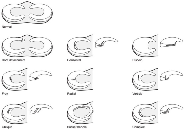
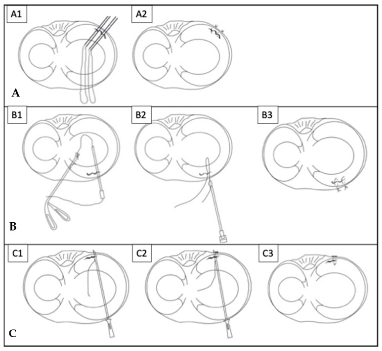
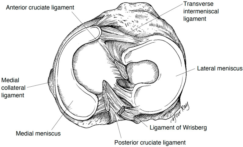
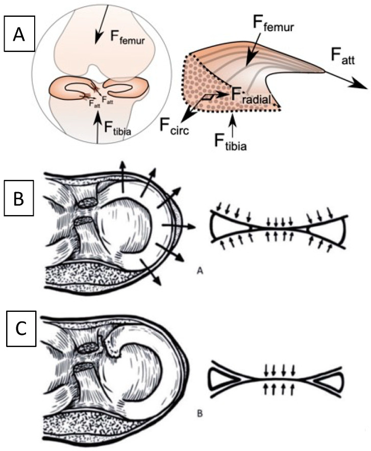
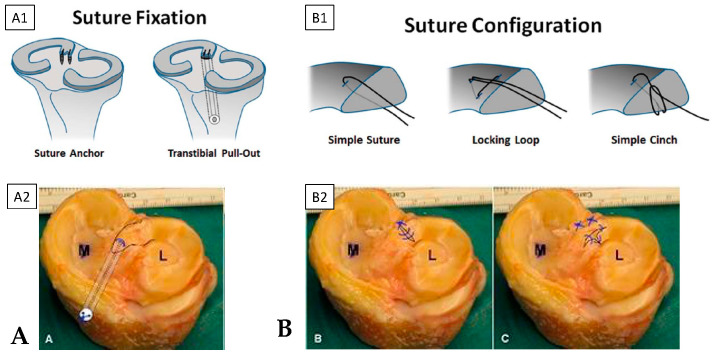
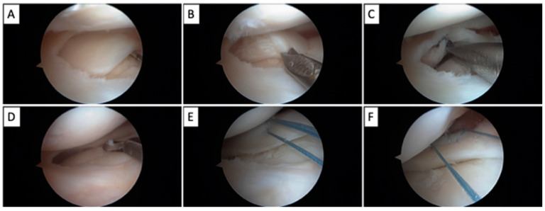
The menisci increase the contact area of load bearing in the knee and thus disperse the mechanical stress via their circumferential tensile fibers. Traumatic meniscus injuries cause mechanical symptoms in the knee, and are more prevalent amongst younger, more active patients, compared to degenerative tears amongst the elderly population. Traumatic meniscus tears typically result from the load-and-shear mechanism in the knee joint. The treatment depends on the size, location, and pattern of the tear. For non-repairable tears, partial or total meniscal resection decreases its tensile stress and increases joint contact stress, thus potentiating the risk of arthritis. A longitudinal vertical tear pattern at the peripheral third red-red zone leads to higher healing potential after repair. The postoperative rehabilitation protocols after repair range from immediate weight-bearing with no range of motion restrictions to non-weight bearing and delayed mobilization for weeks. Pediatric and adolescent patients may require special considerations due to their activity levels, or distinct pathologies such as a discoid meniscus. Further biomechanical and biologic evidence is needed to guide surgical management, postoperative rehabilitation protocols, and future technology applications for traumatic meniscus injuries.

Keywords: knee biomechanics; knee rehabilitation; meniscus repair; meniscus tear; orthopaedics; sports medicine.


Conflict of interest statement

The authors declare no conflict of interest.

Figures

Figure 3
Figure 3
Patterns of meniscus tears; note that vertical tears are also known as longitudinal vertical tears; bucket handle tears result from an extensive vertical tear with or without other associated tear patterns. (Courtesy from Shieh et al. [34]).
Figure 4
Figure 4
Meniscus repair techniques of (A) inside-out repairs, classic technique for posterior horn to mid-body tears: (A1) the sutures are passed from inside of the joint under knee arthroscopy, and (A2) retrieved posterior to the joint capsule accessed via open incision; (B) outside-in repairs, suitable for tears in the anterior horns: (B1) under knee arthroscopy, the sutures are passed from outside of the joint, then (B2) retrieved from a separate portal back to the outside, and (B3) tied down to complete the repair; (C) all-inside repairs with a suture-anchor construct, completed solely under arthroscopy without additional incisions: (C1) passing the first limb of the sutures, (C2) passing the second limb, and (C3) cinching the suture to complete the repair. (Courtesy from Yoon et al. [44]).
Figure 1
Figure 1
Normal anatomy of the menisci, with the C-shaped medial meniscus and O-shaped lateral meniscus, and the inter-meniscal ligament (i.e., “transverse intermeniscal ligament”) anteriorly and menisci roots attachment onto the tibial posteriorly along with ligaments attaching to the posterior cruciate ligament (Ligament of Wrisberg posteriorly, and Ligament of Humphrey anteriorly—not shown). (Courtesy from Greis et al. [4]).
Figure 2
Figure 2
Biomechanical function of the meniscus, where: (A) Axial compressive forces from the femur (Ffemur) and tibia (Ftibia) during joint loading are converted into the circumferential “hoop” direction (Fcirc) that are resisted by the tensile stiffness of the circumferential collagen fibers and are transferred into the horn attachments (Fatt). (B) This load transfer reduces the axial forces on the underlying articular cartilage. (C) With a meniscus root avulsion or large-scale radial tear, the hoop stress is lost and the axial forces are applied directly to the cartilage, the clinical equivalent of a meniscectomy, and leads to cartilage osteoarthritis. (A) Courtesy from Dawn Elliott and Robert Mauck; (B) and (C): (Courtesy from Itthipanichpong et al. [18]).
Figure 5
Figure 5
Surgical treatment of a meniscal root tear by (A) repairing meniscus root avulsion by re-attaching the root to its tibia insertional footprint via a transtibial bone tunnel with suspensory fixation on the bone cortex. (B) Utilizing various suture constructs to repair intrasubstance root tear, or to create the pulling stitch for meniscal root repair as described above. Subpanels: the (A1,B1) schematics, and (A2,B2) cadaveric demonstration photos, of the techniques. (Courtesy from Bonasia et al. 2015 [49], Krych et al. [50]).
Figure 6
Figure 6
Surgical treatment of a discoid lateral meniscus: (A) A complete discoid meniscus that is torn and formed an unstable flap, in addition to the absence of posterior menisco-capsular attachment, probed under knee arthroscopy, (B,C) Debridement and resection of the torn flap using arthroscopic tools (biters and motorized shavers), (D) further saucerization of the meniscus to approach the normal C-shape, (E) stabilizing the meniscus using an all-inside meniscus repair technique, while (F) using the stitch from the first repair construct as a pulling stitch to facilitate the passing of the second all-inside repair construct, to finally achieve a stable meniscus.

Similar articles

Cited by

References

    1. Clark C.R., Ogden J.A. Development of the menisci of the human knee joint. Morphological changes and their potential role in childhood meniscal injury. J. Bone Jt. Surg. Am. 1983;65:538–547. doi: 10.2106/00004623-198365040-00018. - DOI - PubMed
    1. Fithian D.C., Kelly M.A., Mow V.C. Material properties and structure-function relationships in the menisci. Clin. Orthop. Relat. Res. 1990;252:19–31. doi: 10.1097/00003086-199003000-00004. - DOI - PubMed
    1. Fox A.J.S., Bedi A., Rodeo S.A. The Basic Science of Human Knee Menisci: Structure, Composition, and Function. Sports Health. 2012;4:340–351. doi: 10.1177/1941738111429419. - DOI - PMC - PubMed
    1. Greis P.E., Bardana D.D., Holmstrom M.C., Burks R.T. Meniscal Injury: I. Basic Science and Evaluation. J. Am. Acad. Orthop. Surg. 2002;10:168–176. doi: 10.5435/00124635-200205000-00003. - DOI - PubMed
    1. Kumari A., Gupta G., Kumar P., Rani S. Morphological study of the menisci of the knee joint in human cadaver in Jharkhand population. J. Fam. Med. Prim. Care. 2022;11:4723–4729. doi: 10.4103/jfmpc.jfmpc_2416_21. - DOI - PMC - PubMed

LinkOut - more resources