Review
. 2023 Dec 1;10(12):683.
doi: 10.3390/vetsci10120683.

Ultrasound-Based Technologies for the Evaluation of Testicles in the Dog: Keystones and Breakthroughs

Affiliations
Review

Ultrasound-Based Technologies for the Evaluation of Testicles in the Dog: Keystones and Breakthroughs

Claudia Bracco et al. Vet Sci. .

Abstract

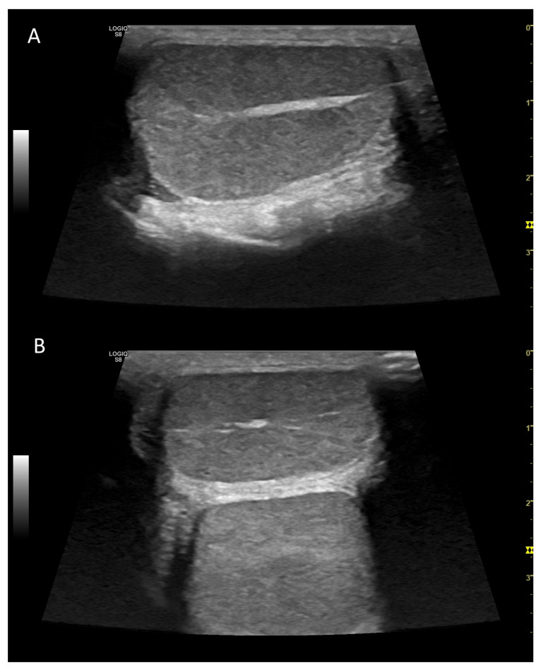
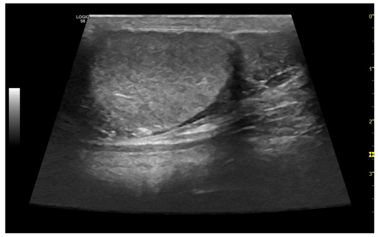
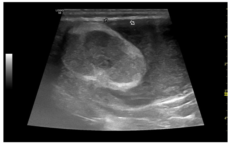
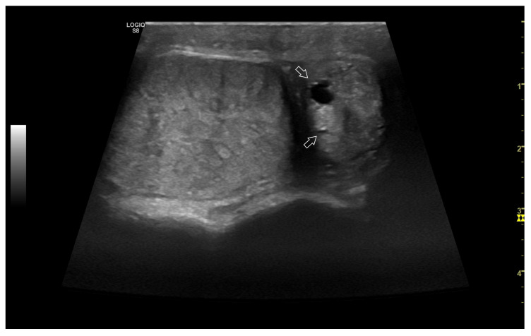
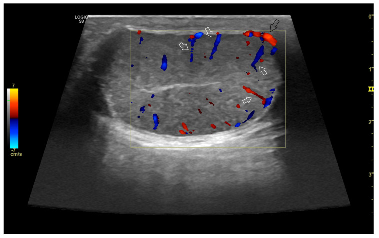
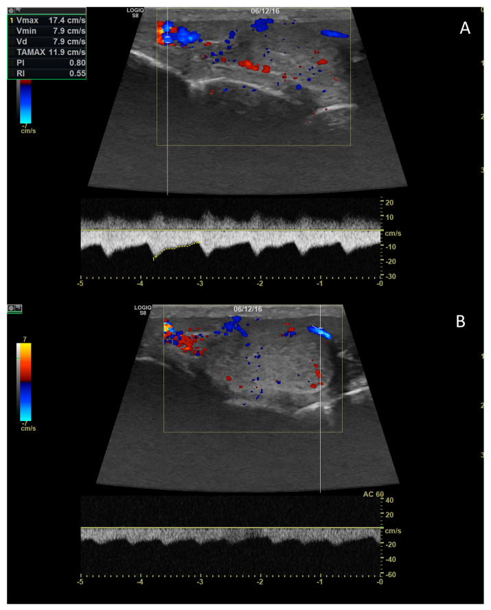
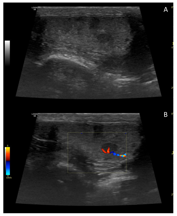
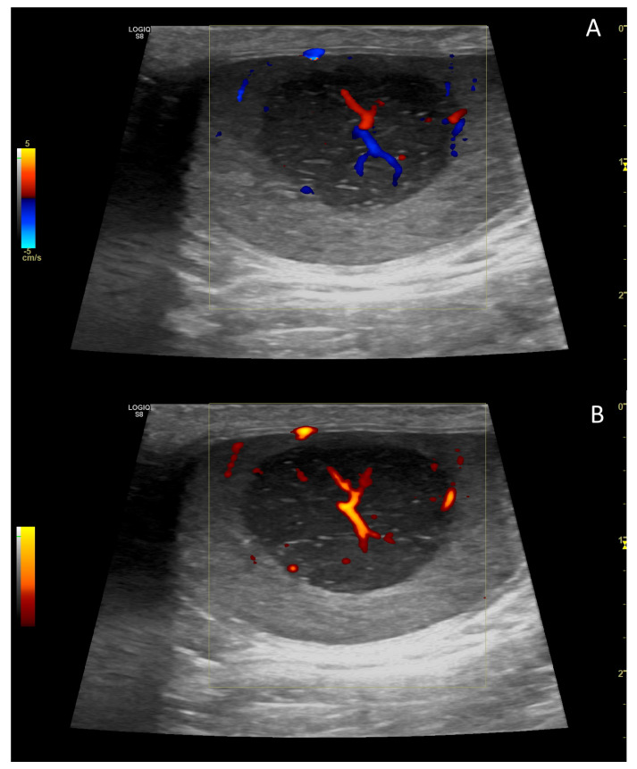
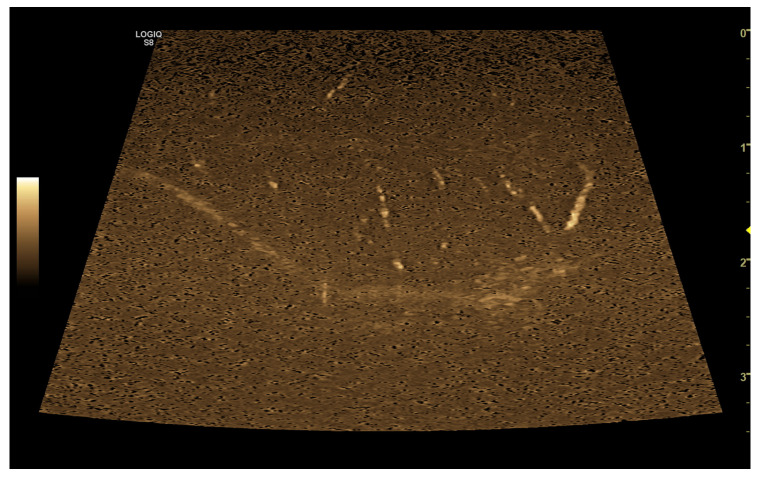
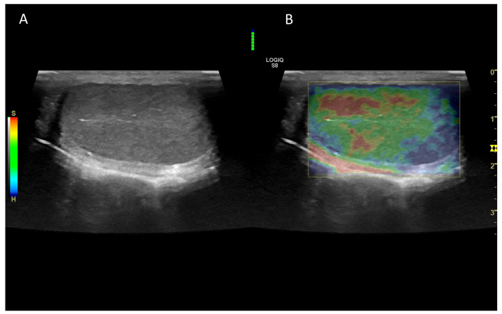
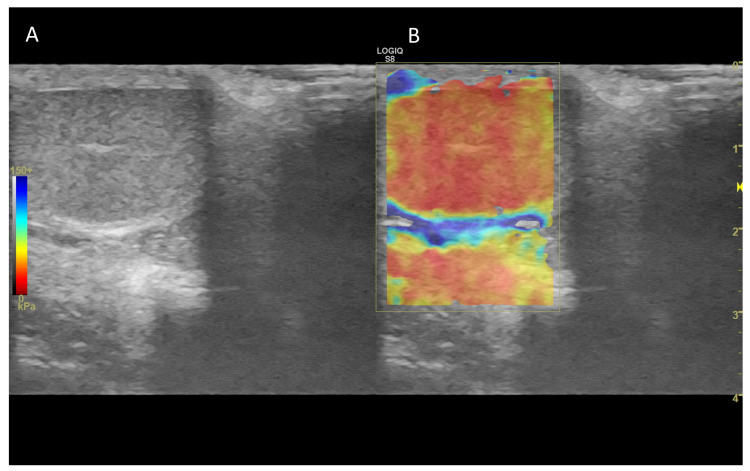
Ultrasonography is a valuable diagnostic tool extensively used in the andrology of human and domestic animals, including dogs. This review aims to provide an overview of various technologies based on ultrasound, from the basic B-Mode ultrasonography to the more recent advancements, such as contrast-enhanced ultrasonography (CEUS) and ultrasound elastography (UEl), all of which are utilized in the evaluation of canine testicles. The review outlines the principles behind each of these technologies and discusses their application in assessing normal and abnormal testicular conditions. B-mode canine testicular ultrasonography primarily focuses on detecting focal lesions but has limitations in terms of objectivity. Other technologies, including Doppler ultrasonography, B-Flow, and CEUS, allow for the characterization of vascular patterns, which could be further measured using specific applications like spectral Doppler or quantitative CEUS. Additionally, ultrasound elastography enables the assessment of parenchyma stiffness both qualitatively and quantitatively. These ultrasound-based technologies play a crucial role in andrology by providing valuable information for evaluating testicular function and integrity, aiding in the identification of pathological conditions that may impact the health and quality of life of male dogs.

Keywords: B-flow; CEUS; dog; doppler; sonoelastography; testis; ultrasonography.


Conflict of interest statement

The authors declare no conflict of interest.

Figures

Figure 1
Figure 1
Example of B-Mode ultrasonography of normal testis in the dog, in longitudinal (A) and transversal (B) scan. The hyperechoic mediastinum is visible as a band (A) or a circular area (B) within the homogeneous parenchyma.
Figure 2
Figure 2
Example of B-Mode ultrasonography of normal epididymis in the dog. The normal tail of the epididymis, isoechoic, can be detected on the right of the image, closely adherent to the testis (on the left).
Figure 3
Figure 3
Ultrasonographic B-mode appearance of a large tumour in the testis (black arrow) compared to the testicular parenchyma (white arrow) in an inguinal cryptorchid testis. Based on histology, the image is consistent with a Sertoli cell tumour.
Figure 4
Figure 4
A case of epididymitis, contextual with orchitis, in the canine testis involving the tail of the epididymis. Note the relative hypertrophy of the tail of the epididymis (on the right) compared to the correspondent testis (on the left). Both organs appear heterogeneous, and small anechoic areas can be detected (white arrows).
Figure 5
Figure 5
Representative sonogram in colour Doppler of canine testis. Note the marginal portion (black arrow) and the intratesticular branches (white arrows) of the testicular artery.
Figure 6
Figure 6
Pulsed-wave Doppler parameters were measured on the supratesticular (A) and marginal (B) portions of the testicular artery.
Figure 7
Figure 7
B-mode (A) and colour Doppler (B) sonogram of a small testicular tumour in the dog. Based on histology, the image referred to an interstitial cell tumour.
Figure 8
Figure 8
Colour (A) and power Doppler (B) sonogram of testicular tumour in the dog. Based on histology, the image referred to a mixed Sertoli cell/seminoma tumour occupying most of the testis.
Figure 9
Figure 9
Representative image of the B-Flow evaluation of canine testis.
Figure 10
Figure 10
Representative elastogramme acquired with the qualitative strain elastography of canine testis (B) compared with B-Mode ultrasonography (A). The colour scale differentiates the hard (blue) and soft (red) area of the testis. The green bar in the centre of the image represents the adequateness of the freehand compression applied by the operator.
Figure 11
Figure 11
Representative elastogramme acquired with the quantitative shear-wave elastography of canine testis (B) compared with B-Mode ultrasonography (A). The technology can quantify the stiffness objectively.

References

    1. Davidson A.P., Baker T.W. Reproductive Ultrasound of the Dog and Tom. Top. Companion Anim. Med. 2009;24:64–70. doi: 10.1053/j.tcam.2008.11.003. - DOI - PubMed
    1. Mantziaras G., Luvoni G.C. Advanced Ultrasound Techniques in Small Animal Reproduction Imaging. Reprod. Dom. Anim. 2020;55:17–25. doi: 10.1111/rda.13587. - DOI - PubMed
    1. Orlandi R., Vallesi E., Boiti C., Polisca A., Bargellini P., Troisi A. Characterization of Testicular Tumor Lesions in Dogs by Different Ultrasound Techniques. Animals. 2022;12:210. doi: 10.3390/ani12020210. - DOI - PMC - PubMed
    1. Lock G., Schmidt C., Helmich F., Stolle E., Dieckmann K.-P. Early Experience with Contrast-Enhanced Ultrasound in the Diagnosis of Testicular Masses: A Feasibility Study. Urology. 2011;77:1049–1053. doi: 10.1016/j.urology.2010.12.035. - DOI - PubMed
    1. Mantziaras G. Imaging of the Male Reproductive Tract: Not so Easy as It Looks Like. Theriogenology. 2020;150:490–497. doi: 10.1016/j.theriogenology.2020.03.009. - DOI - PubMed

LinkOut - more resources