. 2024 Jul 25;230(1):e17-e29.
doi: 10.1093/infdis/jiad590.

Delayed Mucosal Antiviral Responses Despite Robust Peripheral Inflammation in Fatal COVID-19

Collaborators, Affiliations

Delayed Mucosal Antiviral Responses Despite Robust Peripheral Inflammation in Fatal COVID-19

Jasmin K Sidhu et al. J Infect Dis. .

Abstract

Background: While inflammatory and immune responses to severe acute respiratory syndrome coronavirus 2 (SARS-CoV-2) infection in peripheral blood are extensively described, responses at the upper respiratory mucosal site of initial infection are relatively poorly defined. We sought to identify mucosal cytokine/chemokine signatures that distinguished coronavirus disease 2019 (COVID-19) severity categories, and relate these to disease progression and peripheral inflammation.

Methods: We measured 35 cytokines and chemokines in nasal samples from 274 patients hospitalized with COVID-19. Analysis considered the timing of sampling during disease, as either the early (0-5 days after symptom onset) or late (6-20 days after symptom onset) phase.

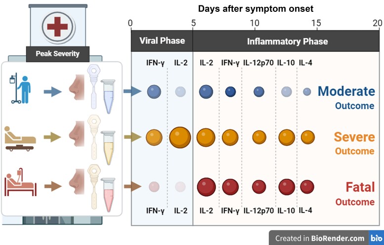
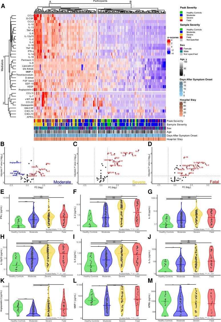
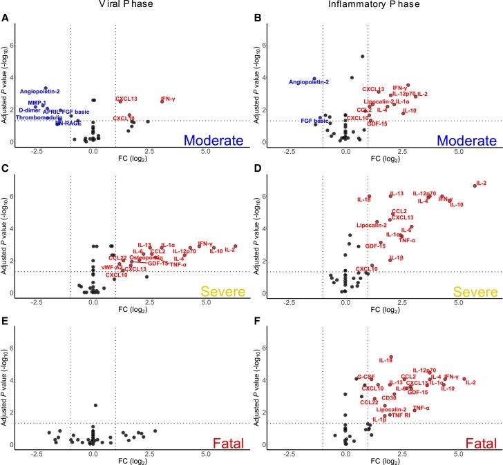
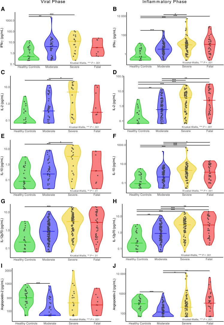
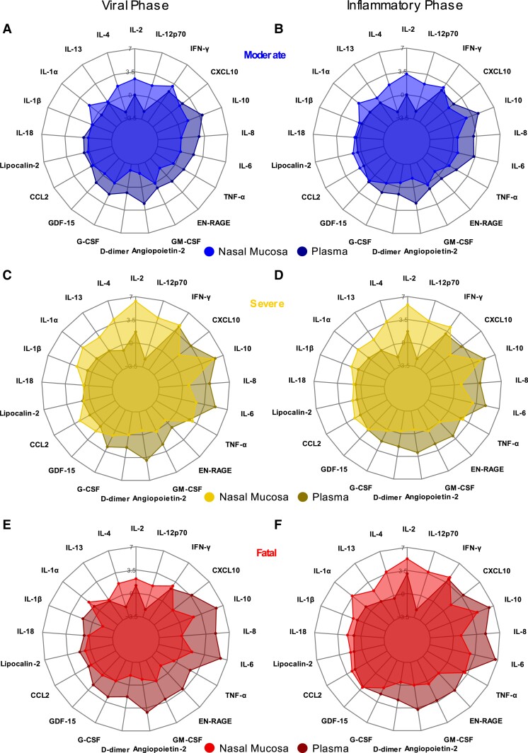
Results: Patients that survived severe COVID-19 showed interferon (IFN)-dominated mucosal immune responses (IFN-γ, CXCL10, and CXCL13) early in infection. These early mucosal responses were absent in patients who would progress to fatal disease despite equivalent SARS-CoV-2 viral load. Mucosal inflammation in later disease was dominated by interleukin 2 (IL-2), IL-10, IFN-γ, and IL-12p70, which scaled with severity but did not differentiate patients who would survive or succumb to disease. Cytokines and chemokines in the mucosa showed distinctions from responses evident in the peripheral blood, particularly during fatal disease.

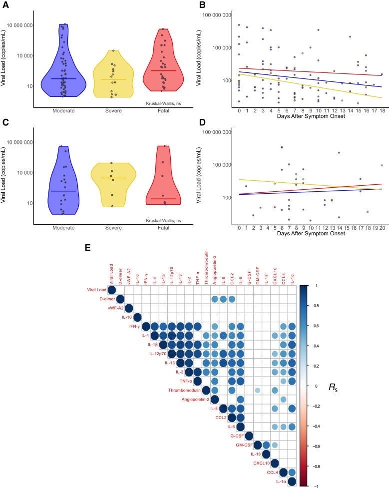
Conclusions: Defective early mucosal antiviral responses anticipate fatal COVID-19 but are not associated with viral load. Early mucosal immune responses may define the trajectory of severe COVID-19.

Keywords: COVID-19; SARS-CoV-2; airway; chemokine; cytokine; lung; mucosa; nose; virus.


Conflict of interest statement

Potential conflicts of interest. P. J. M. O. is the Imperial College Lead Investigator of the EMINENT consortium, supported by the Medical Research Council UK (MR/R502121/1; this grant supports collaborative testing of compounds developed by GlaxoSmithKline in UK universities); is on advisory boards for Affnivax, Oxford Immunotech, Nestle, and Pfizer in relation to immunity to viruses (fees paid to Imperial College London); and is on an advisory board for Janssen/J&J. L.T. has received consulting fees from the Medicines and Healthcare products Regulatory Agency (MHRA); consulting fees from AstraZeneca and Synairgen, paid to the University of Liverpool; speaker’s fees from Eisai, Ltd; and support for conference attendance from AstraZeneca. All other authors report no potential conflicts. All authors have submitted the ICMJE Form for Disclosure of Potential Conflicts of Interest. Conflicts that the editors consider relevant to the content of the manuscript have been disclosed.

Figures

Graphical Abstract
Graphical Abstract
Figure 1.
Figure 1.
COVID-19 severity was associated with clusters of elevated inflammatory markers in the nasal mucosa. A, K-means clustered heatmap of 35 cytokines and chemokines in the nasal mucosa of healthy controls (n = 25) and patients hospitalized with COVID-19 (n = 215). Annotations provide participant peak severity (peak severity), severity at the time of sample collection (sample severity), sex, age, duration of symptoms at the time of sample collection (days after symptom onset), and total duration of hospital stay (hospital stay) in days. BD, Nasal cytokine and chemokine levels in patients hospitalized with COVID-19 (n = 274) shown as volcano plots relative to healthy controls for (B) moderate, (C) severe, and (D) fatal peak COVID-19 severity groups. Data shown are −log10 transformed false discovery rate adjusted Wilcoxon rank-sum P values (horizontal line at cutoff ≥.05) plotted against log2 fold changes (vertical lines at cutoffs ≤ −2 and ≥2) for nasal samples taken at 0–20 days after symptom onset. EM, Individual cytokines and chemokines of particular interest in each COVID-19 peak-severity group and healthy controls: (E) IFN-γ, (F) IL-2, (G) IL-10, (H) IL-12p70, (I) IL-4, (J) IL-1α, (K) angiopoietin-2, (L) MMP-1, and (M) APRIL. Group median levels are shown as lines and significance levels determined using Kruskal-Wallis tests with Dunn's P value correction. BM, Data from the first time point per participant are shown. *P < .05, **P < .01, ***P < .001. Abbreviations: APRIL, A proliferation-inducing ligand; COVID-19, coronavirus disease 2019; FC, fold change; FGF, fibroblast growth factor; G-CSF, granulocyte-colony-stimulating factor; GDF-15, growth differentiation factor 15; GM-CSF, granulocyte-macrophage colony-stimulating factor; IL, interleukin; IFN-γ, interferon-γ; MMP-1, matrix metalloproteinase-1; TNF-α, tumor necrosis factor-α; VEGF, vascular endothelial growth factor.
Figure 2.
Figure 2.
Nasal viral load was not associated with peak COVID-19 severity or nasal marker levels. A, Viral load quantified by RT-qPCR on nasal swabs from patients with COVID-19 (moderate n = 56, severe n = 14, fatal n = 28) taken 0–20 days after symptom onset. B, Viral load data aligned with the duration of symptoms at the time of sampling (days after symptom onset). A subset of these participants (moderate n = 18, severe n = 6, fatal n = 8) had viral load data and time-matched nasosorption cytokine and chemokine data: (C) viral load grouped by peak COVID-19 severity; and (D) viral load data aligned with duration of symptoms. E, Hierarchically clustered correlation matrix of nasal cytokines and chemokines and nasal viral loads, ranked using the Spearman rank correlation coefficient (RS). Correlations were corrected for multiple testing and nonsignificant correlations are denoted by empty boxes. B and D, Linear regression lines for moderate, severe, and fatal peak COVID-19 severity groups. Abbreviations: COVID-19, coronavirus disease 2019; G-CSF, granulocyte-colony-stimulating factor; GM-CSF, granulocyte-macrophage colony-stimulating factor; IFN-γ, interferon-γ; IL, interleukin; ns, not significant; RT-qPCR, reverse transcription quantitative polymerase chain reaction; TNF-α, tumor necrosis factor-α; vWF-A2, von Willebrand factor-A2.
Figure 3.
Figure 3.
A muted early mucosal immune response was associated with fatal COVID-19. Cytokine and chemokine levels in nasal samples were compared to healthy control (n = 25) levels for each COVID-19 patient time group: (A) moderate, viral phase (0–5 days after symptom onset; n = 44); (B) moderate, inflammatory phase (6–20 days after symptom onset; n = 98); (C) severe, viral phase (n = 18); (D) severe, inflammatory phase (n = 74); (E) fatal, viral phase (n = 9); and (F) fatal, inflammatory phase (n = 31). AF, Volcano plots with −log10 transformed false discovery rate adjusted Wilcoxon rank-sum P values (horizontal line at cutoff P ≥ .05) plotted against log2 fold changes relative to healthy controls (vertical lines at cutoffs ≤ -2 and ≥2 fold change). Abbreviations: APRIL, A proliferation-inducing ligand; COVID-19, coronavirus disease 2019; EN-RAGE, Extracellular newly identified receptor for advanced glycation end products/S100A12; FGF, fibroblast growth factor; GDF-15, growth differentiation factor 15; IFN-γ, interferon-γ; IL, interleukin; MMP-1, matrix metalloproteinase-1; TNF-α, tumor necrosis factor-α; vWF-A2, von Willebrand factor-A2.
Figure 4.
Figure 4.
Cytokine and chemokine levels during the viral and inflammatory phases between peak-severity groups. Individual markers’ particular distinctions between each COVID-19 peak-severity group and healthy controls, displayed by viral phase (0–5 days after symptom onset) or inflammatory phase (6–20 days after symptom onset): (A) viral phase IFN-γ; (B) inflammatory phase IFN-γ; (C) viral phase IL-2; (D) inflammatory phase IL-2; (E) viral phase IL-10; (F) inflammatory phase IL-10; (G) viral phase IL-12p70; (H) inflammatory phase IL-12p70; (I) viral phase angiopoietin-2; and (J) inflammatory phase angiopoietin-2. All panels show group median levels as lines and significance levels determined using Kruskal-Wallis tests with Dunn P value correction. *P < .05, **P < .01, ***P < .001. Abbreviations: COVID-19, coronavirus disease 2019; IFN-γ, interferon-γ; IL, interleukin.
Figure 5.
Figure 5.
Pronounced blood plasma responses in the early stages of fatal COVID-19 despite weak mucosal responses. Median log2 fold-change values, relative to healthy control levels, for each cytokine or chemokine were contrasted between nasosorption samples (lighter shades) and plasma (darker shades) for each COVID-19 patient peak-severity group in viral (0–5 days after symptom onset) or inflammatory (6–20 days after symptom onset) phases: (A) moderate, viral phase; (B) moderate, inflammatory phase; (C) severe, viral phase; (D) severe, inflammatory phase; (E) fatal, viral phase; and (F) fatal, inflammatory phase. All panels show median log2 transformed cytokine fold-changes relative to healthy controls (healthy control levels therefore represented as the 0 line) for nasal samples and plasma samples by peak severity. Abbreviations: COVID-19, coronavirus disease 2019; EN-RAGE, Extracellular newly identified receptor for advanced glycation end products/S100A12; G-CSF, granulocyte-colony-stimulating factor; GDF-15, growth differentiation factor 15; GM-CSF, granulocyte-macrophage colony-stimulating factor; IFN-γ, interferon-γ; IL, interleukin; TNF-α, tumor necrosis factor-α.

References

    1. Chiu C, Openshaw PJ. Antiviral B cell and T cell immunity in the lungs. Nat Immunol 2015; 16:18–26. - PMC - PubMed
    1. Akamatsu MA, de Castro JT, Takano CY, Ho PL. Off balance: interferons in COVID-19 lung infections. EBioMedicine 2021; 73:103642. - PMC - PubMed
    1. Diamond MS, Kanneganti TD. Innate immunity: the first line of defense against SARS-CoV-2. Nat Immunol 2022; 23:165–76. - PMC - PubMed
    1. Sette A, Crotty S. Adaptive immunity to SARS-CoV-2 and COVID-19. Cell 2021; 184:861–80. - PMC - PubMed
    1. Nielsen SS, Vibholm LK, Monrad I, et al. SARS-CoV-2 elicits robust adaptive immune responses regardless of disease severity. EBioMedicine 2021; 68:103410. - PMC - PubMed