. 2024 Mar 29;36(4):1159-1181.
doi: 10.1093/plcell/koad320.

CHLOROPLAST UNUSUAL POSITIONING 1 is a plant-specific actin polymerization factor regulating chloroplast movement

Affiliations

CHLOROPLAST UNUSUAL POSITIONING 1 is a plant-specific actin polymerization factor regulating chloroplast movement

Sam-Geun Kong et al. Plant Cell. .

Abstract

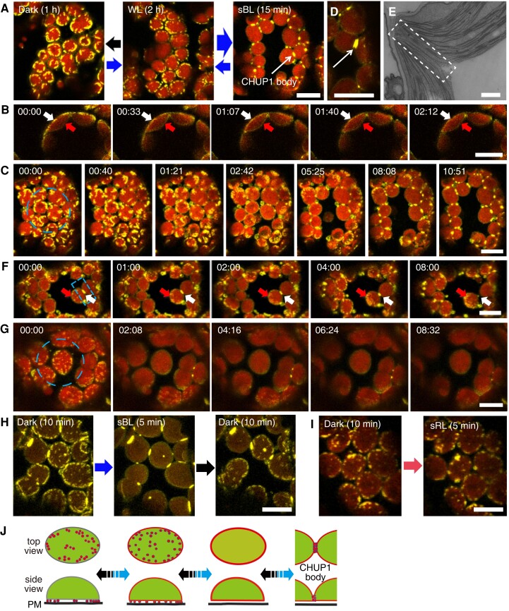
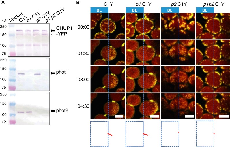
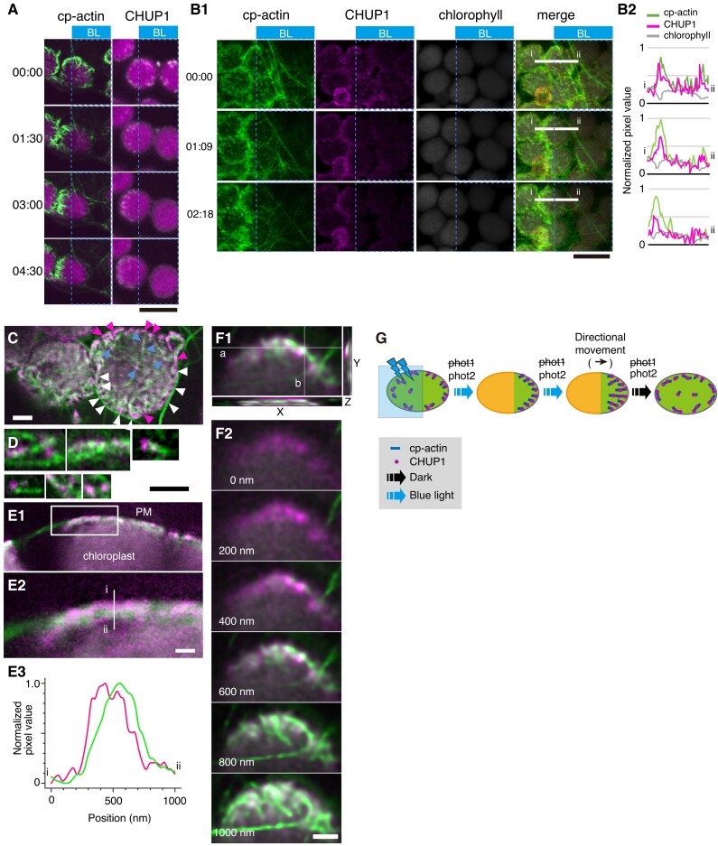
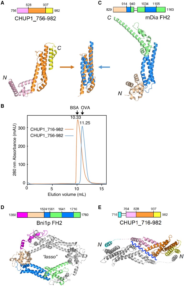
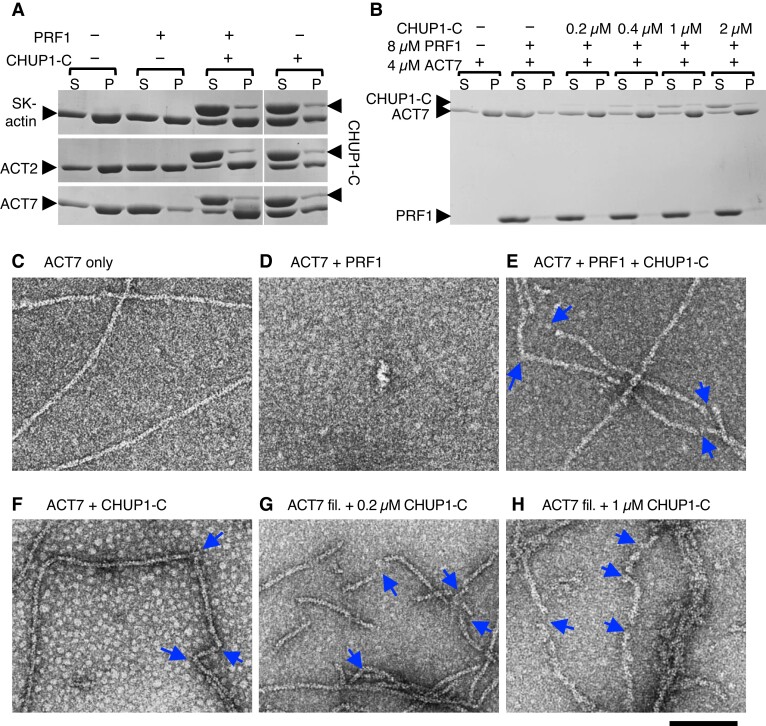
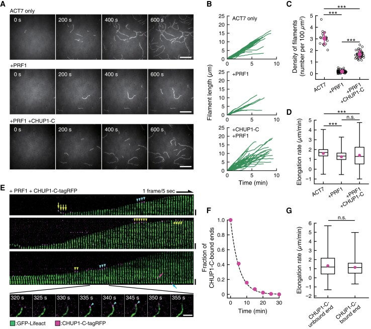
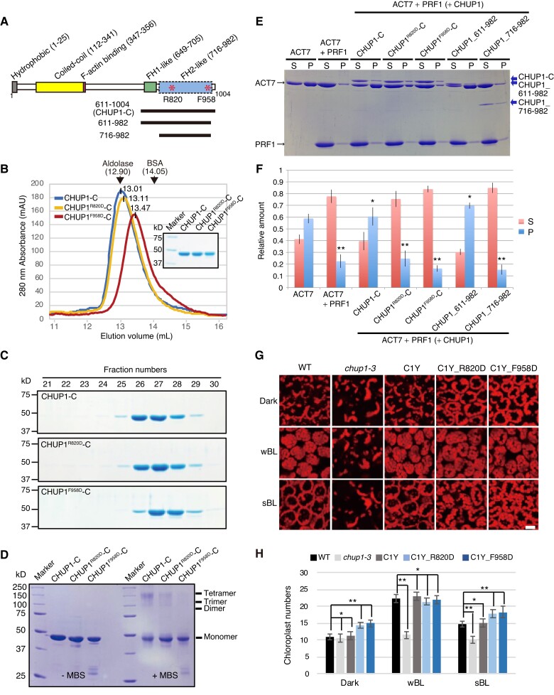
Plants have unique responses to fluctuating light conditions. One such response involves chloroplast photorelocation movement, which optimizes photosynthesis under weak light by the accumulation of chloroplasts along the periclinal side of the cell, which prevents photodamage under strong light by avoiding chloroplast positioning toward the anticlinal side of the cell. This light-responsive chloroplast movement relies on the reorganization of chloroplast actin (cp-actin) filaments. Previous studies have suggested that CHLOROPLAST UNUSUAL POSITIONING 1 (CHUP1) is essential for chloroplast photorelocation movement as a regulator of cp-actin filaments. In this study, we conducted comprehensive analyses to understand CHUP1 function. Functional, fluorescently tagged CHUP1 colocalized with and was coordinately reorganized with cp-actin filaments on the chloroplast outer envelope during chloroplast movement in Arabidopsis thaliana. CHUP1 distribution was reversibly regulated in a blue light- and phototropin-dependent manner. X-ray crystallography revealed that the CHUP1-C-terminal domain shares structural homology with the formin homology 2 (FH2) domain, despite lacking sequence similarity. Furthermore, the CHUP1-C-terminal domain promoted actin polymerization in the presence of profilin in vitro. Taken together, our findings indicate that CHUP1 is a plant-specific actin polymerization factor that has convergently evolved to assemble cp-actin filaments and enables chloroplast photorelocation movement.


Conflict of interest statement

Conflict of interest statement. None declared.

Figures

Figure 1.
Figure 1.
Reorganization of CHUP1-YFP on the chloroplast envelope in response to blue light. A) Reversible changes in the distribution pattern of CHUP1-YFP under the indicated light conditions. CHUP1-YFP in the palisade cells of C1Y transgenic Arabidopsis plants was observed following incubation in darkness for 1 h (black arrow), weak white light (20 μmol m−2 s−1) for 2 h (thin blue arrow), or strong blue light (sBL; 458-nm laser scan with an output power of 2.8 µW) for 15 min (thick blue arrow). The images were captured at a resolution of 512 × 512 pixels using a 4× digital zoom and are presented with false-color indicating YFP (yellow) and chlorophyll (red) fluorescence. The time (min:s) of image acquisition is shown in the upper left corner of each image. B) Redistribution of CHUP1-YFP from the interface between the plasma membrane and the chloroplast envelope (white arrows) to the entire chloroplast envelope, including the vacuole side (red arrows). The whole cell was irradiated with strong blue light. Time-lapse images were captured at 33-s intervals. The image acquisition and presentation are the same as in A). C) Blue light-dependent reorganization of CHUP1-YFP during a strong light-induced avoidance response followed by dark adaptation for 20 min. The dashed blue circle indicates the area that was irradiated. A time-lapse movie of this response can be seen in Supplementary Movie S1. D) A CHUP1 body appears as 2 lines along the interface between 2 chloroplasts after 5 min of strong blue light irradiation. E) Transmission electron microscopy image of the region of contact (at the center of the rectangle) between 2 chloroplasts. F) Redistribution of a CHUP1 body (white arrows) to small dots (red arrows) at the leading edge of a moving chloroplast. G) Redistribution of CHUP1-YFP on an isolated irradiated chloroplast. The dotted CHUP1-YFP signal rapidly disappeared but no CHUP1 body formed. A time-lapse movie of this response can be seen in Supplementary Movie S2. The area irradiated with strong blue light is indicated as a dashed blue circle (20 µm in diameter) in C and G), and a dashed blue rectangle (10 μm × 5 μm) in F) superimposed on the first image (00:00) of each series. Strong blue light was provided by 458-nm laser scans (an output power of 2.8 μW). Time-lapse images were collected at 40-, 33-, 60-, and 30-s intervals in C, F, and G), respectively. The image acquisition and presentation are the same as in A). H and I) Blue light-specific reorganization of CHUP1-YFP. H) CHUP1-YFP was sequentially observed in the same palisade cell of C1Y transgenic Arabidopsis plants following incubation in the dark for 10 min (Dark 10 min), irradiation with strong blue light (sBL; 458-nm laser scans) for 5 min (sBL 5 min), and further incubation in the dark for 10 min (Dark 10 min). I) CHUP1-YFP was sequentially observed in a palisade mesophyll cell following incubation in the dark for 10 min and then irradiated with strong red-light (sRL) by 633-nm laser scans for 5 min. The image acquisition and presentation are the same as in A). Scale bar, 500 nm in E), 10 μm in all other panels. J) Diagram illustrating the reorganization of CHUP1 on the chloroplast envelope. CHUP1 (red) localizes as dots at the interface between the plasma membrane and a chloroplast in darkness. In continuous strong light, CHUP1 disperses to the entire chloroplast envelope, first as dots at the interface and then diffusely throughout the chloroplast outer envelope. Finally, CHUP1 accumulates as a CHUP1 body where 2 chloroplasts are in contact. These steps are reversibly regulated in a blue light-dependent manner. The lower panel shows side views of a chloroplast (side view) and the upper panel shows the chloroplast surface that faces the plasma membrane (top view). Blue arrows indicate phototropin-dependent responses under blue light and black arrows indicate the reverse steps in the dark.
Figure 2.
Figure 2.
Identification of the main photoreceptor regulating CHUP1 localization. A) Immunoblot analysis of CHUP1-YFP abundance in transgenic plants in the WT, phot1, phot2, and phot1 phot2 mutant backgrounds (C1Y, p1 C1Y, p2 C1Y, p1 p2 C1Y). Protein samples (50 μg) were separated on 7.5% SDS-PAGE gels and probed with anti-CHUP1 (top panel), anti-PHOT1 (middle), and anti-PHOT2 (bottom) polyclonal antibodies. Arrows from top to bottom blots indicate CHUP1-YFP, phot1, and phot2, respectively. The band indicated by * most likely represents truncated CHUP1-YFP generated by endogenous proteases, since this band is absent in cells lacking CHUP1-YFP. B) Series of images at the indicated time points (min:s) showing the reorganization of CHUP1-YFP in strong blue light in WT and phot mutant cells. In each image, the region to the left of the blue dotted line was irradiated with strong blue light by 458-nm laser scans with an output power of 2.8 μW at 30-s intervals to induce the avoidance response. The images were captured at a resolution of 512 × 256 pixels using a 4× digital zoom and are presented with false-color indicating YFP (yellow) and chlorophyll (red) fluorescence. The time (min:s) of image acquisition is shown to the left. The paths of individual chloroplasts are indicated in red at the bottom of each image. The centers of each chloroplast were traced. Time-lapse movies of these responses can be seen in Supplementary Movies S3 to S6. Scale bars, 5 μm.
Figure 3.
Figure 3.
Association of CHUP1 with cp-actin filaments. A) Asymmetric distribution of cp-actin filaments (green color, left panels) and CHUP1 (white color right panels) on moving chloroplasts. Time-lapse images were collected at 30-s intervals for 4 min 30 s under strong blue light irradiation at the blue rectangle ROI using 458-nm laser scans (output power of 2.8 μW) in the intervals between image acquisitions. Image acquisition time stamps (min:s) are shown. Fluorescence images were captured at a resolution of 512 × 512 pixels using a 4× digital zoom in GFP-mTalin and CHUP1-YFP transgenic plants, respectively. The fluorescent images show cp-actin (green) or CHUP1 (white) merged with chlorophyll (magenta). The time (min:s) of image acquisition is shown on the left. Scale bar, 10 μm. B) Reorganization of CHUP1 and cp-actin filaments on moving chloroplasts in a CHUP1-tdTomato GFP-mTalin line. Time-lapse images were collected at the indicated time points (min:s). The other details are the same as in A) except that images were acquired at 34-s intervals for 3 min 27 s. Scale bar, 10 µm. A time-lapse movie of this response is shown in Supplementary Movie S7. B1) fluorescence images show cp-actin filaments (cp-actin, green), CHUP1-tdTomato (CHUP1, magenta), chloroplast (chlorophyll, gray), and the merged image of cp-actin filaments, CHUP1, and chlorophyll (merge). B2) fluorescence intensity profiles of CHUP1-tdTomato (magenta) and cp-actin filaments (green) along the white line (i and ii) in B1) (merge). C) Top view of chloroplasts facing the plasma membrane of the periclinal wall of palisade cells. Cp-actin filaments, CHUP1, and chloroplasts are shown with fluorescent images obtained from GFP-mTalin (green), CHUP1-tdTomato (magenta), and chlorophyll (gray), respectively, in a CHUP1-tdTomato GFP-mTalin line. Notably, CHUP1 was predominantly found in the peripheral region of chloroplast (magenta arrowheads). White arrowheads indicate CHUP1 attached to long cytoplasmic actin filaments. Blue arrowheads indicate CHUP1 localized at the tips or along the sides of cp-actin filaments. D) Magnified views of CHUP1 localized close to cp-actin filaments. E) Distribution of CHUP1 and cp-actin filaments within the region between the plasma membrane and the chloroplast envelope. CHUP1 localized to the plasma membrane side and cp-actin filaments along the chloroplast side. E1) An optical slice of a side view of a chloroplast next to the plasma membrane along an anticlinal wall. E2) An enlarged image of the area within the rectangle in E1). E3) Fluorescence intensity profiles of CHUP1-tdTomato (magenta) and cp-actin filaments (green) along the white line (i and ii) in E2) from the plasma membrane side (i, top of the line) to the chloroplast side (ii, bottom of the line). F) Separate distributions of CHUP1 at the plasma membrane side and cp-actin filaments on the chloroplast side. Top views of part of a chloroplast are shown. F1) A Z-series of 6 confocal images of fluorescence of CHUP1-tdTomato (magenta) and GFP-mTalin (green) taken with 200-nm steps from the plasma membrane side to the chloroplast side and superimposed on an XY plane using the maximum intensity projection method. In the XZ plane at white line a and the YZ plane at white line b, the CHUP1-tdTomato and GFP-mTalin fluorescence for the entire Z-series was compiled, confirming that CHUP1 is on the plasma membrane side and cp-actin filaments are on the chloroplast side. The individual images are shown in F2). Scale bars, 1 μm in C, D, and F), and 500 nm in E). G) Diagram illustrating the coordinated phototropin-dependent dynamics of CHUP1 and cp-actin filaments. Clusters of CHUP1 (magenta dots) closely localize with cp-actin filaments. Asymmetric redistribution of CHUP1 and cp-actin filaments is coordinately regulated by phototropin in response to irradiation of part of a chloroplast with strong blue light (blue rectangle). phot2 is the main photoreceptor regulating the strong light-induced avoidance response.
Figure 4.
Figure 4.
Three-dimensional structures of the C-terminal fragments of CHUP1. A) Crystal structure of CHUP1_756–982. B) The elution profiles of CHUP1_756–982 and CHUP1_716–982 were obtained by gel filtration chromatography using a Superdex 75 10/300 column. The elution volumes of BSA (67 kD) and ovalbumin (OVA, 43 kD) used for calibration are presented. The molecular weights of the recombinant proteins were calculated as 42 kD for CHUP1_756–982 and 64 kD for CHUP1_716–982. C) Crystal structure of the mDia1 FH2 domain (PDB 1V9D, Shimada et al. 2004). The central 3-helix bundles of CHUP1_756–982 and FH2 are superimposed in the middle between panels A and C). D) Functional dimer form of Bni1p FH2 (PDB 1UX5, Xu et al. 2004). E) Crystal structure of CHUP1_716–982. This fragment adopts a closed dimeric structure in crystal, which might open along the wavy line to become a functional dimer. In C and E), loops that are missing from the crystal structure due to poor electron density are indicated by thin gray boxes in the amino acid sequences.
Figure 5.
Figure 5.
Interaction of CHUP1-C with actin filaments in vitro. A) Ultracentrifugation assays of the effects of profilin and CHUP1-C on actin polymerization. Rabbit skeletal muscle actin (SK-actin) or Arabidopsis ACT2 or ACT7 was allowed to polymerize in F-buffer 1 in the presence or absence of 8 μM PRF1 and 4 μM CHUP1-C. After ultracentrifugation, the supernatant (S) and pellet (P) fractions were separately run on SDS-PAGE. B) Similar to A), except that only ACT7 was used and the concentration of CHUP1-C varied from 0 to 2 μM. C to F) Electron micrographs of actin polymerized in the presence or absence of profilin and CHUP1-C for 20 min. In C to F), 4 μM ACT7 was allowed to polymerize in F-buffer 1 in the absence of PRF1 and CHUP1-C C), in the presence of 4 μM PRF1 D), in the presence of 4 μM PRF1 and 0.4 μM CHUP1-C E), and in the presence of 0.4 μM CHUP1-C F). Actin solutions were diluted to 0.7 µM ACT7 in EM buffer, and then fixed and negatively stained with uranyl acetate. For particles observed in F), see Supplementary Fig. S7C. G and H) ACT7 filaments polymerized in the absence of PRF1 and CHUP1-C were diluted to 0.7 µM and applied onto an EM grid. After ∼30 s, 0.2 µM G) or 1 µM H) CHUP1-C was added, and the sample was negatively stained within several seconds. Blue arrows indicate the positions of sharp bent or gap of actin filaments. Scale bar, 100 nm.
Figure 6.
Figure 6.
CHUP1-C-dependent polymerization of ACT7 filaments from ACT7–profilin complexes. A) Nucleation and elongation of ACT7 filaments. Monomeric ACT7 at 1.5 μM was allowed to polymerize in F-buffer 2 containing 0.3% (w/v) methylcellulose, 2 mM Trolox, and 0.25 μM GFP-Lifeact (E17K) in a chamber. Prior incubation of ACT7 with a 2-fold molar excess of profilin PRF1 strongly inhibited spontaneous nucleation (middle row). However, the addition of 0.5 µM CHUP1-C-tagRFP partially reversed the effects of PRF1 and allowed nucleation and polymerization of a large number of filaments (bottom row). Scale bars, 10 µm. B) Polymerization kinetics of individual filaments under the 3 conditions in A). C) Quantification of nucleation activities of ACT7 under the 3 conditions in A). Numbers of ACT7 filaments were counted at about 10 min after the induction of polymerization. The means of the densities (the numbers of filaments per 100 µm2) were 3.04 ± 0.54/100 µm2 for ACT7 only (21 ROIs from 2 chambers), 0.19 ± 0.11/100 µm2 for +PRF1 (68 ROIs from 3 chambers) and 1.68 ± 0.31/100 µm2 for +PRF1 +CHUP1-C (40 ROIs from 3 chambers). ***P < 0.001 (Mann–Whitney U-test). D) Boxplot showing the elongation rates during 10-s intervals. The box represents the 25 to 75th percentiles, and the median is indicated by the black line. The whiskers show the complete range from minimum to maximum values. Means (represented by red circle) ± standard deviation (Sd) and the numbers of samples were 1.61 ± 0.61 µm/min for ACT7 only (1,235 intervals of 44 filaments from 2 chambers), 1.27 ± 0.62 µm/min for +PRF1 (347 intervals of 15 filaments from 3 chambers) and 1.41 ± 1.08 µm/min for +PRF1 +CHUP1-C (1,482 intervals of 47 filaments from 3 chambers). ***P < 0.001 (Mann–Whitney U-test). E) Kymographic representation of polymerization of an ACT7 filament in the presence of PRF1 and CHUP1-C-tagRFP. Green shows fluorescence of GFP-Lifeact (E17K), and magenta shows that of CHUP1-C-tagRFP. Raw fluorescence images of a polymerizing ACT7 filament (shown in the bottom row) were straightened and aligned to assemble a kymograph after the background was subtracted using ImageJ software. Spots of CHUP1-C-tagRFP were observed both during the growing phases (cyan arrowheads) and the stationary phases (yellow arrowheads) at the ACT7 filament ends. CHUP1-C-tagRFP spots were also sometimes observed at the end of short ACT7 filaments (yellow arrows), which presumably represent complexes of CHUP1-C-tagRFP and actin filaments at the initial phase of polymerization. Binding to the filament side (magenta arrow) and the less active end (cyan arrow) was also observed, but these frequencies were much lower than that of active-end binding (Supplementary Fig. S7, B and C). Scale bars, 2 µm. F) Dwell time of CHUP1-C-tagRFP at active ends of the filaments. Time-lapse images taken at 5-s intervals were analyzed, and the minimum lifetime of each fluorescent spot were cumulated and plotted (red circles). The dashed line is the exponential fit yielding the dissociation rate of CHUP1-C-tagRFP from the elongating end koff = 0.19 s−1. G) Comparison of the elongation rates between CHUP1-C-unbound ends and CHUP1-C-bound ends. Boxplot shows the elongation rates during 5-s interval of 36 filaments to which CHUP1-C-tagRFP was transiently bound (from 3 chambers), analyzed separately for CHUP1-C-tagRFP-bound phases and unbound phases. The box represents the 25 to 75th percentiles, and the median is indicated by the black line. The whiskers show the complete range from minimum to maximum values. Means (represented by red circle) ± Sd and the numbers of samples were 1.32 ± 1.14 µm/min for unbound ends (2,437 intervals) and 1.14 ± 0.85 µm/min (274 intervals). P = 0.053 (Mann–Whitney U-test).
Figure 7.
Figure 7.
Effects of point mutations at R820 or F958 of CHUP1-C on the structure and activity of CHUP1. A) Diagram of CHUP1 showing its functional domains: the hydrophobic region, the coiled-coil domain, the F-actin-binding domain, the proline-rich FH1-like domain, and the FH2-like domain. Asterisks indicate R820, which is involved in the association with actin, and F958, which is involved in the dimerization of the FH2-like domain. The positions of the 611 to 1,004 fragment (CHUP1-C) and the 611 to 982 and 716 to 982 fragments used for crystallization are indicated by lines below the structure. B and C) Effect of point mutations R820D and F958D on CHUP1-C dimer structure. B) The elution profiles of CHUP1-C, CHUP1R820D-C, and CHUP1F958D-C recombinant proteins were obtained by gel filtration chromatography using a Superdex 200 10/300 column. The elution volumes of aldolase (158 kD) and BSA (67 kD) used for calibration are presented. The molecular weights of the CHUP1-C recombinant proteins were calculated as 151.5 kD for CHUP1-C, 144.7 kD for CHUP1R820D-C, and 122.5 kD for CHUP1F958D-C. C) The protein profiles were confirmed using 10% SDS-PAGE gels. D) Effect of the point mutations R820D and F958D on the multimeric structure of CHUP1-C. Two µM of CHUP1-C, CHUP1R820D-C, and CHUP1F958D-C recombinant proteins was incubated without (left panel) or with (right panel) 20 µM MBS (m-maleimidobenzoyl-N-hydroxysuccinimide ester) for 1 h, and their multimer formations were analyzed using 10% SDS-PAGE. E and F) Effect of deletion and point mutations of CHUP1-C on actin polymerization. E) Ultracentrifugation assay of actin polymerization. ACT7 (4 µM) was allowed to polymerize in F-buffer 1 containing 8 µM PRF1 and 0.4 µM intact or mutant CHUP1-C, and a representative set of supernatant (S) and pellet (P) fractions after ultracentrifugation were analyzed by SDS-PAGE. F) Quantitative analysis of actin polymerization showing the fractions of ACT7 in the pellet and the supernatant under each condition as shown in E). The data are presented as means ± Sd (n = 3). Asterisks indicate statistically significant differences between ACT7 and each line of pellet detected by Student's t-test (* not significant P > 0.05; ** significant P < 0.0001). G) Effect of point mutations at R820D and F958D of CHUP1 on chloroplast positioning. The 4th rosette leaves of WT, chup1-3, C1Y, C1Y_R820D and C1Y_F958D plants were detached after dark adaptation (dark) for 14 h and then were further irradiated with a weak blue light of 2 µmol m−2 s−1 or a strong blue light (sBL) of 50 µmol m−2 s−1 for 2 h, respectively. Chloroplasts were shown with chlorophyll autofluorescence that was captured using confocal microscopy at a resolution of 512 × 512 pixels in a depth of 2 µm. Scale bar, 20 µm. H) Average number of chloroplasts observed on the periclinal side of palisade cells of WT, chup1-3, C1Y, C1Y_R820D, and C1Y_F958D plants. The average numbers of chloroplasts shown in G) were counted from 20 palisade cells from 3 rosette leaves. The data are presented as means ± SE (n = 20). Asterisks indicate statistical differences detected by Student's t-test (* not significant P > 0.05; ** significant P < 0.0001).

References

    1. Adams PD, Afonine PV, Bunkoczi G, Chen VB, Davis IW, Echols N, Headd JJ, Hung LW, Kapral GJ, Grosse-Kunstleve RW, et al. . PHENIX: a comprehensive Python-based system for macromolecular structure solution. Acta Crystallogr D Biol Crystallogr. 2010:66(2):213–221. 10.1107/S0907444909052925 - DOI - PMC - PubMed
    1. Angel CA, Lutz L, Yang X, Rodriguez A, Adair A, Zhang Y, Leisner SM, Nelson RS, Schoelz JE. The P6 protein of Cauliflower mosaic virus interacts with CHUP1, a plant protein which moves chloroplasts on actin microfilaments. Virology. 2013:443(2):363–374. 10.1016/j.virol.2013.05.028 - DOI - PubMed
    1. Belyy A, Merino F, Sitsel O, Raunser S. Structure of the Lifeact–F-actin complex. PLoS Biol. 2020:18(11):e3000925. 10.1371/journal.pbio.3000925 - DOI - PMC - PubMed
    1. Cazzaniga S, Dall’ Osto L, Kong S-G, Wada M, Bassi R. Interaction between avoidance of photon absorption, excess energy dissipation and zeaxanthin synthesis against photooxidative stress in Arabidopsis. Plant J. 2013:76(4):568–579. 10.1111/tpj.12314 - DOI - PubMed
    1. Edelstein A, Amodaj N, Hoover K, Vale R, Stuurman N. Computer control of microscopes using µManager. Curr Protoc Mol Biol. 2010:92(1):14.20.11–14.20.17. 10.1002/0471142727.mb1420s92 - DOI - PMC - PubMed

LinkOut - more resources