. 2024 Jan 4;187(1):44-61.e17.
doi: 10.1016/j.cell.2023.11.027. Epub 2023 Dec 21.

Sensory neurons promote immune homeostasis in the lung

Affiliations

Sensory neurons promote immune homeostasis in the lung

Masato Tamari et al. Cell. .

Abstract

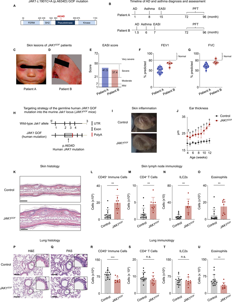
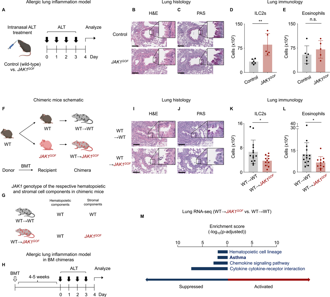
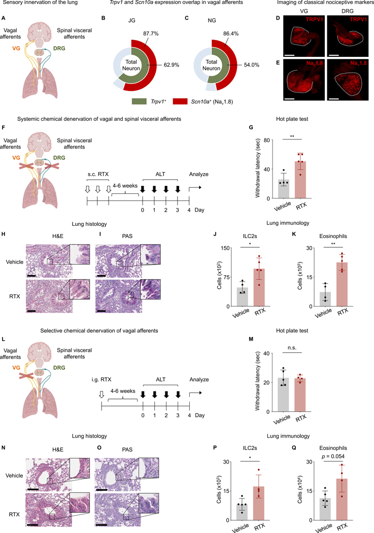
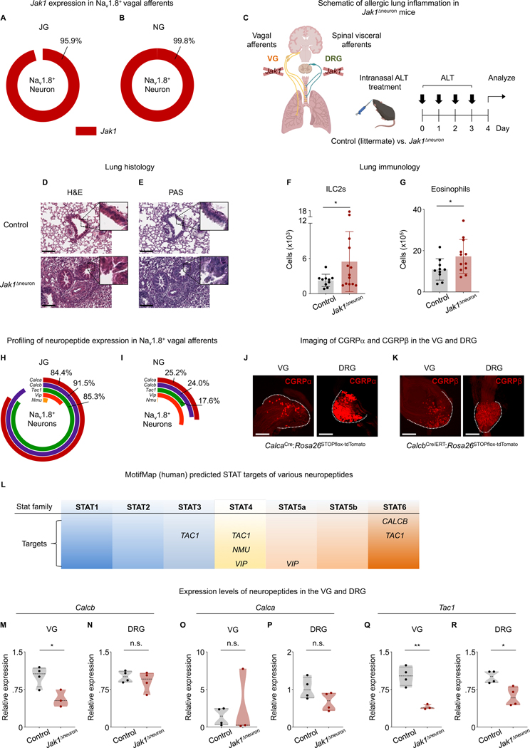
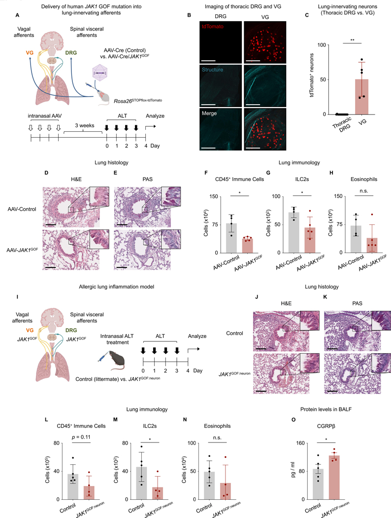
Cytokines employ downstream Janus kinases (JAKs) to promote chronic inflammatory diseases. JAK1-dependent type 2 cytokines drive allergic inflammation, and patients with JAK1 gain-of-function (GoF) variants develop atopic dermatitis (AD) and asthma. To explore tissue-specific functions, we inserted a human JAK1 GoF variant (JAK1GoF) into mice and observed the development of spontaneous AD-like skin disease but unexpected resistance to lung inflammation when JAK1GoF expression was restricted to the stroma. We identified a previously unrecognized role for JAK1 in vagal sensory neurons in suppressing airway inflammation. Additionally, expression of Calcb/CGRPβ was dependent on JAK1 in the vagus nerve, and CGRPβ suppressed group 2 innate lymphoid cell function and allergic airway inflammation. Our findings reveal evolutionarily conserved but distinct functions of JAK1 in sensory neurons across tissues. This biology raises the possibility that therapeutic JAK inhibitors may be further optimized for tissue-specific efficacy to enhance precision medicine in the future.

Keywords: AAV; CGRP; ILC2; JAK1; afferent nerves; allergic lung inflammation; atopic disorders; neuropeptide; sensory neurons; vagus nerve.


Conflict of interest statement

Declaration of interests B.S.K. is founder of KliRNA Biotech; he has served as a consultant for 23andMe, ABRAX Japan, AbbVie, Almirall, Amgen, Arcutis Biotherapeutics, Arena Pharmaceuticals, argenx, AstraZeneca, Boehringer Ingelheim, Bristol-Myers Squibb, Cara Therapeutics, Clexio Biosciences, Eli Lilly and Company, Escient Pharmaceuticals, Evommune, Galderma, Genentech, GlaxoSmithKline, Granular Therapeutics, Incyte Corporation, Innovaderm Research, Janssen, Kiniksa, LEO Pharma, Maruho, Novartis, Pfizer, Recens Medical, Regeneron Pharmaceuticals, Sanofi, Septerna, Triveni Bio, Vial, and WebMD; he has stock in ABRAX Japan, KliRNA Biotech, Locus Biosciences, and Recens Medical; he holds a patent for the use of JAK1 inhibitors for chronic pruritus; and he has a patent pending for the use of JAK inhibitors for interstitial cystitis. D.A. has contributed to scientific advisory boards at Pfizer, Takeda, FARE, and the KRF. D.B. is the founder of Lab11 Therapeutics..

Figures

Figure 1.
Figure 1.. Human germline JAK1 GOF mutation is associated with AD and asthma in patients and promotes spontaneous AD-like disease in mice
(A) Human JAK1 c.1901C>A (p.A634D) GOF mutation within the pseudokinase domain. (B) Timeline of AD and asthma diagnosis, EASI assessment, and PFTs. (C) Baseline visual image of AD skin lesions from Patient A with a JAK1 GOF mutation. (D) Baseline visual image of AD skin lesions from Patient B with a JAK1 GOF mutation. (E) Baseline EASI scores from Patient A and Patient B. (F) Baseline FEV1 of Patient A and Patient B. (G) Baseline FVC of Patient A and Patient B. (H) Schematic of the targeting strategy used to introduce the germline human JAK1 GOF mutation (JAK1GOF) into the murine Jak1 locus. (I) Representative ear skin images of WT control and JAK1GOF mice. (J) Ear thickness measurements of WT control and JAK1GOF mice. (K) Representative skin histology images (H&E) of WT control and JAK1GOF mice. Scale bars indicate 100 μm. (L-O) Flow cytometry of skin lymph nodes from WT control and JAK1GOF mice. Shown are the frequencies of (L) total CD45+ immune cells, (M) CD4+ T cells, (N) ILC2s, and (O) eosinophils. (P-Q) Representative lung histology images of WT control and JAK1GOF mice, obtained by (P) H&E stain and (Q) PAS stain. Scale bars indicate 200 μm. (R-U) Flow cytometry of lung tissue from WT control and JAK1GOF mice. Shown are the frequencies of (R) total CD45+ immune cells, (S) CD4+ T cells, (T) ILC2s, and (U) eosinophils. (J, L-O, R-U) Data were pooled from three independent experiments, n = 8–15 pooled mice per group. p < 0.0001, ***p < 0.001, **p < 0.01, *p < 0.05, and not significant (n.s.) (Unpaired t-test with Welch’s correction). Data are represented as mean ± standard deviation (SD). AD – atopic dermatitis, EASI – eczema area and severity index, FEV1 – forced expiratory volume, FVC – forced vital capacity, GOF – gain-of-function, ILC2s – group 2 innate lymphoid cells, H&E – hematoxylin and eosin, JAK1 – Janus kinase 1, PAS – periodic acid-Schiff, PFT – pulmonary function test, WT – wild-type. See also Figure S1
Figure 2.
Figure 2.. Stroma-intrinsic expression of JAK1GOF is protective in allergic lung inflammation
(A) Schematic of the allergic lung inflammation model using the protease allergen Alternaria alternata (ALT). (B-C) Representative lung histology images of WT control and JAK1GOF mice challenged with intranasal ALT, obtained by (B) H&E stain and (C) PAS stain. (D-E) Flow cytometry of lung tissue from WT control and JAK1GOF mice challenged with intranasal ALT. Shown are the frequencies of (D) ILC2s and (E) eosinophils. Data were obtained from three independent experiments, n = 5 mice per group. (F) Schematic of experimental approach used to generate chimeric mice. BM derived from WT mice was transferred into WT control and experimental JAK1GOF mice. (G) JAK1 genotype of the respective hematopoietic and stromal cell components in chimeric mice (WT→WT and WT→JAK1GOF). (H) Schematic of experimental approach used to generate the allergic lung inflammation model in the BM chimeras. (I-J) Representative lung histology images of chimeric WT→WT and WT→JAK1GOF mice challenged with intranasal ALT, obtained by (I) H&E stain and (J) PAS stain. (K-L) Flow cytometry of lung tissue from chimeric WT→WT and WT→JAK1GOF mice challenged with intranasal ALT. Shown are the frequencies of (K) ILC2s and (L) eosinophils. Data were obtained from three independent experiments, n = 11–14 pooled mice per group. (M) KEGG pathway overrepresentation analysis of RNA-seq data obtained from the lung tissues of chimeric WT→JAK1GOF vs. WT→WT mice (n = 2 mice/group). Overrepresentation analysis used Fisher’s exact test to determine the significance of enrichment. All terms shown were identified as the top four significantly suppressed pathways in terms of adjusted p-value (p-adjusted < 0.05, Benjamini-Hochberg procedure). **p < 0.01, *p < 0.05, and not significant (n.s.) (Unpaired t-test with Welch’s correction). Data are represented as mean ± SD. Scale bars indicate 200 μm. (A, F-G) Figure adapted from an image created with BioRender.com. ALT- Alternaria alternata, BM – Bone marrow, BMT – Bone marrow transplantation. See also Figure S1
Figure 3.
Figure 3.. Chemical denervation of TRPV1+ sensory neurons exacerbates allergic lung inflammation
(A) Visual representation of the sensory innervation of the lung arising from the VG and DRG. (B-C) Expression of Trpv1 and Scn10a (the gene encoding Nav1.8) within the VG, specifically the (B) JG, and (C) NG. Trpv1 expression is shown as the percentage of total neurons sequenced. Scn10a expression is shown as the percentage of Trpv1-expressing neurons. The full scRNA-seq dataset is available in Kupari et al. (D-E) Imaging of classical nociceptive markers (TRPV1 and Nav1.8) in the VG and DRG using Rosa26STOPflox-tdTomato mice crossed with the respective Trpv1Cre and Scn10aCre mice. (D) TRPV1 in the VG and DRG, (E) Nav1.8 in the VG and DRG. (F) Schematic of the allergic lung inflammation model generated following systemic denervation of vagal and spinal visceral afferents. (G) Withdrawal latency from noxious heat (hot plate test). Mice treated with s.c. vehicle control or RTX were placed on a hot plate set at 50 degrees Celsius, and withdrawal latency was measured as the time until the appearance of paw withdrawal behavior. (H-I) Representative lung histology images from mice treated with s.c. vehicle control or RTX challenged with intranasal ALT, obtained by (H) H&E stain and (I) PAS stain. (J-K) Flow cytometry of lung tissue from mice treated with s.c. vehicle control or RTX and challenged with intranasal ALT. Shown are the frequencies of (J) ILC2s and (K) eosinophils. Data were obtained from three independent experiments, n = 4–5 mice per group. (L) Schematic of the allergic lung inflammation model generated following selective denervation of vagal afferents. (M) Withdrawal latency from noxious heat (hot plate test). Mice treated with intraganglionic (i.g.) vehicle control or RTX were placed on a hot plate set at 50 degrees Celsius, and withdrawal latency was measured as the time until the appearance of paw withdrawal behavior. (N-O) Representative lung histology images of mice treated with i.g. vehicle control or RTX challenged with intranasal ALT, obtained by (N) H&E stain and (O) PAS stain. (P-Q) Flow cytometry of lung tissue from mice treated with i.g. vehicle control or RTX challenged with intranasal ALT. Shown are the frequencies of (P) ILC2s and (Q) eosinophils. Data were obtained from two independent experiments, n = 4–5 mice per group. **p < 0.01, *p < 0.05, and not significant (n.s.) (Unpaired t-test with Welch’s correction). Data are represented as mean ± SD. Scale bars indicate 200 μm. (A, F, L) Figure adapted from an image created with BioRender.com. DRG – dorsal root ganglia, i.g. – intraganglionic, JG – jugular ganglia, NG – nodose ganglia, RTX – resiniferatoxin, s.c. – subcutaneous VG – vagal ganglia See also Figures S1 and S2
Figure 4.
Figure 4.. Disruption of sensory neuron-intrinsic Jak1 exacerbates allergic lung inflammation and alters the expression levels of neuropeptides
(A-B) Jak1 expression in Nav1.8+ vagal afferents, (A) JG and (B) NG. Gene expression is shown as the percentage of Nav1.8+ neurons that express Jak1. The full scRNA-seq dataset is available in Kupari et al. (C) Schematic of the allergic lung inflammation model using littermate control (Jak1flox) and experimental Jak1Δneuron mice. (D-E) Representative lung histology images of control and Jak1Δneuron mice challenged with intranasal ALT, obtained by (D) H&E stain and (E) PAS stain. (F-G) Flow cytometry of lung tissue from control and Jak1Δneuron mice challenged with intranasal ALT. Shown are the frequencies of (F) ILC2s and (G) eosinophils. Data were obtained from two independent experiments, n = 10–12 pooled mice per group. (H-I) Profiling of neuropeptide expression in Nav1.8+ vagal afferents; Calca, Calcb, Tac1, Vip, and Nmu were evaluated in the (H) JG and (I) NG. Gene expression is shown as the percentage of Nav1.8+ neurons that express the indicated gene. The full scRNA-seq dataset is available in Kupari et al. (J-K) Imaging of the classical neuropeptides CGRPα and CGRPβ in the VG and DRG using Rosa26STOPflox-tdTomato mice crossed with CalcaCre and CalcbCre/ERT mice, respectively. Shown are (J) CGRPα in the VG and DRG and (K) CGRPβ in the VG and DRG. (L) MotifMap (human)-predicted neuropeptide targets of STAT family members. Data were analyzed using the MotifMap Predicted Transcription Factor Targets dataset (see Methods). (M-R) Quantitative PCR (qPCR) of transcripts from the VG and DRG from control and Jak1Δneuron mice challenged with intranasal ALT. Shown are (M, N) Calcb, (O, P) Calca, and (Q, R) Tac1 transcript levels. Data were obtained from two independent experiments, n = 3–4 mice per group. **p < 0.01, *p < 0.05, and not significant (n.s.) (Unpaired t-test with Welch’s correction). Data are represented as mean ± SD. Scale bars indicate 200 μm. (C) Figure adapted from an image created with BioRender.com. See also Figures S1 and S3–5
Figure 5.
Figure 5.. CGRPβ suppresses type 2 cytokine production from lung ILC2s and allergic lung inflammation
(A) Schematic of the lung ILC2 suppression assay. (B-C) Type 2 cytokine measurement from lung ILC2s stimulated with IL-33, SP, and/or CGRPβ. Shown are data for (B) IL-5 and (C) IL-13. (D) Schematic of the induction of allergic lung inflammation in Rag1−/− mice. (E-F) Flow cytometry of lung tissue from Rag1−/− mice challenged with intranasal ALT as well as vehicle control or CGRPβ or CGRPβ 8–37. Shown are the frequencies of (E) ILC2s and (F) eosinophils. Data were obtained from two independent experiments, n = 4–5 mice per group. (G-H) Representative lung histology images of Rag1−/− mice challenged with intranasal ALT as well as vehicle control or CGRPβ or CGRPβ 8–37, obtained by (G) H&E stain and (H) PAS stain. ***p < 0.001, **p < 0.01, *p < 0.05 (Unpaired t-test with Welch’s correction). Data are represented as mean ± SD. Scale bars indicate 200 μm. (A, D) Figure adapted from an image created with BioRender.com. ELISA – enzyme-linked immunosorbent assay, IL – interleukin, SP – substance P See also Figures S1 and S6
Figure 6.
Figure 6.. Expression of human JAK1GOF in lung sensory neurons suppresses allergic inflammation
(A) Schematic of the allergic lung inflammation model generated by delivery of AAV-Cre (control) or AAV-Cre/JAK1 GOF (experimental) into the airways of Rosa26STOPflox-tdTomato mice. (B) Imaging of tdTomato in the DRG and VG following AAV-assisted gene delivery into lung-innervating sensory neurons. Shown are representative images of DRG and VG from mice treated with AAV-Cre/JAK1GOF. (C) Numbers of tdTomato+ neurons in DRG (thoracic segments 4–8) and VG, n = 5 mice per group. Data were analyzed in mice treated with AAV-Cre/JAK1 GOF. (D-E) Representative lung histology images of mice challenged with intranasal ALT following AAV-Cre control and AAV-Cre/JAK1GOF infection, obtained by (D) H&E stain and (E) PAS stain. (F-H) Flow cytometry of lung tissue from mice challenged with intranasal ALT following AAV-Cre control and AAV-Cre/JAK1GOF infection. Shown are the frequencies of (F) total CD45+ immune cells, (G) ILC2s, and (H) eosinophils. Data were obtained from two independent experiments, n = 4–5 mice per group. (I) Schematic of the allergic lung inflammation model generated by conditional insertion of JAK1GOF variant into sensory neurons. (J-K) Representative lung histology images of mice challenged with intranasal ALT obtained by (J) H&E stain and (K) PAS stain. (L-N) Flow cytometry of lung tissue from mice challenged with intranasal ALT. Shown are the frequencies of (L) total CD45+ immune cells, (M) ILC2s, and (N) eosinophils. (O) Protein levels of CGRPβ in the bronchoalveolar lavage fluid (BALF). Data were obtained from two independent experiments, n = 4–5 pooled mice per group. *p < 0.05, **p < 0.01, and not significant (n.s.) (Unpaired t-test with Welch’s correction). Data are represented as mean ± SD. Scale bars indicate 200 μm. (A, I) Figure adapted from an image created with BioRender.com. AAV – adeno-associated virus; specifically, AAV2-retro, BALF – Bronchoalveolar lavage fluid See also Figure S1, Video S1, and S2

References

    1. Philips RL, Wang Y, Cheon H, Kanno Y, Gadina M, Sartorelli V, Horvath CM, Darnell JE, Stark GR, and O’Shea JJ (2022). The JAK-STAT pathway at 30: Much learned, much more to do. Cell 185, 3857–3876. 10.1016/j.cell.2022.09.023. - DOI - PMC - PubMed
    1. Bieber T, Simpson EL, Silverberg JI, Thaçi D, Paul C, Pink AE, Kataoka Y, Chu C-Y, DiBonaventura M, Rojo R, et al. (2021). Abrocitinib versus Placebo or Dupilumab for Atopic Dermatitis. New England Journal of Medicine 384, 1101–1112. 10.1056/NEJMoa2019380. - DOI - PubMed
    1. Guttman-Yassky E, Teixeira HD, Simpson EL, Papp KA, Pangan AL, Blauvelt A, Thaçi D, Chu C-Y, Hong H.C. h., Katoh N, et al. (2021). Once-daily upadacitinib versus placebo in adolescents and adults with moderate-to-severe atopic dermatitis (Measure Up 1 and Measure Up 2): results from two replicate double-blind, randomised controlled phase 3 trials. The Lancet 397, 2151–2168. 10.1016/S0140-6736(21)00588-2. - DOI - PubMed
    1. Reich K, Teixeira HD, de Bruin-Weller M, Bieber T, Soong W, Kabashima K, Werfel T, Zeng J, Huang X, Hu X, et al. (2021). Safety and efficacy of upadacitinib in combination with topical corticosteroids in adolescents and adults with moderate-to-severe atopic dermatitis (AD Up): results from a randomised, double-blind, placebo-controlled, phase 3 trial. The Lancet 397, 2169–2181. 10.1016/S0140-6736(21)00589-4. - DOI - PubMed
    1. Reich K, Thyssen JP, Blauvelt A, Eyerich K, Soong W, Rice ZP, Hong HC-H, Katoh N, Valenzuela F, DiBonaventura M, et al. (2022). Efficacy and safety of abrocitinib versus dupilumab in adults with moderate-to-severe atopic dermatitis: a randomised, double-blind, multicentre phase 3 trial. Lancet 400, 273–282. 10.1016/S0140-6736(22)01199-0. - DOI - PubMed

Publication types