. 2023 Dec 22;11(12):e007705.
doi: 10.1136/jitc-2023-007705.

Scalable generation of functional human iPSC-derived CAR-macrophages that efficiently eradicate CD19-positive leukemia

Affiliations

Scalable generation of functional human iPSC-derived CAR-macrophages that efficiently eradicate CD19-positive leukemia

Shifaa M Abdin et al. J Immunother Cancer. .

Abstract

Background: Macrophages have recently become attractive therapeutics in cancer immunotherapy. The potential of macrophages to infiltrate and influence solid malignancies makes them promising targets for the chimeric antigen receptor (CAR) technology to redirect their stage of polarization, thus enhancing their anticancer capacities. Given the emerging interest for CAR-macrophages, generation of such cells so far mainly depends on peripheral blood monocytes, which are isolated from the respective donor prior to genetic manipulation. This procedure is time-intensive and cost-intensive, while, in some cases, insufficient monocyte amounts can be recovered from the donor, thus hampering the broad applicability of this technology. Hence, we demonstrate the generation and effectiveness of CAR-macrophages from various stem cell sources using also modern upscaling technologies for next generation immune cell farming.

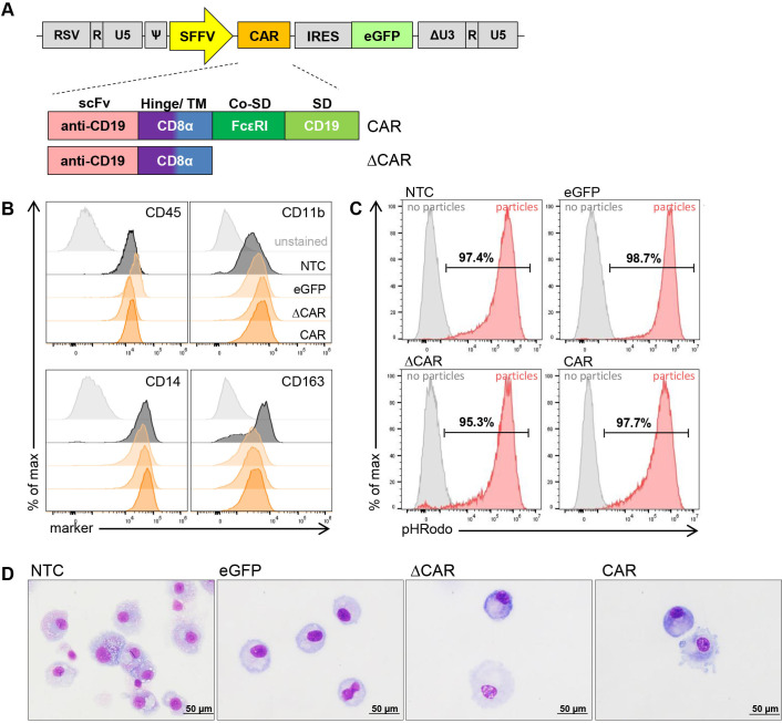
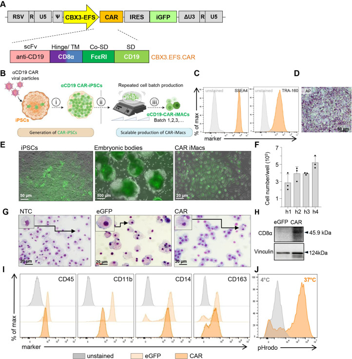
Methods: Primary human hematopoietic stem and progenitor cells and induced pluripotent stem cells were used to derive anti-CD19 CAR-macrophages. Anticancer activity of the cells was demonstrated in co-culture systems, including primary material from patients with leukemia. Generation of CAR-macrophages was facilitated by bioreactor technologies and single-cell RNA (scRNA) sequencing was used to characterize in-depth response and behavior of CAR-macrophages.

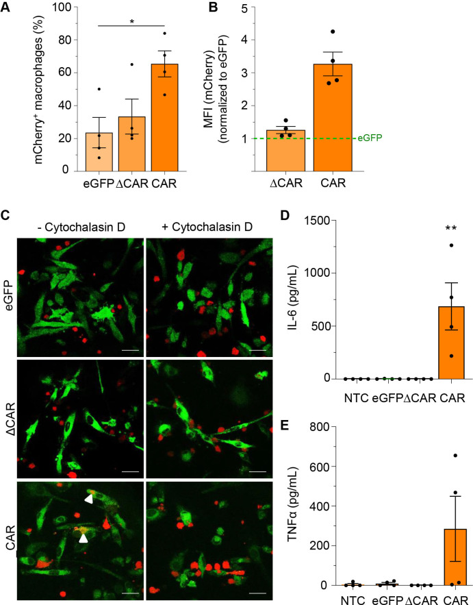
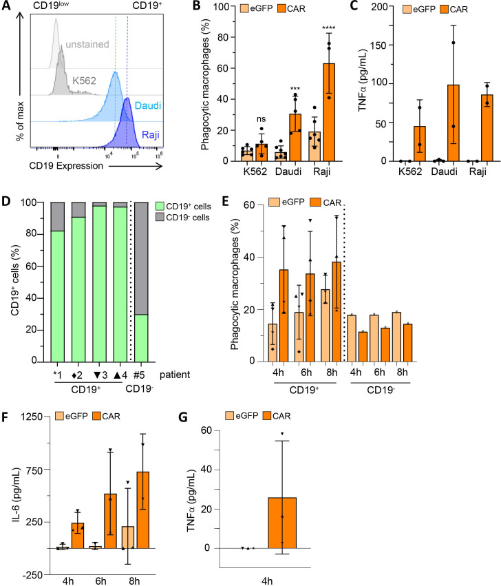
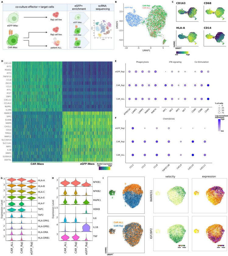
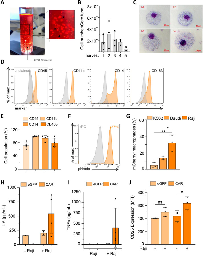
Results: Irrespective of the stem-cell source, CAR-macrophages exhibited enhanced and antigen-dependent phagocytosis of CD19+ target cancer cells with increased pro-inflammatory responses. Phagocytic capacity of CAR-macrophages was dependent on target cell CD19 expression levels with superior function of CAR-macrophages against CD19+ cancer cell lines and patient-derived acute lymphocytic leukemia cancer cells. scRNA sequencing revealed CAR-macrophages to be distinct from eGFP control cells after co-culture with target cells, which includes the activation of pro-inflammatory pathways and upregulation of chemokines and cytokines associated with adaptive immune cell recruitment, favoring the repolarization of CAR-macrophages to a pro-inflammatory state. Taken together, the data highlight the unique features of CAR-macrophages in combination with the successful upscaling of the production pipeline using a three-dimensional differentiation protocol and intermediate scale bioreactors.

Conclusion: In summary, our work provides insights into the seminal use and behavior of CAR-macrophages which are derived from various sources of stem cells, while introducing a unique technology for CAR-macrophage manufacturing, all dedicated to the clinical translation of CAR-macrophages within the field of anticancer immunotherapies.

Keywords: immunotherapy; macrophages; phagocytosis; receptors, chimeric antigen.


Conflict of interest statement

Competing interests: NL, MA and TM have filled a patent application on the generation of human iPSC-derived macrophages. All other authors declare no conflict of interest.

Figures

Figure 1
Figure 1
Characterization of cord blood-derived anti-CD19 CAR-macrophages. (A) CAR scheme depicting the composition of the anti-CD19 CAR (CAR) and the truncated version (ΔCAR) that lacks the internal signaling domain. (B) Surface marker expression profile by flow cytometric analyses. Controls include unstained control, non-transduced macrophage controls (NTC), truncated CAR-macrophages (ΔCAR) and eGFP-expressing macrophages (eGFP) (n=1). (C) Basic phagocytic activity against Escherichia coli-coupled particles as shown by flow cytometry (n=1). (D) Microscopic analyses of May-Grünwald-stained cytospins (using a fluorescence microscope Olympus IX71, 40× magnification, scale bar: 50 µm). CAR, chimeric antigen receptor; Co-SD, co-stimulatory signaling domain; eGFP, enhanced green fluorescent protein; IRIS,internal ribosome entery site; NTC, non-transduced control; scFv, single chain variable fragment; TM, transmembrane domain.
Figure 2
Figure 2
Evaluation of phagocytosis and cytokine secretion of cord blood-derived CAR-macrophages after co-culture with CD19+ Raji cells. Flow cytometric analysis of (A) percentage (%) of phagocytic macrophages and (B) geometric mean of mCherry signal intensity inside eGFP+ Macs after co-culture with target cells. Data are represented as the mean±SEM of n=4 biological replicates. (C) Confocal microscopy images showing phagocytosis of CD19+ Raji cells (mCherry) by macrophages (eGFP)±cytochalasin D treatment (63× oil immersion objective of the Leica DMi8 confocal microscope, scale bar: 20 µm). Secretion of (D) IL-6 and (E) TNF-α after 24 hours of co-culture of macrophages and Raji cells. Data are represented as the mean±SEM of n=4 biological replicates. Statistical significance was calculated with one-way analysis of variance using multiple comparisons. CAR, chimeric antigen receptor; eGFP enhanced green fluorescent expressing control macrophages; IL, interleukin; MFI, mean fluorescence intensity; NTC, non-transduced control; TNF, tumor necrosis factor.
Figure 3
Figure 3
Characterization of anti-CD19 CAR-iPSCs and thereof derived CAR-iMacs. (A) CAR scheme demonstrating the composition of the subcloned anti-CD19 CAR (CAR). (B) Illustration of the work flow to generate CAR-iMacs. (C) Flow cytometry analyses of CAR hiPSC expression of SSEA4 and TRA160 in comparison to unstained controls. (D) Bright field microscopic images of alkaline phosphatase stained CAR hiPSCs (scale bar 50 µm). (E) Representative fluorescent microscopic images of the different steps of the hematopoietic differentiation of hiPSCs to iMacs. (F) Quantities of macrophages harvested from the supernatants at different weeks from various differentiation cultures (n=3 independent experiments, mean±SD). (G) Photographs of cytospins prepared from non-transduced iMacs (NTC), eGFP iMacs (eGFP), and the anti-CD19 CAR-iMacs (CAR) (scale bar 20 µm). (H) Western blot analysis of CD8α expression (45.9 kDa) in eGFP and CAR-iMacs, Vinculin (124 kDa) served as a control. (I) Flow cytometric analyses of macrophage surface markers (representative data of n=3, (online supplemental figure 3A)). (J) Analyses of pHrodo-Escherichia coli bioparticles phagocytosis via flow cytometry (representative data of n=5, (online supplemental figure 3B)). CAR, chimeric antigen receptor; Co-SD, co-stimulatory signaling domain;eGFP, enhanced green fluorescent protein expressing control iMacs; hiPSC, human induced pluripotent stem cell; scFv, single chain variable fragment; TM, transmembrane domain.
Figure 4
Figure 4
Assessment of CAR-mediated phagocytosis in human induced pluripotent stem cells-derived CAR-macrophages after co-culture with CD19+ Raji cells. (A) Schematic showing co-culture of CAR or eGFP iMacs with CD19+ Raji cells. (B) Representative fluorescent microscopic images of CAR-iMacs in co-culture with the target Raji cells (20×, scale bar: 20 µm). (C) Representative image from the acquired gallery using an image-based flow cytometer to analyze the co-localization of the target cells inside the co-cultured CAR-iMacs. (D) Flow cytometric analyses of percentage (%) of mCherry-positive Raji cells inside the eGFP+ macrophages (eGFP/CAR-iMacs) or the CD14 FITC stained non-transduced control cells (NTC) after co-culture with target cells for 4 hours or after increased incubation periods to 6 and 8 hours (E). ELISA assays of IL-6 (F) and TNF-α (G) secretion by the indicated iMacs in mono-cultures versus co-cultures with Raji cells. Data are represented as the mean±SD of n=2–3 biological replicates. Statistical significance was calculated with one-way analysis of variance using multiple comparisons. CAR, chimeric antigen receptor; eGFP, enhanced green fluorescent protein expressing control iMacs; IL, interleukin; TNF, tumor necrosis factor.
Figure 5
Figure 5
Assessment of CAR-mediated antigen specificity in iPSC-derived macrophages against CD19 and patient with ALL samples. (A) MFI of CD19 expression on leukemia target cells (representative for n=2, (online supplemental figure 3D)). (B) Percentage (%) of phagocytic macrophages after 4 hours co-culture with CD19low or CD19+ target cells. Statistical significance was calculated with one-way analysis of variance using multiple comparisons. (C) TNF-α secretion from iMacs after co-culture with target cells (n=2–3). (D) Percentage (%) of CD19 expression on cells from five different primary ALL patient samples. (E) Percentage (%) of phagocytic macrophages after co-culture with CD19low or CD19+ primary ALL samples at the different indicated time points (n=4–5). ELISA assays of IL-6 (F) and TNF-α (G) secretion by the indicated iMacs after the co-culture with the different primary ALL samples (n=2–3). ALL, acute lymphocytic leukemia; CAR, chimeric antigen receptor; eGFP, enhanced green fluorescent expressing control iMacs; IL, interleukin; TNF, tumor necrosis factor.
Figure 6
Figure 6
CAR-iMacs show pro-inflammatory phenotype after co-culture with CD19+ target cells. (A) Scheme showing co-culture of CAR-iMacs or eGFP-iMacs with CD19+ Raji cells or ALL patient samples followed by subsequent scRNA-seq analysis. (B) UMAP plots illustrating the phenotypic distinction of eGFP-macrophages and CAR-macrophages after co-culture with CD19+ Raji or ALL cells colored according to cell lineages. (C) UMAP plots of CAR-macrophages and eGFP-macrophages after culturing with CD19+ Raji cells, showing the relative expression of typical macrophage genes within the populations colored according to gene expression. (D) Top 15 differentially-expressed genes of CAR-macrophage population after co-culture with Raji or ALL cells compared with eGFP-macrophage population. (E) and (F) Clustered dot plots showing differentially-expressed genes of eGFP-macrophages and CAR-macrophages after co-culture with Raji cells. (G) and (H) Violin plots illustrating differentially-expressed genes in eGFP-macrophages and CAR-macrophages in the context of antigen presentation and NF-κB signaling. (I) RNA velocity analysis and representative gene expression showing the progression of subsets inside the CAR-macrophage population. ALL, acute lymphocytic leukemia; CAR, chimeric antigen receptor;eGFP, enhanced green fluorescent expressing control iMacs; NF-κB, nuclear factor kappa B; scRNA-seq, single-cell RNA sequencing; UMAP, Uniform Manifold Approximation and Projection.
Figure 7
Figure 7
Continuous upscaled production of CAR-iMacs in a CERO bioreactor. (A) Photo illustrating the ongoing differentiation of anti-CD19 CAR-induced pluripotent stem cells in the 50 mL bioreactor tube, highlighting the formation of the hemanoids, which are continuously shedding the CAR-iMacs. (B) Quantities of CAR-iMacs harvested from the supernatants at different weeks from the ongoing bioreactor differentiation (n=3). (C) Representative cytospin images of the generated CAR-iMacs from every harvest. (D) Histograms of the flow cytometric analysis of macrophage surface markers compared with unstained control cells, (representative data of n=4, (online supplemental figure 6D)). (E) Bar chart quantifying the percentage (%) of surface marker expression from the stained CAR-iMacs from every harvest (different symbols reflect different harvests). (F) Flow cytometry analysis of pHrodo-Escherichia coli bioparticle phagocytosis by iMacs, (representative data of n=5, (online supplemental figure 3B)). (G) Bar chart quantifying the phagocytosis of mCherry+-labeled target cells inside the GFP+ CAR-iMacs via flow cytometry. LEGENDplex analysis of (H) IL-6 and (I) TNF-α secretion by CERO-bioreactor-generated iMacs in mono-cultures versus co-cultures with Raji cells. (J) Flow cytometric analyses of T-cell activation (CD25 expression) after 24 hours of incubation with supernatants from co-cultures (eGFP/CAR-iMacs and Raji cells for 4 hours). Data are represented as the mean±SD of n=3–4 biological replicates. Statistical significance was calculated with one-way analysis of variance using multiple comparisons. CAR, chimeric antigen receptor;eGFP,enhanced green fluorescent protein expressing control iMacs; IL, interleukin; MFI, mean fluorescence intensity; TNF, tumor necrosis factor.

References

    1. Pan K, Farrukh H, Chittepu VCSR, et al. . CAR race to cancer Immunotherapy: from CAR T, CAR NK to CAR macrophage therapy. J Exp Clin Cancer Res 2022;41:119. 10.1186/s13046-022-02327-z - DOI - PMC - PubMed
    1. Williams BA, Law AD, Routy B, et al. . A phase I trial of NK-92 cells for refractory hematological malignancies relapsing after autologous hematopoietic cell transplantation shows safety and evidence of efficacy. Oncotarget 2017;8:89256–68. 10.18632/oncotarget.19204 - DOI - PMC - PubMed
    1. Albinger N, Hartmann J, Ullrich E. Current status and perspective of CAR-T and CAR-NK cell therapy trials in Germany. Gene Ther 2021;28:513–27. 10.1038/s41434-021-00246-w - DOI - PMC - PubMed
    1. Wagner J, Wickman E, DeRenzo C, et al. . CAR T cell therapy for solid tumors: bright future or dark reality? Mol Ther 2020;28:2320–39. 10.1016/j.ymthe.2020.09.015 - DOI - PMC - PubMed
    1. Wrona E, Borowiec M, Potemski P. CAR-NK cells in the treatment of solid tumors. Int J Mol Sci 2021;22:5899. 10.3390/ijms22115899 - DOI - PMC - PubMed

Publication types

Substances