. 2023 Dec 22;14(1):8525.
doi: 10.1038/s41467-023-44179-x.

Epithelial SIRT6 governs IL-17A pathogenicity and drives allergic airway inflammation and remodeling

Affiliations

Epithelial SIRT6 governs IL-17A pathogenicity and drives allergic airway inflammation and remodeling

Jingyun Quan et al. Nat Commun. .

Abstract

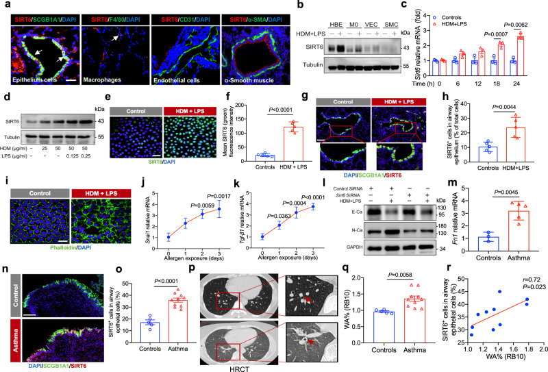
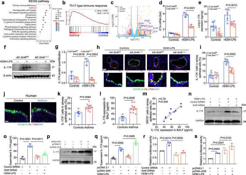
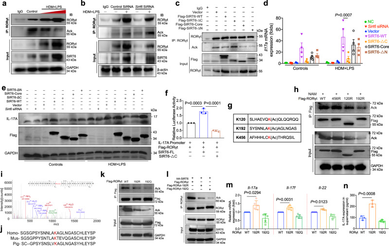
Dysregulation of IL-17A is closely associated with airway inflammation and remodeling in severe asthma. However, the molecular mechanisms by which IL-17A is regulated remain unclear. Here we identify epithelial sirtuin 6 (SIRT6) as an epigenetic regulator that governs IL-17A pathogenicity in severe asthma. Mice with airway epithelial cell-specific deletion of Sirt6 are protected against allergen-induced airway inflammation and remodeling via inhibiting IL-17A-mediated inflammatory chemokines and mesenchymal reprogramming. Mechanistically, SIRT6 directly interacts with RORγt and mediates RORγt deacetylation at lysine 192 via its PPXY motifs. SIRT6 promotes RORγt recruitment to the IL-17A gene promoter and enhances its transcription. In severe asthma patients, high expression of SIRT6 positively correlates with airway remodeling and disease severity. SIRT6 inhibitor (OSS_128167) treatment significantly attenuates airway inflammation and remodeling in mice. Collectively, these results uncover a function for SIRT6 in regulating IL-17A pathogenicity in severe asthma, implicating SIRT6 as a potential therapeutic target for severe asthma.


Conflict of interest statement

The authors declare no competing interests.

Figures

Fig. 1
Fig. 1. Increased SIRT6 expression is positively related to asthma severity.
a Schematic illustrating an established HDM/LPS-induced acute severe asthma (ASA) mouse model. b, c qRT-PCR analysis of the expression of the SIRT family (left margin) in mouse lung tissues. Results were normalized to those of the gene encoding β-actin. d, e Western blot analysis of the SIRT family (SIRT1, SIRT3, SIRT5, and SIRT6). Quantification of SIRT6 expression was analyzed by using Image J software. f, g mRNA levels of Sirt6 in peripheral blood from healthy participants (n = 11) and asthmatic patients (n = 33). h, i Asthmatic patients were divided into mild to moderate asthma (n = 16) and severe asthma (n = 17) according to their disease severity. mRNA and protein levels of Sirt6 in peripheral blood were assessed. j, k Postbronchodilator forced expiratory volume in 1 second (FEV1) was assessed. Correlation between Sirt6 expression in peripheral blood and FEV1 was determined by Spearman analysis. l, m Asthma control test (ACT) score was analyzed. Correlation between Sirt6 expression in peripheral blood and ACT score was determined by Spearman analysis. n, o Percentage of neutrophils in peripheral blood was analyzed. Correlation between Sirt6 expression and neutrophils percentage in peripheral blood was determined by Spearman analysis. Data are shown as means ± SEM and three or more independent experiments were performed. Significance was calculated by Two-tailed unpaired Student’s t-test for (aef, gl); one-way ANOVA followed by Tukey’s post-hoc test for (hjn).
Fig. 2
Fig. 2. SIRT6 was highly expressed in airway epithelium cells and promoted mesenchymal transition.
a IF staining for SIRT6 with SCGB1A1, F4/80, CD31, or α-SMA. Scale bars, 100 μm. b the expression of SIRT6 in HBE cells, M0, VEC, and SMC as determined by Western blot. c, d Sirt6 epxression in allergen-induced HBE cells for different times and different doses was analyzed. e, f Representative IF staining of SIRT6 in HBE cells stimulated with allergen (n = 5). Scale bars, 20 μm. g, h Representative IF staining of SIRT6 (Red) in the airway epithelium (SCGB1A1, Green) from asthmatic and control mice (n = 5). Quantification was done using Image J software. Scale bars, 100 μm. i IF micrographs of HBE cells incubated in the absence or presence of HDM/LPS for 15 days. Cells were stained with Alexa Fluor 488-conjugated phalloidin (green color) and DAPI (blue color). Scale bars, 20 μm. j, k qRT-PCR for the EMT regulators Snai1, Tgf-β1 in HBE cells stimulated with HDM/LPS. l HBE cells were transfected with Sirt6 siRNA for 24 h and then treated with HDM/LPS for another 48 h. The expression of E-Ca and N-Ca was measured using western blot. m qRT-PCR for bronchial mucosal biopsy specimens from healthy participants (n = 3) or patients with asthma (n = 5) for the EMT regulator Fn1 mRNA expression. n, o Representative of SIRT6 expression in bronchial biopsy specimens (Controls n = 5; Asthma n = 10). p Representative of high-resolution CT (HRCT) images of bronchial wall thickening in control participants (n = 5) and asthmatic patients (n = 10). q Single-slice airway measurements were collected in the apical bronchus of the right lower lobe (RB10) (Controls n = 5; Asthma n = 10). r Correlation between SIRT6 expression in airway epithelium and airway wall thickness in asthmatic patients was investigated using Spearman analysis. Data are shown as means ± SEM and three or more independent experiments were performed. Significance was calculated by Two-tailed unpaired Student’s t-test for (cfhmoq); one-way ANOVA followed by Tukey’s post-hoc test for (j, k).
Fig. 3
Fig. 3. SIRT6 deficiency protects against airway remodeling in mouse model of asthma.
a Schematic illustrating the genetic approach used to generate airway epithelium cell–conditional knockout of Sirt6 (AE-Sirt6Δ/Δ) mice. b SIRT6 deficiency was confirmed by assessing genomic DNA. cf Representative periodic acid-Schiff (PAS) staining and Masson of lung sections from AE-Sirt6fl/fl and AE-Sirt6Δ/Δ mice treated with HDM/LPS in a chronic severe asthma (CSA) model and their quantification (n = 5 per group per study). Scale bars, 100 μm. g Western blot analysis of the EMT markers N-Ca, vimentin or the myofibroblast markers α-SMA and Collagen I in lung homogenate from AE-Sirt6fl/fl and AE-Sirt6Δ/Δ mice treated with HDM/LPS in CSA model (n = 5 per group per study). h, i qRT-PCR analysis of the EMT regulators Fn1 and Snai1 in lung homogenate from AE-Sirt6fl/fl and AE-Sirt6Δ/Δ mice treated with HDM/LPS in CSA model (n = 5 per group per study). jm Total BAL fluid (BALF) cells, differential cell counts, and histologic analysis of the lung sections were performed with hematoxylin and eosin staining to visualize inflammatory cell recruitment from AE-Sirt6fl/fl and AE-Sirt6Δ/Δ mice treated with HDM/LPS in ASA model (AE-Sirt6fl/fl-Control, AE-Sirt6Δ/Δ-Control, AE-Sirt6fl/fl-Asthma n = 6; AE-Sirt6Δ/Δ-Asthma n = 7). Data are shown as means ± SEM and three or more independent experiments were performed. Significance was calculated by one-way ANOVA followed by Tukey’s post-hoc test for (dfhkm).
Fig. 4
Fig. 4. SIRT6 deficiency inhibites IL-17A expression in airway epithelium.
a KEGG enrichment analysis in the lung tissues of WT mice in ASA model compared to control. b Gene expression of Th17 type immune response was assessed by Gene set enrichment analysis (GSEA). c A volcano plot showed Il-17A and other upregulated genes encoding inflammatory products (red, downregulated genes; brown, downregulated genes). dg qRT-PCR (n = 3), ELISA (AE-Sirt6fl/fl-Control, AE-Sirt6Δ/Δ-Control, AE-Sirt6Δ/Δ-Asthma n = 6; AE-Sirt6fl/fl-Asthma n = 5) and Western blot (n = 3) analysis of IL-17A in lung homogenates from AE-Sirt6fl/fl and AE-Sirt6Δ/Δ mice treated with HDM/LPS in ASA model. h, i Representative of IL-17A expression (Red) in airway epithelium cells (SCGB1A1, Green) of AE-Sirt6fl/fl and AE-Sirt6Δ/Δ mice treated with HDM/LPS in ASA model. Quantification was done using Image J software (n = 5). j, k Representative of IL-17A expression (Red) in airway epithelium cells (SCGB1A1, Green) of bronchial biopsy samples from control donors (n = 5) and asthmatic patients (n = 10). Quantification was analyzed by using Image J software. l, m The levels of IL-17A in the BALF from control donors (n = 5) and asthmatic patients (n = 10) were detected using ELISA. The correlation between SIRT6 expression in airway epithelium and IL-17A levels in BALF was investigated using Spearman analysis. nq HBE cells were transfected with Sirt6 small interfering RNA (siRNA) or Sirt6 plasmid for 24 h and were then treated with HDM/LPS for another 24 h. The expression of IL-17A was studied by using Western blot and ELISA analysis. r, s HBE cells were pretreated with Sirt6 siRNA or Sirt6 plasmid for 24 h and then transfected with the IL17A-reporter plasmid. Luciferase activity was then measured in HDM/LPS-treated and untreated cells. Data are shown as means ± SEM and three or more independent experiments were performed. Significance was calculated by one-way ANOVA followed by Tukey’s post-hoc test for (d, egik, loq, r, s).
Fig. 5
Fig. 5. SIRT6 binds directly to RORγt and regulates RORγt expression.
a Experimental flow chart of RORγt discovery by mass spectrometry (MS). b, c Endogenous SIRT6 or RORγt in HBE cells was analyzed using Immunoblot (IB). d SIRT6 or RORγt in HBE cells transfected with HA-tagged SIRT6 alone or together with Flag-tagged RORγt was analyzed using Immunoblot (IB). e IB analysis of SIRT6 and RORγt interaction in a GST pull-down assay. f SIRT6-RORγt molecular interactions in HBE cells was analyzed using Proximity ligation assay (PLA). Foci of interactions are amplified in green (Scale bars, 20 μm). g Sequence alignment of RORγt; red indicates the conserved PPXY motif. h IB analysis of lysates of HEK293T cells transfected with various combinations (above lanes) of plasmids, followed by immunoprecipitation (IP) with anti-Flag and immunoblot analysis with anti-HA or anti-Flag. i Schematic of SIRT6 deletion and point mutants used in this study. j Representative IF staining of GFP-tagged SIRT6 WT and truncations (SIRT6-Core, SIRT6-ΔN, and SIRT6-ΔC) are shown (green) (Scale bars, 400 μm). k IB analysis of HEK293T cells transiently transfected with Flag-tagged SIRT6 or Flag-tagged mutant SRIT6-∆C domain plus HA-tagged RORγt and assessed 24 h later before (input) or after IP with antibody to Flag. l IL-17A expression in HEK293T cells transfected with Flag-tagged SIRT6 WT or Flag-tagged SIRT6 truncations (SIRT6-Core, SIRT6-ΔN, and SIRT6-ΔC) was studied by using Western blot. m Representative confocal IF image of the bronchial tissues from control donor and asthmatic patient stained for RORγt (red), SIRT6 (green), IL-17A (pink), and DAPI (blue) (Scale bars, 100 μm). n The expression of RORγt in the lung homogenate of HDM/LPS-exposed AE-Sirt6fl/fl and AE-Sirt6Δ/Δ mice was analyzed by using Western blot. o SIRT6, RORγt, and IL-17A expressions in HDM/LPS-treated HBE cells were determined by Western blot. p, q Western blot analysis of RORγt in cytoplasmic and nuclear extracts of HBE cells transfected with Sirt6 siRNA for 24 h and then treated with HDM/LPS for another 24 h. GAPDH and Lamin B1 were used as the cytoplasmic and nuclear controls, respectively. Data are representative of three independent experiments with similar results.
Fig. 6
Fig. 6. SIRT6 targetes RORγt for deacetylation.
a IB analysis of RORγt acetylation (Ack) in HBE cells treated with HDM/LPS and assessed 24 h later before (input) or after IP with antibody to RORγt and Ack. b HBE cells were pretreated with Sirt6 siRNA for 24 h and assessed 24 h later before (input) or after IP with antibody to RORγt and Ack. c IB analysis of RORγt Ack in HEK293T cells transfected with WT SIRT6 or SIRT6 truncations (SIRT6-Core, SIRT6-ΔN, and SIRT6-ΔC), assessed before (input) or after IP with antibody to RORγt and Ack. d, e qRT-PCR and western blot analysis of Il17a expression in HEK293T cells transfected with WT SIRT6 or SIRT6 truncations (SIRT6-Core, SIRT6-ΔN, and SIRT6-ΔC) (n = 4). f HBE cells were pretreated with transfected with WT SIRT6 or SIRT6-ΔC together with RORγt for 24 h and then transfected with the IL17A-reporter plasmid. Luciferase activity was then measured. g Identification of acetylated RORγt peptides by mass spectrometry. h Analysis of acetylation of individual RORγt mutants. The acetylated lysine (K) sites were mutated to arginine (R, mimics deacetylation) or glutamine (Q, mimics acetylation). i Tandem mass spectrometry of RORγt peptide modified with methylation on lysine 192 residue. j Alignment of protein sequences surrounding K192 of RORγt from different organisms. Homo: human, Mus: mouse. k Mutation of K192 decreases RORγt acetylation. l IB analysis of RORγt Ack in HEK293T cells transfected with HA-tagged SIRT6 WT, Flag-tagged RORγt WT or Flag-tagged RORγt mutant plasmids (K192R and K192Q), assessed before (input) or after IP with antibody to RORγt and Ack. m qRT-PCR analysis of Il17a, Il17f, and Il22 expression in HEK293T cells transfected with K192R and K192Q mutant plasmids (n = 5). n The K192R and K192Q mutant plasmids were transfected into HBE cells for 24 h and were then treated with HDM/LPS for 24 h. IL-17A protein were measured using ELISA. Data are shown as means ± SEM and three or more independent experiments were performed. Significance was calculated by one-way ANOVA followed by Tukey’s post-hoc test for (dfm).
Fig. 7
Fig. 7. The SIRT6 inhibitor OSS_128167 (OSS) preventes airway remodeling in asthmatic mice.
a Chemical Structure of OSS_128167 (OSS) is shown. b Schematic overview of experimental design for (bj) in an ASA model. c Representative photomicrographs of lung inflammation expression are shown (Control n = 4; HDM + LPS n = 5, HDM + LPS + OSS n = 6). Scale bars, 100 μm. d, e The expression of RORγt, SIRT6, and IL-17A in the lung homogenate from mice treated with HDM/LPS or mice treated with HDM/LPS and SIRT6 inhibitor OSS_128167 (OSS) was analyzed by using Western blot. Quantification was analyzed by Image J software. f, g ELISA and qRT-PCR analysis of IL-17A in lung homogenate of mice. hj The expression of inflammatory cytokines in the lung homogenate of mice was analyzed by using qRT-PCR or ELISA analysis. k Schematic overview of experimental design for (L-O) in a chronic severe asthma (CSA) model. (Control n = 4; HDM + LPS n = 5, HDM + LPS + OSS n = 5). l The expression of N-ca, Muc5ac, Collagen I, α-Sma, and Mmp9 expression in the lung homogenate of mice was analyzed by using qRT-PCR. m Representative periodic acid-Schiff (PAS) staining, Masson, and α-SMA of lung sections from WT mice treated with HDM/LPS or HDM/LPS plus SIRT6 inhibitor OSS. Scale bars, 100 μm. n, o The expression of N-Ca, Collagen I, and α-SMA in the lung homogenate of mice were analyzed by using Western blot. Quantification was analyzed by Image J software. Data are shown as means ± SEM and three or more independent experiments were performed. Significance was calculated by one-way ANOVA followed by Tukey’s post-hoc test for (ejlo).

References

    1. Reddel HK, et al. Global initiative for asthma strategy 2021: executive summary and rationale for key changes. Eur. Respir. J. 2021;59:2102730. doi: 10.1183/13993003.02730-2021. - DOI - PMC - PubMed
    1. Brusselle GG, Koppelman GH. Biologic therapies for severe ssthma. N. Engl. J. Med. 2022;386:157–171. doi: 10.1056/NEJMra2032506. - DOI - PubMed
    1. Busse WW, et al. Randomized, double-blind, placebo-controlled study of brodalumab, a human anti-IL-17 receptor monoclonal antibody, in moderate to severe asthma. Am. J. Respir. Crit. Care Med. 2013;188:1294–1302. doi: 10.1164/rccm.201212-2318OC. - DOI - PubMed
    1. Krishnamoorthy N, et al. Neutrophil cytoplasts induce TH17 differentiation and skew inflammation toward neutrophilia in severe asthma. Sci. Immunol. 2018;3:eaao4747. doi: 10.1126/sciimmunol.aao4747. - DOI - PMC - PubMed
    1. Tian B, et al. Mucosal bromodomain-containing protein 4 mediates aeroallergen-induced inflammation and remodeling. J. Allergy Clin. Immunol. 2019;143:1380–1394.e1389. doi: 10.1016/j.jaci.2018.09.029. - DOI - PMC - PubMed

Publication types