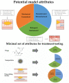
Cardiovascular Tissue Engineering Models for Atherosclerosis Treatment Development
- PMID: 38135964
- PMCID: PMC10740643
- DOI: 10.3390/bioengineering10121373
Cardiovascular Tissue Engineering Models for Atherosclerosis Treatment Development
Abstract
In the early years of tissue engineering, scientists focused on the generation of healthy-like tissues and organs to replace diseased tissue areas with the aim of filling the gap between organ demands and actual organ donations. Over time, the realization has set in that there is an additional large unmet need for suitable disease models to study their progression and to test and refine different treatment approaches. Increasingly, researchers have turned to tissue engineering to address this need for controllable translational disease models. We review existing and potential uses of tissue-engineered disease models in cardiovascular research and suggest guidelines for generating adequate disease models, aimed both at studying disease progression mechanisms and supporting the development of dedicated drug-delivery therapies. This involves the discussion of different requirements for disease models to test drugs, nanoparticles, and drug-eluting devices. In addition to realistic cellular composition, the different mechanical and structural properties that are needed to simulate pathological reality are addressed.
Keywords: atherosclerosis; disease models; drug-coated balloons; drug-eluting stents; nanoparticles; tissue engineering.
Conflict of interest statement
The authors declare no conflict of interest.
Figures


Similar articles
-
Disease-inspired tissue engineering: Investigation of cardiovascular pathologies.ACS Biomater Sci Eng. 2020 May 11;6(5):2518-2532. doi: 10.1021/acsbiomaterials.9b01067. Epub 2019 Oct 29. ACS Biomater Sci Eng. 2020. PMID: 32974421 Free PMC article. Review.
-
Progress in drug-delivery systems in cardiovascular applications: stents, balloons and nanoencapsulation.Nanomedicine (Lond). 2022 Feb;17(5):325-347. doi: 10.2217/nnm-2021-0288. Epub 2022 Jan 21. Nanomedicine (Lond). 2022. PMID: 35060758 Review.
-
BAlloon versus Stenting in severe Ischaemia of the Leg-3 (BASIL-3): study protocol for a randomised controlled trial.Trials. 2017 May 19;18(1):224. doi: 10.1186/s13063-017-1968-6. Trials. 2017. PMID: 28526046 Free PMC article. Clinical Trial.
-
The future of Cochrane Neonatal.Early Hum Dev. 2020 Nov;150:105191. doi: 10.1016/j.earlhumdev.2020.105191. Epub 2020 Sep 12. Early Hum Dev. 2020. PMID: 33036834
-
Assessing the influence of atherosclerosis on drug coated balloon therapy using computational modelling.Eur J Pharm Biopharm. 2021 Jan;158:72-82. doi: 10.1016/j.ejpb.2020.09.016. Epub 2020 Oct 17. Eur J Pharm Biopharm. 2021. PMID: 33075477
Cited by
-
A Cross-Stage Partial Network and a Cross-Attention-Based Transformer for an Electrocardiogram-Based Cardiovascular Disease Decision System.Bioengineering (Basel). 2024 May 29;11(6):549. doi: 10.3390/bioengineering11060549. Bioengineering (Basel). 2024. PMID: 38927785 Free PMC article.
-
Transcriptomic Approaches to Cardiomyocyte-Biomaterial Interactions: A Review.ACS Biomater Sci Eng. 2024 Jul 8;10(7):4175-4194. doi: 10.1021/acsbiomaterials.4c00303. Epub 2024 Jun 27. ACS Biomater Sci Eng. 2024. PMID: 38934720 Free PMC article. Review.
References
Publication types
Grants and funding
LinkOut - more resources
Full Text Sources
Miscellaneous

