Review
. 2023 Dec 6;10(12):1393.
doi: 10.3390/bioengineering10121393.

fNIRS-EEG BCIs for Motor Rehabilitation: A Review

Affiliations
Review

fNIRS-EEG BCIs for Motor Rehabilitation: A Review

Jianan Chen et al. Bioengineering (Basel). .

Abstract

Motor impairment has a profound impact on a significant number of individuals, leading to a substantial demand for rehabilitation services. Through brain-computer interfaces (BCIs), people with severe motor disabilities could have improved communication with others and control appropriately designed robotic prosthetics, so as to (at least partially) restore their motor abilities. BCI plays a pivotal role in promoting smoother communication and interactions between individuals with motor impairments and others. Moreover, they enable the direct control of assistive devices through brain signals. In particular, their most significant potential lies in the realm of motor rehabilitation, where BCIs can offer real-time feedback to assist users in their training and continuously monitor the brain's state throughout the entire rehabilitation process. Hybridization of different brain-sensing modalities, especially functional near-infrared spectroscopy (fNIRS) and electroencephalography (EEG), has shown great potential in the creation of BCIs for rehabilitating the motor-impaired populations. EEG, as a well-established methodology, can be combined with fNIRS to compensate for the inherent disadvantages and achieve higher temporal and spatial resolution. This paper reviews the recent works in hybrid fNIRS-EEG BCIs for motor rehabilitation, emphasizing the methodologies that utilized motor imagery. An overview of the BCI system and its key components was introduced, followed by an introduction to various devices, strengths and weaknesses of different signal processing techniques, and applications in neuroscience and clinical contexts. The review concludes by discussing the possible challenges and opportunities for future development.

Keywords: brain–computer interface; electroencephalography; functional near-infrared spectroscopy; motor imagery; motor rehabilitation; multimodal.


Conflict of interest statement

The authors declare no conflict of interest.

Figures

Figure 2
Figure 2
(A) Examples of some fNIRS devices. (A1) NIRScout (NIRx GmbH, Berlin, Germany). This figure is taken with permission from [57]. (A2) LABNIRS system (Shimadzu Corporation, Kyoto, Japan). This figure is taken with permission from [58]. (A3) LUMO (Gowerlabs Ltd., London, UK). This figure is taken with permission from [56]. (B) Examples of some fNIRS-EEG designs. (B1) An NIRScout cap completely mounted (with EEG electrodes, fNIRS sources, and detectors). This figure is taken with permission from [44]. (B2) A NIRx fNIRS/EEG cap using collocated passive EEG electrodes with a fNIRS probe. This figure is taken with permission from [59]. (B3) g.GAMMAcap fNIRS system: designed to mount g.SENSOR fNIRS together with g.SCARABEO electrodes or g.SAHARA hybrid electrodes (g.Tec medical engineering GmbH, Austria). This figure is taken with permission from [60]. (B4) Wearable Sensing’s wireless DSI-EEG + fNIRS system: a total of 8 sensor pods on the system, and each pod has 1 dry EEG electrode in the middle, 4 emitters, and 4 detectors surrounding. (Wearable Sensing, San Diego, CA, USA). This figure is taken with permission from [61].
Figure 1
Figure 1
Typical hybrid—BCI system (HbO—oxyhemoglobin; HbR—deoxyhemoglobin; SMR—sensory motor rhythm; ERPs—event-related potentials; LDA—linear discriminant analysis; SVM—support vector machines; ANN—artificial neural network).
Figure 3
Figure 3
Examples of some fNIRS-EEG configurations in BCI studies. (A) A cohesive configuration of optodes and electrodes. This figure is taken with permission from [44]. (B) Electrode and optode placement of EEG (for visual frequency change) and NIRS (for left and right motor imageries). Numbers indicates fNIRS channels. This figure is taken with permission from [40]. (C) Illustration of a BCI–robot system. The subfigure on the left illustrates 16 EEG electrodes used in experiments. The subfigure on the right illustrates a fNIRS configuration with 12 channels. The probes are located over the prefrontal cortex (bilateral cortex near FPZ), the primary motor cortex (M1) (bilateral). This figure is taken with permission from [53].
Figure 4
Figure 4
Examples of some signal processing pipelines in BCI systems. (A) Comparison of time-series fNIRS signal processing of conventional ML and DL algorithms. This figure is taken and modified with permission from [39], with an application for classifying walking and rest tasks in gait rehabilitation. (B) The procedure of sequential data processing in hybrid BCIs. Reprinted/adapted with permission from Ref. [42]. 2023, Elsevier.
Figure 5
Figure 5
Examples of combining fNIRS and EEG measurements in BCI systems for upper limb rehabilitation. (A) Configuration of fNIRS-EEG with FES. The standard FES electrodes were about 10 cm apart placed at the right wrist and middle position of the forearm. Reprinted/adapted with permission from Ref. [47]. 2023, IEEE. (B) Ball-catching task as shown in the VR video: subjects passively watched a VR video which displayed a right hand repeatedly grasping an incoming ball (13 actions, approx. 0.86 Hz, 20 s). This figure is taken and modified with permission from [29]. (C) Photographs of a motor imagery brain–computer interface (MI-BCI) upper limb rehabilitation training system. The subfigures illustrate the process of using the EEG-BCI system for rehabilitation training (left) and using fNIRS to evaluate the training performance of the EEG-BCI system (right). This figure is taken with permission from [54].
Figure 6
Figure 6
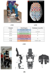
Examples of implementing fNIRS measurement in BCI systems for lower limb rehabilitation. (A) The pilot study of fNIRS-BCI gait rehabilitation conducted by Rea et al. Experimental setup. (A1) Mechanical pedals used to execute active hip movements while sitting on an armchair. EMG electrodes were positioned along muscle fibers of the femoris quadriceps and sartorius muscles of both legs. (A2,A3) Representation of optodes’ location and the corresponding anatomical location of each channel. These figures are taken with permission from [33]. (B) The implementation (left) and the mechanical design (right) of the Atalante®exoskeleton (Wandercraft company, Paris, France). This figure is taken and modified with permission from [64].

References

    1. Cervera M.A., Soekadar S.R., Ushiba J., Millán J.d.R., Liu M., Birbaumer N., Garipelli G. Brain-Computer Interfaces for Post-Stroke Motor Rehabilitation: A Meta-Analysis. Ann. Clin. Transl. Neurol. 2018;5:651–663. doi: 10.1002/acn3.544. - DOI - PMC - PubMed
    1. Cieza A., Causey K., Kamenov K., Hanson S.W., Chatterji S., Vos T. Global Estimates of the Need for Rehabilitation Based on the Global Burden of Disease Study 2019: A Systematic Analysis for the Global Burden of Disease Study 2019. Lancet. 2020;396:2006–2017. doi: 10.1016/S0140-6736(20)32340-0. - DOI - PMC - PubMed
    1. Hatem S.M., Saussez G., della Faille M., Prist V., Zhang X., Dispa D., Bleyenheuft Y. Rehabilitation of Motor Function after Stroke: A Multiple Systematic Review Focused on Techniques to Stimulate Upper Extremity Recovery. Front. Hum. Neurosci. 2016;10:422. doi: 10.3389/fnhum.2016.00442. - DOI - PMC - PubMed
    1. Khan M.A., Das R., Iversen H.K., Puthusserypady S. Review on Motor Imagery Based BCI Systems for Upper Limb Post-Stroke Neurorehabilitation: From Designing to Application. Comput. Biol. Med. 2020;123:103843. doi: 10.1016/j.compbiomed.2020.103843. - DOI - PubMed
    1. Ang K.K., Guan C. Brain-Computer Interface in Stroke Rehabilitation. J. Comput. Sci. Eng. 2013;7:139–146. doi: 10.5626/JCSE.2013.7.2.139. - DOI

LinkOut - more resources