. 2023 Dec 14;10(12):1422.
doi: 10.3390/bioengineering10121422.

Anti-Apoptosis Therapy for Meniscal Avascular Zone Repair: A Proof-of-Concept Study in a Lapine Model

Affiliations

Anti-Apoptosis Therapy for Meniscal Avascular Zone Repair: A Proof-of-Concept Study in a Lapine Model

Wenqiang Yan et al. Bioengineering (Basel). .

Abstract

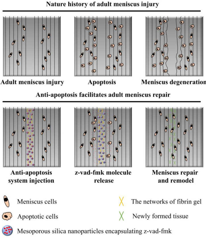
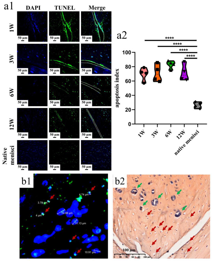
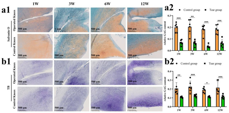
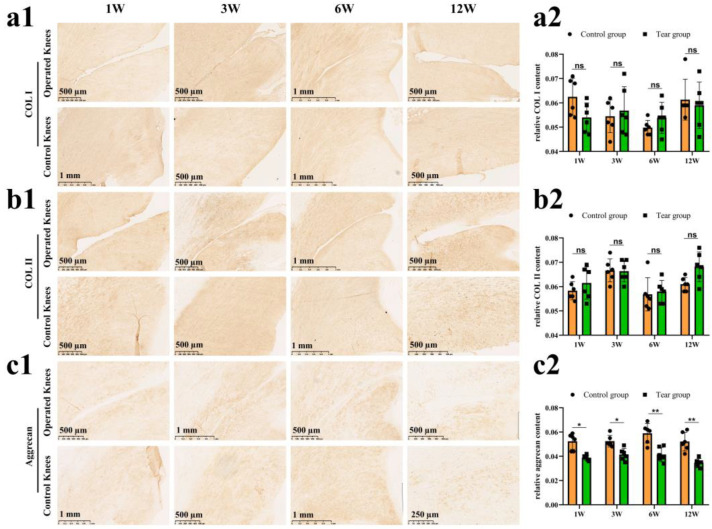
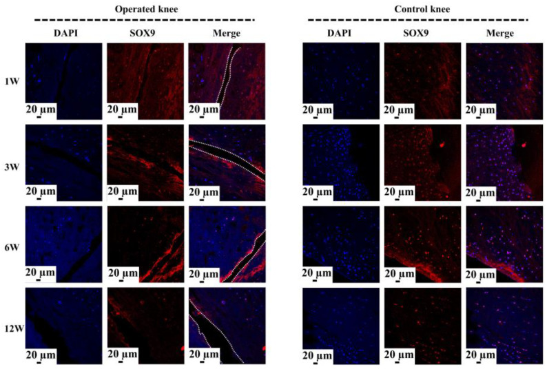
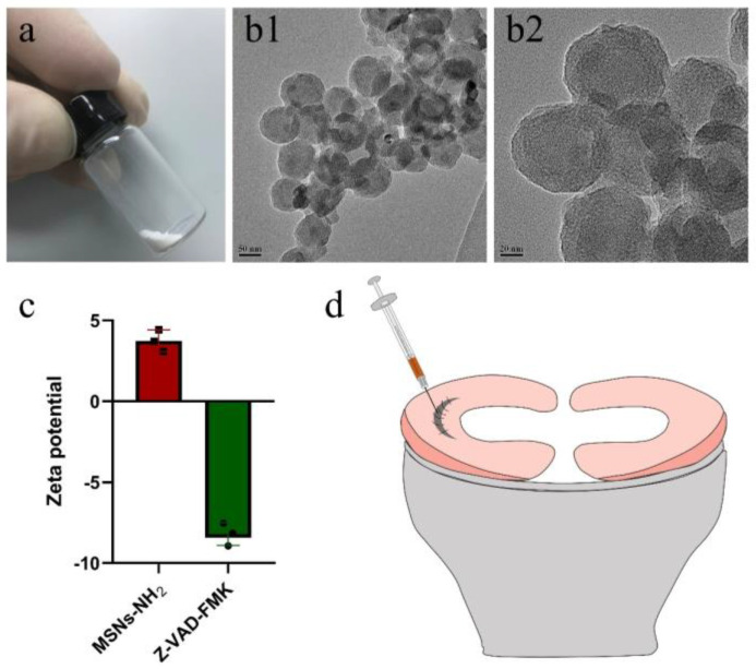
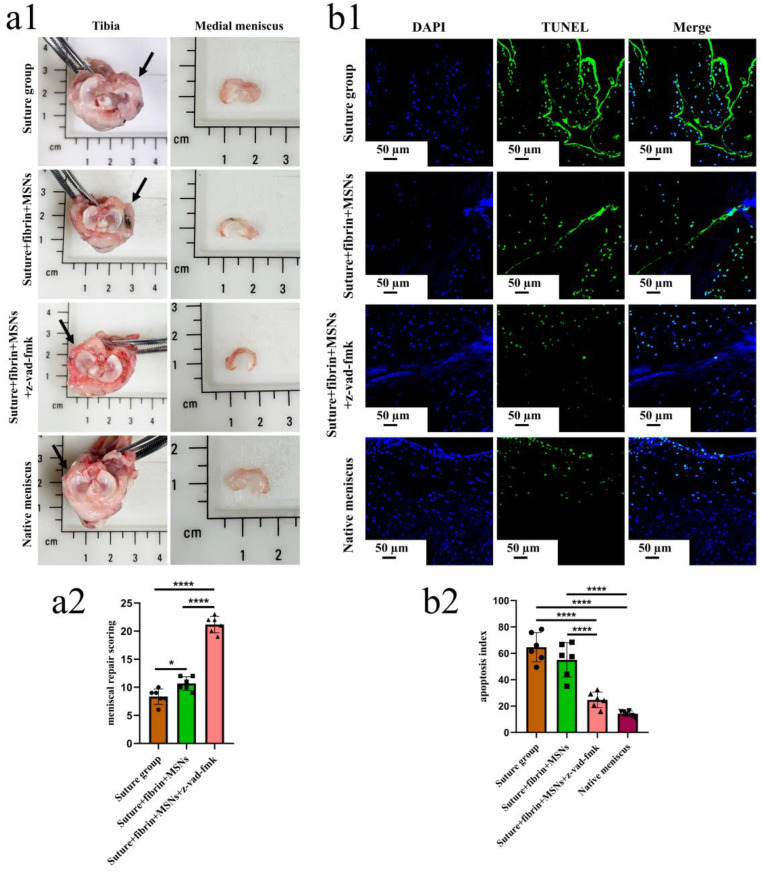
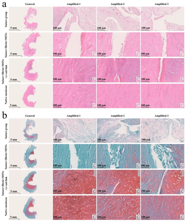
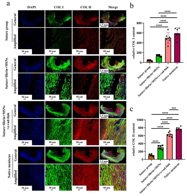
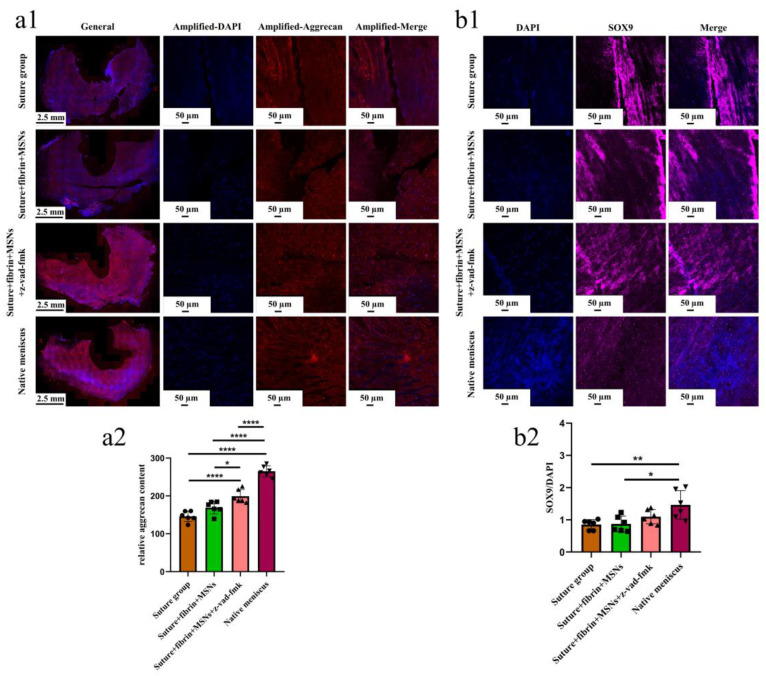
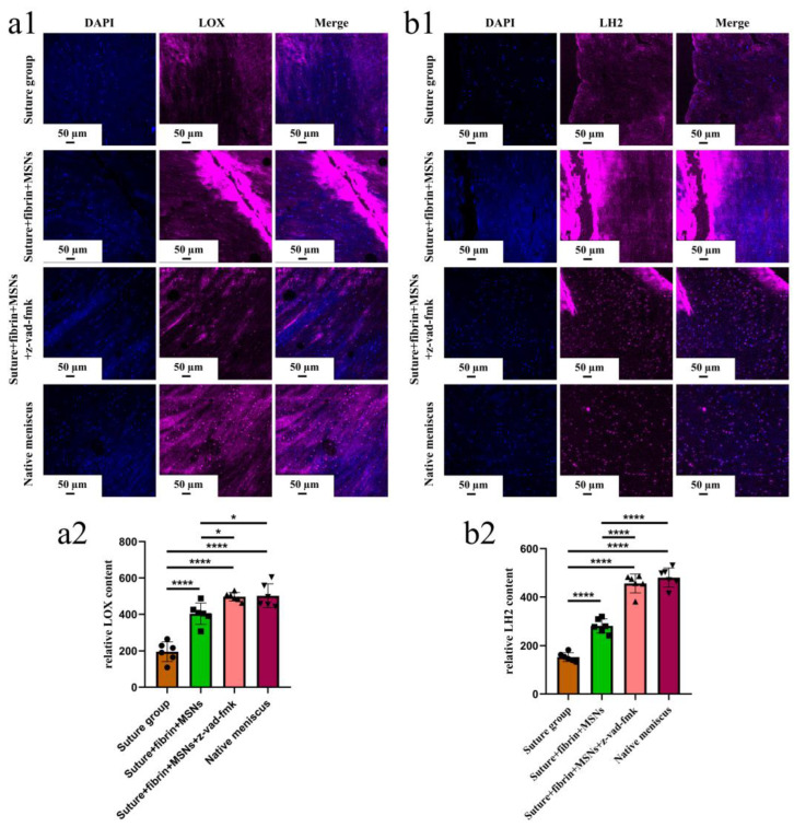
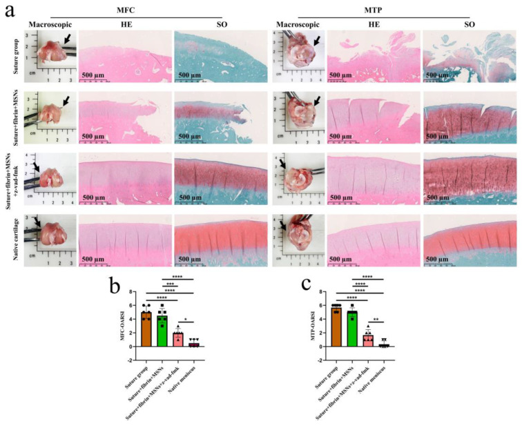
In the present study, 24 rabbits were firstly used to evaluate the apoptosis index and matrix degeneration after untreated adult meniscal tears. Vertical tears (0.25 cm in length) were prepared in the avascular zone of the anterior horn. Specimens were harvested at 1, 3, 6, 12 weeks postoperatively. The apoptosis index around tear sites stayed at a high level throughout the whole follow-up period. The depletion of glycosaminoglycans (GAG) and aggrecan at the tear site was observed, while the deposition of COL I and COL II was not affected, even at the last follow-up of 12 weeks after operation. The expression of SOX9 decreased significantly; no cellularity was observed at the wound interface at all timepoints. Secondly, another 20 rabbits were included to evaluate the effects of anti-apoptosis therapy on rescuing meniscal cells and enhancing meniscus repair. Longitudinal vertical tears (0.5 cm in length) were made in the meniscal avascular body. Tears were repaired by the inside-out suture technique, or repaired with sutures in addition to fibrin gel and blank silica nanoparticles, or silica nanoparticles encapsulating apoptosis inhibitors (z-vad-fmk). Samples were harvested at 12 months postoperatively. We found the locally administered z-vad-fmk agent at the wound interface significantly alleviated meniscal cell apoptosis and matrix degradation, and enhanced meniscal repair in the avascular zone at 12 months after operation. Thus, local administration of caspase inhibitors (z-vad-fmk) is a promising therapeutic strategy for alleviating meniscal cell loss and enhancing meniscal repair after adult meniscal tears in the avascular zone.

Keywords: apoptosis; caspases inhibitor; meniscal repair; meniscal tear; z-vad-fmk.


Conflict of interest statement

The authors declare no conflict of interest.

Figures

Figure 1
Figure 1
The schematics of anti-apoptosis therapy for adult meniscal repair. Apparent meniscal cell apoptosis could be observed at the tear interface following matrix degeneration. The apoptosis inhibitor (z-vad-fmk) was administered locally at the tear interface through silica nanoparticles and fibrin gel delivery system. Meniscal cell apoptosis was alleviated significantly. Finally, robust repair and integration of adult meniscal tear was achieved after remodeling.
Figure 2
Figure 2
The presence of meniscal cell apoptosis at the tear site after untreated adult meniscal tears in the avascular zone. (a1) meniscal cell apoptosis at the tear site at different timepoints; green fluorescence indicates apoptotic cells, and W represents week; (a2) violin plots of apoptosis index, n = 4, one-way ANOVA, ****, p < 0.0001; (b1) pyknosis reflected by TUNEL assay and DAPI. Red arrows represent apoptotic cells; (b2) pyknosis reflected by hematoxylin. Red arrows represent apoptotic cells, and green arrows represent relative normal meniscal fibrochondrocytes.
Figure 3
Figure 3
The evaluation of proteoglycan degradation after untreated adult meniscal tears. (a1) proteoglycan degradation evaluated by safranin O–fast green staining; (a2) semi-quantitative analysis of GAG content reflected by safranin O–fast green staining, n = 6, and a pairwise comparison test of two-way ANOVA, **, p < 0.01, ***, p < 0.0005); (b1) proteoglycan degradation evaluated by toluidine blue staining; (b2), semi-quantitative analysis of GAG content reflected by toluidine blue staining, n = 6, and a pairwise comparison test of two-way ANOVA, *, p < 0.05, **, p < 0.01, ***, p < 0.0005), the circle symbol represents control group, the cube symbol represents tear group.
Figure 4
Figure 4
The evaluation of COL I, COL II and aggrecan after untreated adult meniscal tears. (a1) IHC staining for COL I; (a2) semi-quantitative analysis of COL I content, n = 6, and a pairwise comparison test of two-way ANOVA. ns refers to no significant difference; (b1) IHC staining for COL II; (b2) semi-quantitative analysis of COL II content, n = 6, and a pairwise comparison test of two-way ANOVA; (c1) IHC staining for aggrecan; (c2) semi-quantitative analysis of aggrecan content, n = 6, and a pairwise comparison test of two-way ANOVA). *, p < 0.05, **, p < 0.01, ns, no significance), the circle symbol represents control group, the cube symbol represents tear group.
Figure 5
Figure 5
The analysis of SOX9 expression at the tear site after untreated adult meniscal tears. The white dotted lines indicate the tear edge. The SOX9 positively expressed cells have a pink color in the merged images.
Figure 6
Figure 6
The characterization of aminated mesoporous silica nanoparticles (MSNs–NH2) and z-vad-fmk. The preparation of meniscal tears in a lapine model. (a) MSNs–NH2 powder; (b1) transmission electron microscopic evaluation of MSNs–NH2 at low magnification; (b2) transmission electron microscopic evaluation of MSNs–NH2 at high magnification; (c) the zeta potential of MSNs–NH2 and z-vad-fmk solution at pH = 7.0, n = 3, the cube symbol represents MSNs–NH2, the circle symbol represents z-vad-fmk; (d) the preparation and repair of a longitudinal vertical tear in the avascular body of the medial meniscus. The fibrin gel containing MSNs–NH2–z-vad-fmk is injected into the wound interface, then the suture was knotted to close the tear.
Figure 7
Figure 7
The macroscopic evaluation of meniscal repair and meniscal repair scoring. The apoptosis evaluation around the tear edge in the repaired menisci. (a1), the macroscopic analysis of meniscal repair; black arrows represent the medial meniscus; (a2) Meniscal repair scoring of different groups, n = 6, one-way ANOVA, *, p < 0.05, ****, p < 0.0001, the circle symbol represents the suture group, the cube symbol represents the suture + fibrin + MSNs group, the triangle symbol represents the suture + fibrin + MSNs + z-vad-fmk group; (b1), the presence of apoptosis around the tear site evaluated by TUNEL assay in the repaired menisci; (b2), the apoptosis index at the tear site in different groups, n = 6, one-way ANOVA, ****, p < 0.0001, the circle symbol represents the suture group, the cube symbol represents the suture + fibrin + MSNs group, the upper triangle symbol represents the suture + fibrin + MSNs + z-vad-fmk group, the lower triangle symbol represents the native meniscus.
Figure 8
Figure 8
Histological analysis of meniscal repair in different groups. (a) H&E staining; (b) safranin O–fast green staining. In H&E staining, the meniscus matrix is stained with red color, and the nucleus of the meniscus cell is stained with blue color. In safranin O–fast green staining, the GAG matrix is stained with red color. GAG represents glycosaminoglycans.
Figure 9
Figure 9
The evaluation of COL I and COL II deposition in repaired menisci. (a) Immunofluorescent co-staining of COL I and COL II in the repaired menisci and native menisci; (b) semi-quantitative analysis of COL I content in different groups, n = 6, one-way ANOVA; (c) semi-quantitative analysis of COL II content in different groups, n = 6, one-way ANOVA. ***, p < 0.0005, ****, p < 0.0001, the circle symbol represents the suture group, the cube symbol represents the suture + fibrin + MSNs group, the upper triangle symbol represents the suture + fibrin + MSNs + z-vad-fmk group, the lower triangle symbol represents the native meniscus.
Figure 10
Figure 10
The evaluation of aggrecan and SOX9 expression in repaired menisci. (a1) immunofluorescent staining of aggrecan; (a2) the semi-quantitative analysis of aggrecan content in different groups, n = 6, one-way ANOVA; (b1) the immunofluorescent staining of SOX9; (b2) the semi-quantitative analysis of SOX9 expression in different groups, n = 6, one-way ANOVA. *, p < 0.05, **, p < 0.01, ****, p < 0.0001, the circle symbol represents the suture group, the cube symbol represents the suture + fibrin + MSNs group, the upper triangle symbol represents the suture + fibrin + MSNs + z-vad-fmk group, the lower triangle symbol represents the native meniscus.
Figure 11
Figure 11
The evaluation of LOX and LH2 in the repaired and native menisci. (a1) the immunofluorescent staining of LOX; (a2) semi-quantitative analysis of LOX expression in different groups, n = 6, one-way ANOVA; (b1) immunofluorescent staining of LH2; (b2) semi-quantitative analysis of LH2 expression in different groups, n = 6, one-way ANOVA. *, p < 0.05, ****, p < 0.0001, the circle symbol represents the suture group, the cube symbol represents the suture + fibrin + MSNs group, the upper triangle symbol represents the suture + fibrin + MSNs + z-vad-fmk group, the lower triangle symbol represents the native meniscus.
Figure 12
Figure 12
The assessment of knee joint cartilage degeneration. (a) macroscopic and histological analysis of cartilage degeneration in MFC and MTP. Black arrows represent the medial side. MFC, medial femoral condyle; MTP, medial tibial plateau; SO, safranin O–fast green; (b) semi-quantitative OARSI scores of cartilage degeneration in MFC, n = 6, one-way ANOVA; (c) semi-quantitative OARSI scores of cartilage degeneration in MTP, n = 6, one-way ANOVA. OARSI, osteoarthritis cartilage histopathology assessment (Osteoarthritis Research Society International [OARSI] system. *, p < 0.05, **, p < 0.01, ***, p < 0.0005, ****, p < 0.0001, the circle symbol represents the suture group, the cube symbol represents the suture + fibrin + MSNs group, the upper triangle symbol represents the suture + fibrin + MSNs + z-vad-fmk group, the lower triangle symbol represents the native meniscus.

Similar articles

Cited by

References

    1. Makris E.A., Hadidi P., Athanasiou K.A. The knee meniscus: Structure-function, pathophysiology, current repair techniques, and prospects for regeneration. Biomaterials. 2011;32:7411–7431. doi: 10.1016/j.biomaterials.2011.06.037. - DOI - PMC - PubMed
    1. Kim S., Bosque J., Meehan J.P., Jamali A., Marder R. Increase in outpatient knee arthroscopy in the United States: A comparison of National Surveys of Ambulatory Surgery, 1996 and 2006. J. Bone Jt. Surg. Am. 2011;93:994–1000. doi: 10.2106/JBJS.I.01618. - DOI - PubMed
    1. Logerstedt D.S., Scalzitti D.A., Bennell K.L., Hinman R.S., Silvers-Granelli H., Ebert J., Hambly K., Carey J.L., Snyder-Mackler L., Axe M.J., et al. Knee Pain and Mobility Impairments: Meniscal and Articular Cartilage Lesions Revision 2018. J. Orthop. Sports Phys. Ther. 2018;48:A1–A50. doi: 10.2519/jospt.2018.0301. - DOI - PubMed
    1. Gee S.M., Tennent D.J., Cameron K.L., Posner M.A. The Burden of Meniscus Injury in Young and Physically Active Populations. Clin. Sports Med. 2020;39:13–27. doi: 10.1016/j.csm.2019.08.008. - DOI - PubMed
    1. Mitchell J., Graham W., Best T.M., Collins C., Currie D.W., Comstock R.D., Flanigan D.C. Epidemiology of meniscal injuries in US high school athletes between 2007 and 2013. Knee Surg. Sports Traumatol. Arthrosc. 2016;24:715–722. doi: 10.1007/s00167-015-3814-2. - DOI - PMC - PubMed