

Hyperbaric Oxygen Therapy Alleviates Memory and Motor Impairments Following Traumatic Brain Injury via the Modulation of Mitochondrial-Dysfunction-Induced Neuronal Apoptosis in Rats
- PMID: 38136154
- PMCID: PMC10740762
- DOI: 10.3390/antiox12122034
Hyperbaric Oxygen Therapy Alleviates Memory and Motor Impairments Following Traumatic Brain Injury via the Modulation of Mitochondrial-Dysfunction-Induced Neuronal Apoptosis in Rats
Abstract
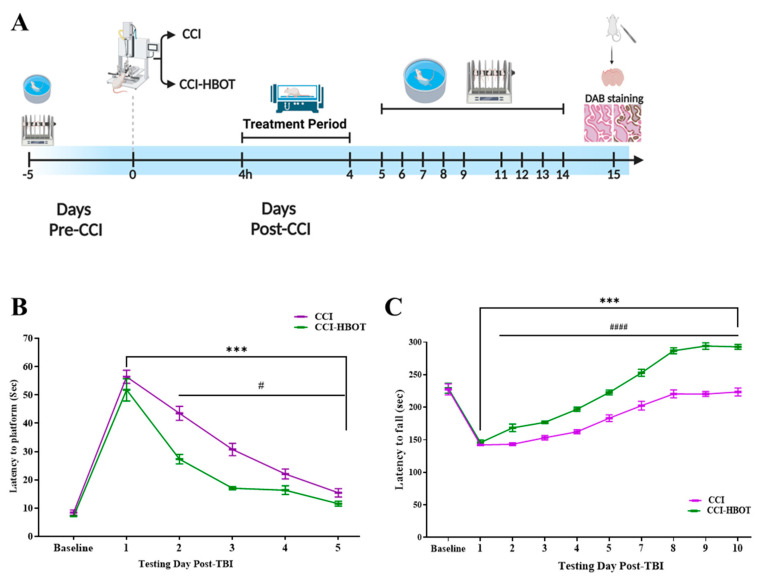
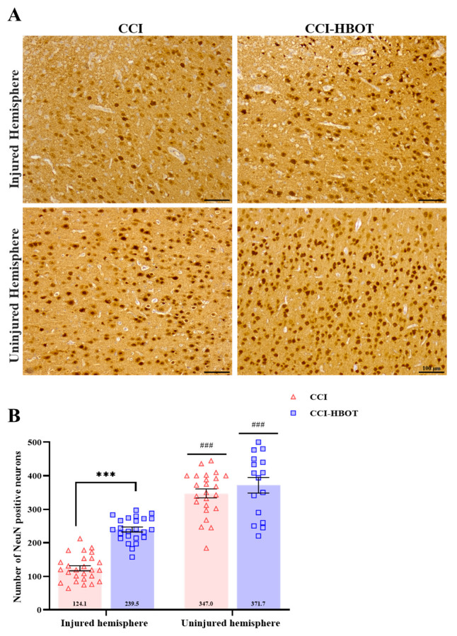
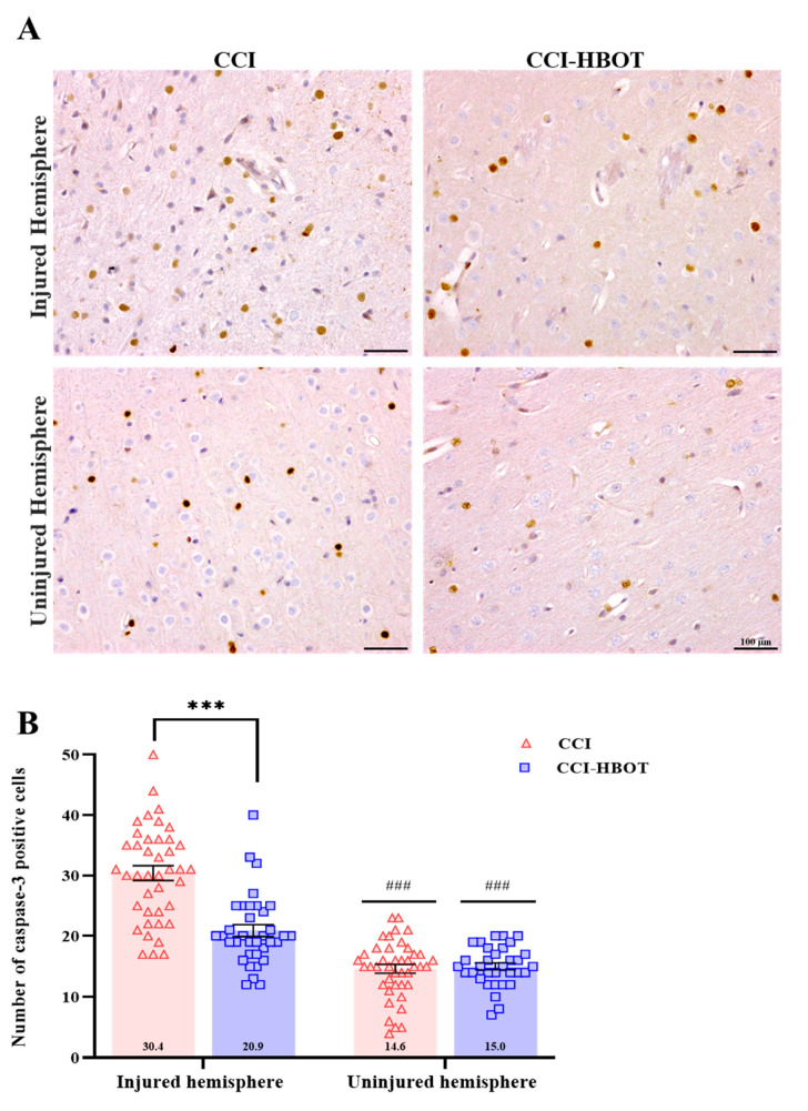
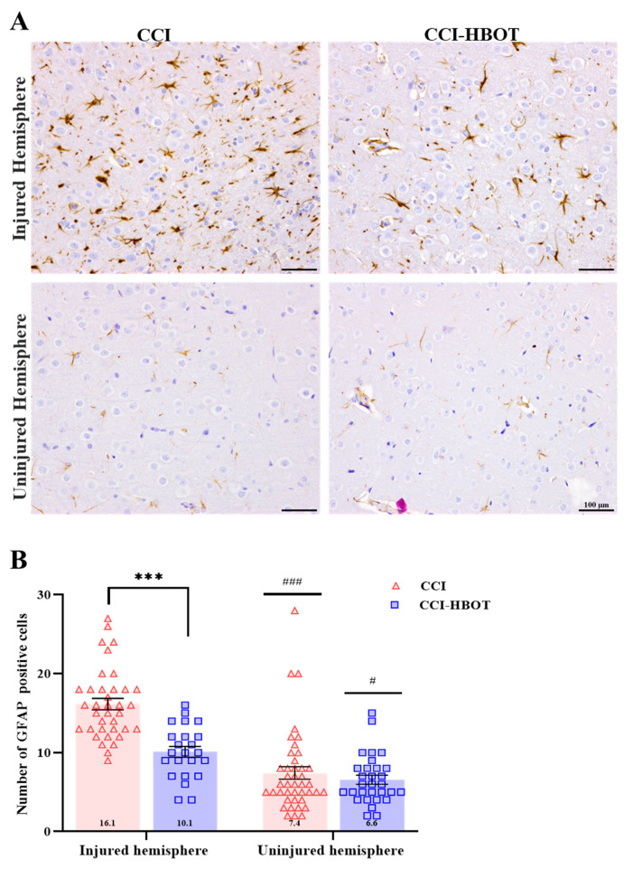
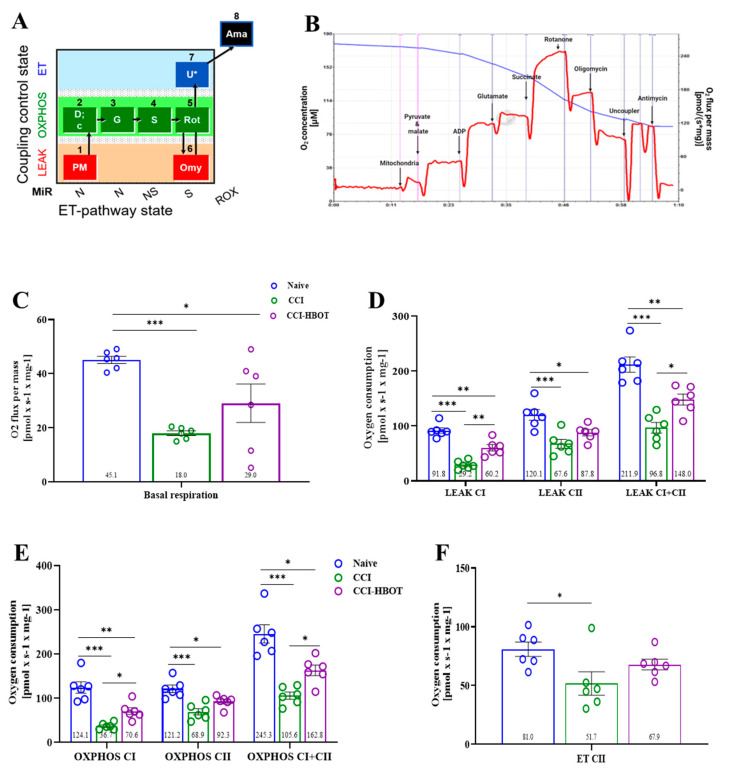
Traumatic brain injury (TBI) is a leading cause of morbidity and mortality in young adults, characterized by primary and secondary injury. Primary injury is the immediate mechanical damage, while secondary injury results from delayed neuronal death, often linked to mitochondrial damage accumulation. Hyperbaric oxygen therapy (HBOT) has been proposed as a potential treatment for modulating secondary post-traumatic neuronal death. However, the specific molecular mechanism by which HBOT modulates secondary brain damage through mitochondrial protection remains unclear. Spatial learning, reference memory, and motor performance were measured in rats before and after Controlled Cortical Impact (CCI) injury. The HBOT (2.5 ATA) was performed 4 h following the CCI and twice daily (12 h intervals) for four consecutive days. Mitochondrial functions were assessed via high-resolution respirometry on day 5 following CCI. Moreover, IHC was performed at the end of the experiment to evaluate cortical apoptosis, neuronal survival, and glial activation. The current result indicates that HBOT exhibits a multi-level neuroprotective effect. Thus, we found that HBOT prevents cortical neuronal loss, reduces the apoptosis marker (cleaved-Caspase3), and modulates glial cell proliferation. Furthermore, HBO treatment prevents the reduction in mitochondrial respiration, including non-phosphorylation state, oxidative phosphorylation, and electron transfer capacity. Additionally, a superior motor and spatial learning performance level was observed in the CCI group treated with HBO compared to the CCI group. In conclusion, our findings demonstrate that HBOT during the critical period following the TBI improves cognitive and motor damage via regulating glial proliferation apoptosis and protecting mitochondrial function, consequently preventing cortex neuronal loss.
Keywords: apoptosis; hyperbaric oxygen therapy (HBOT); mitochondria respiration; secondary brain injury; traumatic brain injury.
Conflict of interest statement
The authors declare that they have no competing interest.
Figures
Similar articles
-
HBO treatment enhances motor function and modulates pain development after sciatic nerve injury via protection the mitochondrial function.J Transl Med. 2023 Aug 15;21(1):545. doi: 10.1186/s12967-023-04414-x. J Transl Med. 2023. PMID: 37582750 Free PMC article.
-
Effect of Hyperbaric oxygen on myelin injury and repair after hypoxic-ischemic brain damage in adult rat.Neurosci Lett. 2023 Jan 18;794:137015. doi: 10.1016/j.neulet.2022.137015. Epub 2022 Dec 13. Neurosci Lett. 2023. PMID: 36526030
-
Immediate and delayed hyperbaric oxygen therapy as a neuroprotective treatment for traumatic brain injury in mice.Mol Cell Neurosci. 2017 Sep;83:74-82. doi: 10.1016/j.mcn.2017.06.004. Epub 2017 Jul 8. Mol Cell Neurosci. 2017. PMID: 28690173
-
Hyperbaric oxygen therapy: A new look on treating stroke and traumatic brain injury.Brain Circ. 2019 Sep 30;5(3):101-105. doi: 10.4103/bc.bc_31_19. eCollection 2019 Jul-Sep. Brain Circ. 2019. PMID: 31620655 Free PMC article. Review.
-
Hyperbaric oxygen therapy applied research in traumatic brain injury: from mechanisms to clinical investigation.Med Gas Res. 2014 Dec 4;4:18. doi: 10.1186/2045-9912-4-18. eCollection 2014. Med Gas Res. 2014. PMID: 25905012 Free PMC article. Review.
Cited by
-
High-temperature requirement serine protease A2 inhibitor UCF-101 ameliorates damaged neurons in traumatic brain-injured rats by the AMPK/NF-κB pathway.Open Life Sci. 2025 Mar 6;20(1):20220971. doi: 10.1515/biol-2022-0971. eCollection 2025. Open Life Sci. 2025. PMID: 40059880 Free PMC article.
-
Hyperbaric Oxygen Improves Long-Term Learning and Memory Impairment by Attenuating Neuronal Apoptosis in aMCI Rats.J Inflamm Res. 2024 May 15;17:3043-3055. doi: 10.2147/JIR.S455155. eCollection 2024. J Inflamm Res. 2024. PMID: 38770175 Free PMC article.
-
Hyperbaric oxygen therapy alleviates intestinal dysfunction following traumatic brain injury via m6A regulation.Int J Med Sci. 2024 Aug 19;21(12):2272-2284. doi: 10.7150/ijms.97682. eCollection 2024. Int J Med Sci. 2024. PMID: 39310263 Free PMC article.
-
Hyperbaric oxygen therapy alleviates intestinal and brain damage in experimental necrotizing enterocolitis.Med Gas Res. 2025 Dec 1;15(4):471-477. doi: 10.4103/mgr.MEDGASRES-D-24-00108. Epub 2025 Apr 29. Med Gas Res. 2025. PMID: 40300882 Free PMC article.
References
Grants and funding
LinkOut - more resources
Full Text Sources
Research Materials
