. 2023 Nov 30;12(12):2060.
doi: 10.3390/antiox12122060.

Contribution of Mitochondrial Reactive Oxygen Species to Chronic Hypoxia-Induced Pulmonary Hypertension

Affiliations

Contribution of Mitochondrial Reactive Oxygen Species to Chronic Hypoxia-Induced Pulmonary Hypertension

Simin Yan et al. Antioxidants (Basel). .

Abstract

Pulmonary hypertension (PH) resulting from chronic hypoxia (CH) occurs in patients with chronic obstructive pulmonary diseases, sleep apnea, and restrictive lung diseases, as well as in residents at high altitude. Previous studies from our group and others demonstrate a detrimental role of reactive oxygen species (ROS) in the pathogenesis of CH-induced PH, although the subcellular sources of ROS are not fully understood. We hypothesized that mitochondria-derived ROS (mtROS) contribute to enhanced vasoconstrictor reactivity and PH following CH. To test the hypothesis, we exposed rats to 4 weeks of hypobaric hypoxia (PB ≈ 380 mmHg), with control rats housed in ambient air (PB ≈ 630 mmHg). Chronic oral administration of the mitochondria-targeted antioxidant MitoQ attenuated CH-induced decreases in pulmonary artery (PA) acceleration time, increases in right ventricular systolic pressure, right ventricular hypertrophy, and pulmonary arterial remodeling. In addition, endothelium-intact PAs from CH rats exhibited a significantly greater basal tone compared to those from control animals, as was eliminated via MitoQ. CH also augmented the basal tone in endothelium-disrupted PAs, a response associated with increased mtROS production in primary PA smooth muscle cells (PASMCs) from CH rats. However, we further uncovered an effect of NO synthase inhibition with Nω-nitro-L-arginine (L-NNA) to unmask a potent endothelial vasoconstrictor influence that accentuates mtROS-dependent vasoconstriction following CH. This basal tone augmentation in the presence of L-NNA disappeared following combined endothelin A and B receptor blockade with BQ123 and BQ788. The effects of using CH to augment vasoconstriction and PASMC mtROS production in exogenous endothelin 1 (ET-1) were similarly prevented by MitoQ. We conclude that mtROS participate in the development of CH-induced PH. Furthermore, mtROS signaling in PASMCs is centrally involved in enhanced pulmonary arterial constriction following CH, a response potentiated by endogenous ET-1.

Keywords: hypoxia; mitochondria; pulmonary hypertension; reactive oxygen species; vasoconstriction.


Conflict of interest statement

The authors declare no conflict of interest.

Figures
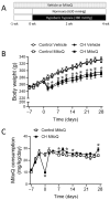
Figure 1
Figure 1
Chronic MitoQ administration. (A) Schematic protocol. MitoQ (500 μM) or vehicle was delivered via drinking water beginning 1 week prior to placement of animals in the hypobaric chamber and continuously during the 4-week CH or ambient air exposure period. (B) Body weight and (C) MitoQ consumption were monitored throughout the study. n = 7–8 animals/group; * p < 0.05 vs. respective control; analyzed using two-way ANOVA, followed by Tukey’s post hoc test (panel B) or unpaired t-test (panel C).
Figure 2
Figure 2
MitoQ limits the effect of CH to reduce PAAT. Pulmonary artery acceleration time (PAAT) was monitored by echocardiography as an estimation of pulmonary arterial pressure (inverse relationship). Animals were studied under anesthesia by 2% isoflurane inhalation. Measurements were taken before and after 2 or 4 weeks of normoxic or hypoxic exposure. (A) Representative traces of PAAT. (B) PAAT in rats treated with vehicle or MitoQ (500 µM) in drinking water. n = 5–8 animals/group; * p < 0.05 vs. respective control, and # p < 0.05 vs. CH vehicle; analyzed using two-way ANOVA, followed by Tukey’s post hoc test.
Figure 3
Figure 3
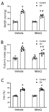
MitoQ attenuates CH-induced increases in RVSP and right ventricular hypertrophy. (A) Peak RVSP, (B) Fulton’s index (right ventricle wt. normalized to left ventricle plus septum wt. (RV/LV+S)), and (C) hematocrit (Hct) in control and CH rats treated with vehicle or MitoQ. RVSP was measured with an indwelling catheter in isoflurane-anesthetized rats. n = 5–8 animals/group; * p < 0.05 vs. respective control, and # p < 0.05 vs. CH vehicle; analyzed using two-way ANOVA, followed by Tukey’s post hoc test.
Figure 4
Figure 4
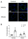
MtROS mediate the pulmonary arterial remodeling response to CH. (A) Representative immunofluorescence images of small pulmonary arteries (green = smooth muscle α-actin; blue = TOPRO3) and (B) % muscularization of arteries from control and CH rats administered vehicle or MitoQ (mean diameter of ~50 μm from 20–30 vessels/animal/group). n = 4–6 animals/group; * p < 0.05 vs. control vehicle, and # p < 0.05 vs. CH vehicle; analyzed using two-way ANOVA, followed by Tukey’s post hoc test.
Figure 5
Figure 5
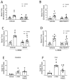
MtROS are responsible for CH-induced increases in pulmonary arterial tone. Basal tone (% passive (Ca2+-free) i.d.) in isolated small pulmonary arteries with intact endothelium. Experiments were performed in the absence (A,B) and presence of the NO synthase inhibitor L-NNA (300 μM, C,D). The contribution of mtROS to basal tone was studied by utilizing the mitochondria-targeted antioxidants MitoQ (1 μM, panels A,C) or MitoTEMPO (200 μM, panels B,D). n = 7–8 animals/group; * p < 0.05 vs. control vehicle, and # p < 0.05 vs. CH vehicle; analyzed using two-way ANOVA, followed by Tukey’s post hoc test. Panels (E,F) depict the reorganization of data from panels (AC), showing that basal tone was augmented by acute NOS inhibition, a response dependent on mtROS in arteries from CH rats but not control arteries. n = 7–8 animals/group; * p < 0.05 vs. vehicle, and # p < 0.05 vs. L-NNA; analyzed using one-way ANOVA, followed by Tukey’s post hoc test.
Figure 6
Figure 6
CH increases PASMC mtROS generation leading to greater basal pulmonary arterial tone. (A) Basal tone in endothelium-disrupted small pulmonary arteries from control and CH rats in the presence of MitoQ (1 μM) or vehicle. n = 5–6 animals/group. (B) Representative images of MitoSOX (5 μM) fluorescence in primary cultures of PASMCs from control and CH rats (top), and MitoSOX mean fluorescence intensity (MFI) (bottom) from cells treated with the mitochondria-targeted antioxidant MitoQ (1 μM) or the mitochondrial superoxide scavenger MitoTEMPO (200 μM). n = 6–7 animals/group; * p < 0.05 vs. control vehicle, and # p < 0.05 vs. CH vehicle; analyzed using two-way ANOVA, followed by Tukey’s post hoc test.
Figure 7
Figure 7
CH increases MtROS levels in acutely isolated pulmonary artery endothelial cell sheets. (A) Sheets were positive for the endothelial marker lectin (in blue). Nuclei were labeled with SYTOX Green. (B) Representative images of MitoSOX fluorescence and (C) MitoSOX mean fluorescence intensity (MFI) in pulmonary artery endothelial cell sheets isolated from intrapulmonary arteries (3rd–5th order, 100-to-400 μm i.d.) of control and CH rats. Experiments were conducted in the presence and absence of MitoQ (1 μM) or L-NNA (300 μM). n = 9 animals/group; * p < 0.05 vs. control vehicle, and # p < 0.05 vs. CH vehicle; analyzed using two-way ANOVA, followed by Tukey’s post hoc test.
Figure 8
Figure 8
Endogenous ET-1 contributes to enhanced mtROS-dependent basal tone following CH. Basal tone in endothelium-intact small pulmonary arteries in the combined presence or absence of ETA and ETB receptor antagonists (BQ123 and BQ788, respectively; 10 μM each). Experiments were performed in the presence of L-NNA (300 μM). n = 5–6 animals/group; * p < 0.05 vs. control vehicle, and # p < 0.05 vs. CH vehicle; analyzed using two-way ANOVA, followed by Tukey’s post hoc test.
Figure 9
Figure 9
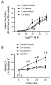
ET-1 stimulates mtROS-dependent vasoconstriction in endothelium-disrupted pulmonary arteries and mtROS production in PASMCs from CH rats. (A) Vasoconstrictor responses (% baseline i.d.) to ET-1 (10−11–10−7 M) in endothelium-disrupted, pressurized pulmonary arteries from control and CH rats in the presence of MitoQ (1 μM) or vehicle. n = 5–6 animals/group; * p < 0.05 vs. control vehicle, and # p < 0.05 vs. CH vehicle; analyzed via two-way ANOVA, followed by Tukey’s post hoc test, at each concentration. (B) Time-dependent changes in MitoSOX (10 μM) fluorescence (% change from baseline) in response to ET-1 (10−9 M) or vehicle (administered at time zero) in primary cultures of PASMCs from control and CH rats. n = 6 animals/group. * p < 0.05 vs. control ET-1, # p < 0.05 vs. CH vehicle; analyzed by two-way ANOVA followed by Tukey’s post hoc test at each time point.

References

    1. Galiè N., Humbert M., Vachiery J.-L., Gibbs S., Lang I., Torbicki A., Simonneau G., Peacock A., Vonk Noordegraaf A., Beghetti M., et al. 2015 ESC/ERS Guidelines for the diagnosis and treatment of pulmonary hypertension. Eur. Respir. J. 2015;46:903–975. doi: 10.1183/13993003.01032-2015. - DOI - PubMed
    1. Jernigan N.L., Naik J.S., Weise-Cross L., Detweiler N.D., Herbert L.M., Yellowhair T.R., Resta T.C. Contribution of reactive oxygen species to the pathogenesis of pulmonary arterial hypertension. PLoS ONE. 2017;12:e0180455. doi: 10.1371/journal.pone.0180455. - DOI - PMC - PubMed
    1. Shimoda L.A., Sham J.S., Sylvester J.T. Altered pulmonary vasoreactivity in the chronically hypoxic lung. Physiol. Res. 2000;49:549–560. - PubMed
    1. Nagaoka T., Morio Y., Casanova N., Bauer N., Gebb S., McMurtry I., Oka M. Rho/Rho kinase signaling mediates increased basal pulmonary vascular tone in chronically hypoxic rats. Am. J. Physiol. Lung Cell. Mol. Physiol. 2004;287:L665–L672. doi: 10.1152/ajplung.00050.2003. - DOI - PubMed
    1. Undem C., Luke T., Shimoda L.A. Contribution of elevated intracellular calcium to pulmonary arterial myocyte alkalinization during chronic hypoxia. Pulm. Circ. 2016;6:93–102. doi: 10.1086/685053. - DOI - PMC - PubMed

LinkOut - more resources