. 2023 Dec 15;15(24):5866.
doi: 10.3390/cancers15245866.

Acid Ceramidase Inhibitor LCL-805 Antagonizes Akt Signaling and Promotes Iron-Dependent Cell Death in Acute Myeloid Leukemia

Affiliations

Acid Ceramidase Inhibitor LCL-805 Antagonizes Akt Signaling and Promotes Iron-Dependent Cell Death in Acute Myeloid Leukemia

Johnson Ung et al. Cancers (Basel). .

Abstract

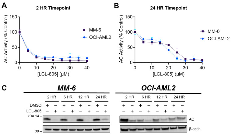
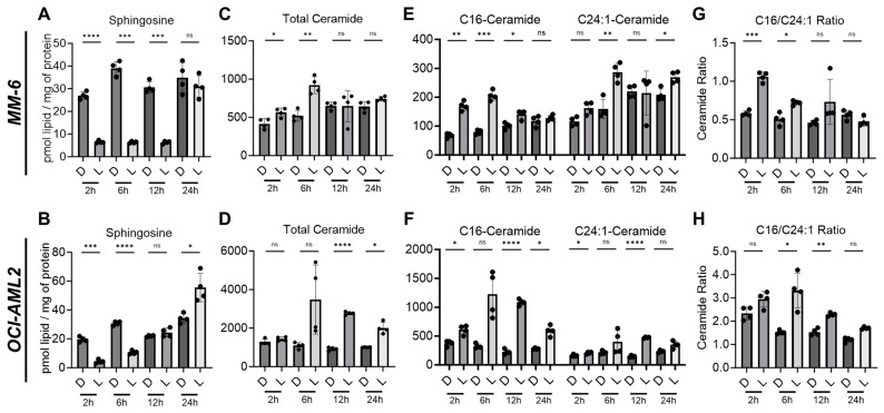
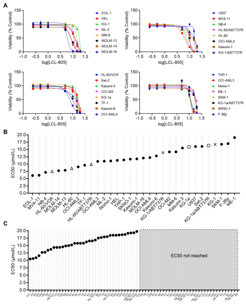
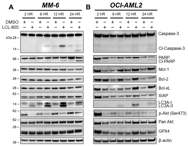
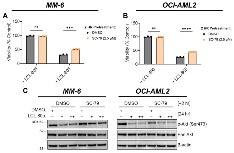
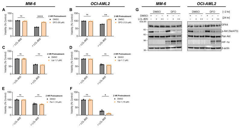
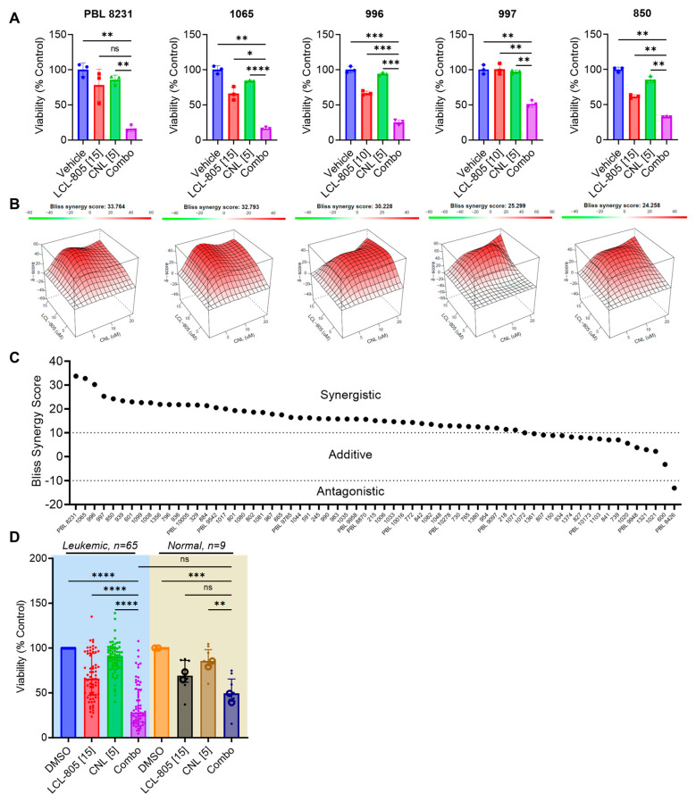
Acute myeloid leukemia (AML) is an aggressive hematologic malignancy requiring urgent treatment advancements. Ceramide is a cell-death-promoting signaling lipid that plays a central role in therapy-induced cell death. We previously determined that acid ceramidase (AC), a ceramide-depleting enzyme, is overexpressed in AML and promotes leukemic survival and drug resistance. The ceramidase inhibitor B-13 and next-generation lysosomal-localizing derivatives termed dimethylglycine (DMG)-B-13 prodrugs have been developed but remain untested in AML. Here, we report the in vitro anti-leukemic efficacy and mechanism of DMG-B-13 prodrug LCL-805 across AML cell lines and primary patient samples. LCL-805 inhibited AC enzymatic activity, increased total ceramides, and reduced sphingosine levels. A median EC50 value of 11.7 μM was achieved for LCL-805 in cell viability assays across 32 human AML cell lines. As a single agent tested across a panel of 71 primary AML patient samples, a median EC50 value of 15.8 μM was achieved. Exogenous ceramide supplementation with C6-ceramide nanoliposomes, which is entering phase I/II clinical trial for relapsed/refractory AML, significantly enhanced LCL-805 killing. Mechanistically, LCL-805 antagonized Akt signaling and led to iron-dependent cell death distinct from canonical ferroptosis. These findings elucidated key factors involved in LCL-805 cytotoxicity and demonstrated the potency of combining AC inhibition with exogenous ceramide.

Keywords: Akt; C6-ceramide nanoliposome; LCL-805; acid ceramidase; acute myeloid leukemia; ceramide; dimethylglycine-B-13 prodrug; iron chelation; sphingolipids.


Conflict of interest statement

D.J.F. has received research funding, honoraria, and/or stock options from AstraZeneca, Dren Bio, Recludix Pharma, and Kymera Therapeutics. T.P.L. has received Scientific Advisory Board membership, consultancy fees, honoraria, and/or stock options from Keystone Nano, Flagship Labs 86, Dren Bio, Recludix Pharma, Kymera Therapeutics, and Prime Genomics. M.C.C. owns shares in Keystone Nano. There are no conflicts of interest with the work presented in this manuscript. Other authors declare no competing interests. The funders had no role in the design of this study; in the collection, analyses, or interpretation of data; in the writing of this manuscript; or in the decision to publish the results.

Figures

Figure 1
Figure 1
LCL-805 inhibited AC in a concentration- and time-dependent manner. (A,B) Fluorogenic AC activity assay conducted in MM-6 and OCI-AML2 human AML cell lines. Cells were treated with vehicle (DMSO) or the indicated concentration of LCL-805 for the indicated times. Error bars represent +/− standard deviation. (C) Immunoblotting of AC protein levels (α-subunit) in MM-6 and OCI-AML2 cells treated with DMSO or LCL-805 (15 µM) for the indicated times. Data presented are representative experiments with at least three technical replicates. Whole, unedited western blot figures can be found in the supplementary materials.
Figure 2
Figure 2
LCL-805 increased cellular ceramide and decreased sphingosine levels. Sphingolipid profiling of MM-6 and OCI-AML2 cells treated with vehicle (DMSO) or LCL-805 (15 µM) for the indicated times. Levels of sphingosine (A,B), total ceramides (C,D), C16- and C24:1-ceramides (E,F), and C16-/C24:1-ceramide ratios (G,H) were evaluated with liquid chromatography–mass spectrometry. Bars represent the average of four technical replicates from a representative experiment. Error bars represent +/− standard deviation. Statistical analyses represent Welch’s ANOVA with Dunnett’s T3 multiple comparisons test. * p < 0.05, ** p < 0.005, *** p < 0.001, **** p < 0.0001, ns = non-significant. D = DMSO; L = LCL-805. C## = fatty acid chain length. Total = sum of sphingolipid species.
Figure 3
Figure 3
LCL-805 reduced viability of human AML cell lines and primary patient samples. (A) Cell viability of 32 human AML cell lines treated with vehicle (DMSO) or the indicated concentrations of LCL-805 for 48 h. (B,C) Calculated LCL-805 EC50 values across AML cell lines (B) or primary AML patient samples (C). Legend for parental and drug-resistant cell line pairs: HL-60 (△); KG-1 (×); KG-1a (□); ABT737R = ABT737-resistant; VCR = vincristine-resistant. Data represent the average of three technical replicates from a representative experiment. Error bars represent +/− standard deviation.
Figure 4
Figure 4
LCL-805 increased phosphatidylserine externalization and triggered mitochondrial depolarization. (A,B) Annexin V and 7-AAD flow cytometry in MM-6 and OCI-AML2 treated for 24 h with vehicle (DMSO) or the indicated concentrations of LCL-805 (in µM, shown in brackets). (C,D) Mitochondrial membrane potential in MM-6 and OCI-AML2 treated for 24 h with DMSO or the indicated concentrations of LCL-805. Bars represent the average of three technical replicates from a representative experiment. Error bars represent +/− standard deviation. Statistical analyses represent two-way ANOVA with Dunnett’s multiple comparisons test (A,B) and Welch’s ANOVA with Dunnett’s multiple comparisons test (C,D). ** p < 0.005, **** p < 0.0001, ns = non-significant.
Figure 5
Figure 5
LCL-805 induced caspase-3 and PARP cleavage, LC3A processing, and reduced Akt phosphorylation. Immunoblotting of MM-6 (A) and OCI-AML2 cells (B) treated with DMSO or LCL-805 (15 µM) for the indicated times. Data are from a representative experiment. Whole, unedited western blot figures can be found in the supplementary materials.
Figure 6
Figure 6
Inhibition of caspases or autophagy did not rescue LCL-805-mediated cell death. (A,B) Cell viability of MM-6 and OCI-AML2 cells pretreated with vehicle (DMSO) or zVAD-FMK (25 µM) for 2 h and subsequently treated with DMSO or LCL-805 (15 µM) for 24 h. (C,D) Cell viability of MM-6 and OCI-AML2 cells pretreated with vehicle (DMSO) or chloroquine (10 µM) for 2 h and subsequently treated with DMSO or LCL-805 (C = 20 µM; D = 17.5 µM) for 24 h. (E,F) Cell viability of MM-6 and OCI-AML2 cells pretreated with vehicle (DMSO) or bafilomycin A1 (500 nM) for 2 h and subsequently treated with DMSO or LCL-805 (E = 20 µM; F = 17.5 µM) for 24 h. (G) Immunoblotting of AC (α-subunit), ATG5, and ATG7 protein levels in non-targeting control (NT CT), ATG5-deficient (ATG5 KO), or ATG7-deficient (ATG7 KO) MV4-11 cells. (H) Cell viability of NT CT, ATG5 KO, or ATG7 KO MV4-11 cells treated with vehicle (DMSO) or LCL-805 (10 µM) for 24 h. Bars represent the average of three technical replicates from a representative experiment. Error bars represent +/− standard deviation. Statistical analyses represent unpaired Welch’s t-tests with Holm–Šídák’s multiple comparisons test (AF) and two-way ANOVA with Dunnett’s multiple comparisons test (H). * p < 0.05, *** p < 0.001, ns = non-significant. Whole, unedited western blot figures can be found in the supplementary materials.
Figure 7
Figure 7
Akt reactivation rescued LCL-805-induced cell death. (A,B) Cell viability of MM-6 and OCI-AML2 cells pretreated with vehicle (DMSO) or SC-79 (2.5 µM) for 2 h and subsequently treated with DMSO or LCL-805 (A = 20 µM; B = 17.5 µM) for 24 h. (C) Immunoblotting of MM-6 and OCI-AML2 cells pretreated with SC-79 (2.5 µM) for 2 h then treated with DMSO or LCL-805 (MM-6: + = 17.5 µM, ++ = 20 µM; OCI-AML2: + = 12.5 µM, ++ = 15 µM) for 24 h. Bars represent the average of three technical replicates from a representative experiment. Error bars represent +/− standard deviation. Statistical analyses represent unpaired Welch’s t-tests and Holm–Šídák’s multiple comparisons test. *** p < 0.001, **** p < 0.0001, ns = non-significant. Whole, unedited western blot figures can be found in the supplementary materials.
Figure 8
Figure 8
Iron chelation rescued LCL-805-induced cell death. (A,B) Cell viability of MM-6 and OCI-AML2 cells pretreated with vehicle (DMSO) or the indicated concentration of DFO for 2 h and subsequently treated with DMSO or LCL-805 (15 µM) for 24 h. (C,D) Cell viability of MM-6 and OCI-AML2 cells pretreated with vehicle (DMSO) or the indicated concentration of liproxstatin-1 (Lip-1) for 2 h and subsequently treated with DMSO or LCL-805 (C = 20 µM; D = 17.5 µM) for 24 h. (E,F) Cell viability of MM-6 and OCI-AML2 cells pretreated with vehicle (DMSO) or the indicated concentration of ferrostatin-1 (Fer-1) for 2 h and subsequently treated with DMSO or LCL-805 (E = 15 µM; F = 15 µM) for 24 h. (G) Immunoblotting of MM-6 and OCI-AML2 cells pretreated with DFO for 2 h (DFO; MM-6 = 50 µM; OCI-AML2 = 12.5 µM) then treated with DMSO or LCL-805 (MM-6: + = 15 µM, ++ = 17.5 µM; OCI-AML2: + = 12.5 µM, ++ = 15 µM) for 24 h. Bars represent the average of three technical replicates from a representative experiment. Error bars represent +/− standard deviation. Statistical analyses represent unpaired Welch’s t-tests and Holm–Šídák’s multiple comparisons test. * p < 0.05, ** p < 0.005, **** p < 0.0001, ns = non-significant. Whole, unedited western blot figures can be found in the supplementary materials.
Figure 9
Figure 9
C6-ceramide nanoliposome (CNL) supplementation increased LCL-805 toxicity. Cell viability was measured in primary AML patient samples treated with multiple concentrations of LCL-805 (5, 10, or 15 µM) and CNL (5, 10, or 20 µM). (A) Cell viability and Bliss synergy scores (B) of the top five AML patient samples with the highest synergy scores treated with vehicle (DMSO), LCL-805, CNL, or the combination. Drug concentration (in µM) is indicated in brackets within the x-axis labels. (C) Bliss synergy scores of primary AML patient samples treated with LCL-805 and CNL. (D) Cell viability of primary AML patient samples or normal controls (7 PBMC samples (•) and 2 CD34+ bone marrow samples (o)) treated with DMSO, LCL-805, CNL, or the combination. Drug concentration (in µM) is indicated in brackets within the x-axis labels. Bars represent the average of three technical replicates from a representative experiment. Error bars represent +/− standard deviation. Statistical analyses represent Welch ANOVA with Dunnett’s multiple comparisons test. * p < 0.05, ** p < 0.005, *** p < 0.001, **** p < 0.0001, ns = non-significant.

Update of

References

    1. Siegel R.L., Miller K.D., Jemal A. Cancer statistics, 2020. CA Cancer J. Clin. 2020;70:7–30. doi: 10.3322/caac.21590. - DOI - PubMed
    1. De Kouchkovsky I., Abdul-Hay M. Acute myeloid leukemia: A comprehensive review and 2016 update. Blood Cancer J. 2016;6:e441. doi: 10.1038/bcj.2016.50. - DOI - PMC - PubMed
    1. Sasaki K., Ravandi F., Kadia T.M., DiNardo C.D., Short N.J., Borthakur G., Jabbour E., Kantarjian H.M. De novo acute myeloid leukemia: A population-based study of outcome in the United States based on the Surveillance, Epidemiology, and End Results (SEER) database, 1980 to 2017. Cancer. 2021;127:2049–2061. doi: 10.1002/cncr.33458. - DOI - PMC - PubMed
    1. Kantarjian H., Kadia T., DiNardo C., Daver N., Borthakur G., Jabbour E., Garcia-Manero G., Konopleva M., Ravandi F. Acute myeloid leukemia: Current progress and future directions. Blood Cancer J. 2021;11:41. doi: 10.1038/s41408-021-00425-3. - DOI - PMC - PubMed
    1. Bose R., Verheij M., Haimovitz-Friedman A., Scotto K., Fuks Z., Kolesnick R. Ceramide synthase mediates daunorubicin-induced apoptosis: An alternative mechanism for generating death signals. Cell. 1995;82:405–414. doi: 10.1016/0092-8674(95)90429-8. - DOI - PubMed