Review
. 2023 Nov 24;13(12):1705.
doi: 10.3390/biom13121705.

Phosphoinositide Signaling in Immune Cell Migration

Affiliations
Review

Phosphoinositide Signaling in Immune Cell Migration

Ruchi Kakar et al. Biomolecules. .

Abstract

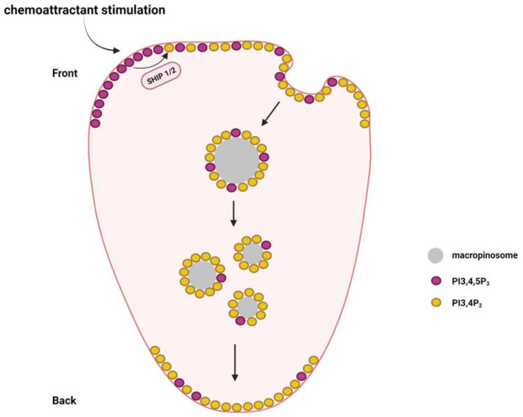
In response to different immune challenges, immune cells migrate to specific sites in the body, where they perform their functions such as defense against infection, inflammation regulation, antigen recognition, and immune surveillance. Therefore, the migration ability is a fundamental aspect of immune cell function. Phosphoinositide signaling plays critical roles in modulating immune cell migration by controlling cell polarization, cytoskeletal rearrangement, protrusion formation, and uropod contraction. Upon chemoattractant stimulation, specific phosphoinositide kinases and phosphatases control the local phosphoinositide levels to establish polarized phosphoinositide distribution, which recruits phosphoinositide effectors to distinct subcellular locations to facilitate cell migration. In this Special Issue of "Molecular Mechanisms Underlying Cell Adhesion and Migration", we discuss the significance of phosphoinositide production and conversion by phosphoinositide kinases and phosphatases in the migration of different types of immune cells.

Keywords: B cell; T cell; immune cells; macrophage; migration; neutrophil; phosphatase; phosphoinositide kinase; phosphoinositides; polarization.


Conflict of interest statement

The authors declare no conflict of interest.

Figures

Figure 1
Figure 1
Phosphoinositide signaling pathway. (A) Schematic representation of the phosphoinositide (PI) family. The enzymes participating in the conversion of one form of PI to another are shown. (B) Schematic representation of regulation of phosphoinositides on chemoattractant stimulation is depicted. Phosphoinositide kinases and phosphatases control the specific phosphoinositide levels in the presence of a chemoattractant, which regulates the recruitment of phosphoinositide effectors to carry out targeted biological functions.
Figure 2
Figure 2
Function of PI3K family kinases in immune cell migration. (A) Different classes of the PI3K family kinases. Specific catalytic and regulatory subunits of different classes of PI3K are shown. (B) Chemoattractants stimulate PI3K activation at the leading edge, where the production of PI3,4,5P3 recruits its effectors such as Akt, DOCK2, Rac, and CDC42 to modulate cell polarization, protrusion formation, and cytoskeletal rearrangement. At the cell rear, PTEN hydrolyses PI3,4,5P3 to PI4,5P2 to maintain the PI3,4,5P3 front-to-rear gradient.
Figure 3
Figure 3
Function of PIPKIγi2 and PIPKIβ at the uropods in neutrophil migration. PIPKIγi2 and PIPKIβ are localized at the uropods during neutrophil migration. By producing PI4,5P2, PIPKIγi2 is required for neutrophil adhesion, integrin activation, and RhoA activation. By producing PI4,5P2 and interacting with EBP50, PIPKIβ enables ERM–RhoGDI interaction to trigger RhoA activation.
Figure 4
Figure 4
The establishment of a PI3,4P2 back-to-front gradient during neutrophil migration. SHIP1 and SHIP2 are required for the production of PI3,4P2 via dephosphorylation of PI3,4,5P3. The PI3,4P2 in the leading edge membrane can be transported to the rear by the formation of macropinosomes, which are sorted from the front-to-rear end.

References

    1. Lauffenburger D.A., Horwitz A.F. Cell migration: A physically integrated molecular process. Cell. 1996;84:359–369. doi: 10.1016/S0092-8674(00)81280-5. - DOI - PubMed
    1. Liu Y.J., Le Berre M., Lautenschlaeger F., Maiuri P., Callan-Jones A., Heuzé M., Takaki T., Voituriez R., Piel M. Confinement and low adhesion induce fast amoeboid migration of slow mesenchymal cells. Cell. 2015;160:659–672. doi: 10.1016/j.cell.2015.01.007. - DOI - PubMed
    1. Moreau H.D., Piel M., Voituriez R., Lennon-Duménil A.M. Integrating Physical and Molecular Insights on Immune Cell Migration. Trends Immunol. 2018;39:632–643. doi: 10.1016/j.it.2018.04.007. - DOI - PubMed
    1. Vesperini D., Montalvo G., Qu B., Lautenschläger F. Characterization of immune cell migration using microfabrication. Biophys. Rev. 2021;13:185–202. doi: 10.1007/s12551-021-00787-9. - DOI - PMC - PubMed
    1. Renkawitz J., Schumann K., Weber M., Lämmermann T., Pflicke H., Piel M., Polleux J., Spatz J.P., Sixt M. Adaptive force transmission in amoeboid cell migration. Nat. Cell Biol. 2009;11:1438–1443. doi: 10.1038/ncb1992. - DOI - PubMed

Publication types

MeSH terms

Substances

LinkOut - more resources