. 2023 Dec 12;11(12):3290.
doi: 10.3390/biomedicines11123290.

Genomic Landscape Comparison of Cardiac versus Extra-Cardiac Angiosarcomas

Affiliations

Genomic Landscape Comparison of Cardiac versus Extra-Cardiac Angiosarcomas

Livia Gozzellino et al. Biomedicines. .

Abstract

Angiosarcomas (ASs) are rare malignant vascular entities that can affect several regions in our body, including the heart. Cardiac ASs comprise 25-40% of cardiac sarcomas and can cause death within months of diagnosis. Thus, our aim was to identify potential differences and/or similarities between cardiac and extra-cardiac ASs to enhance targeted therapies and, consequently, patients' prognosis. Whole-transcriptome analysis of three cardiac and eleven extra-cardiac non-cutaneous samples was performed to investigate differential gene expression and mutational events between the two groups. The gene signature of cardiac and extra-cardiac non-cutaneous ASs was also compared to that of cutaneous angiosarcomas (n = 9). H/N/K-RAS and TP53 alterations were more recurrent in extra-cardiac ASs, while POTE-gene family overexpression was peculiar to cardiac ASs. Additionally, in vitro functional analyses showed that POTEH upregulation conferred a growth advantage to recipient cells, partly supporting the cardiac AS aggressive phenotype and patients' scarce survival rate. These features should be considered when investigating alternative treatments.

Keywords: bioinformatics; cardiac angiosarcomas; extra-cardiac angiosarcomas; whole-transcriptome sequencing.


Conflict of interest statement

The authors declare no conflict of interest.

Figures

Figure 1
Figure 1
Histological findings representing the L359 and L360 samples. (A) L359: the sheet-like proliferation of round to spindle cells associated with focal necrosis and collagen stroma (hematoxylin–eosin, 200×); (B) L360: epithelioid pattern with high cell proliferation prevalently made up of epithelioid cells with marked nuclear pleomorphism and intracytoplasmic lumina (hematoxylin–eosin, 200×); and (C) L360: immunohistochemistry of neoplastic cells diffusely positive for endothelial marker CD34 (200×).
Figure 2
Figure 2
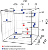
Unsupervised principal component analysis (PCA) on cardiac angiosarcomas (n = 3) versus extra-cardiac non-cutaneous angiosarcomas (n = 11), showing a close localization of cardiac ASs.
Figure 3
Figure 3
Hierarchical clustering of the most overexpressed gene markers in cardiac (n = 3) versus extra-cardiac non-cutaneous (n = 11) angiosarcomas.
Figure 4
Figure 4
GSEA analysis of enriched pathways in cardiac versus extra-cardiac non-cutaneous angiosarcomas applying the following gene sets: Hallmarks, Reactome, and KEGG. ES: Enrichment Score; NES: Normalized Enrichment Scores; FDR q-val: False Discovery Rate q-value.
Figure 5
Figure 5
(A) GFP expression levels in POTEH-transfected cells (red) versus mock- (green) and pcDNA3.1-transfected (blue) cells. (B) Higher POTEH mRNA expression levels in the POTEH-transfected cells compared with pcDNA3.1-transfected cells (RT-PCR). (C) MTT assay results showing cell viability in pcDNA3.1- and POTEH-transfected cells 48 and 72 h after seeding transfected cells in complete medium (** p < 0.01; *** p < 0.001).
Figure 6
Figure 6
Mutational events classified according to their consequences and occurring in at least 3 samples among cardiac (n = 4), visceral (n = 11), and cutaneous (n = 9) angiosarcomas.

References

    1. Fayette J., Martin E., Piperno-Neumann S., le Cesne A., Robert C., Bonvalot S., Ranchère D., Pouillart P., Coindre J.M., Blay J.Y. Angiosarcomas, a Heterogeneous Group of Sarcomas with Specific Behavior Depending on Primary Site: A Retrospective Study of 161 Cases. Ann. Oncol. 2007;18:2030–2036. doi: 10.1093/annonc/mdm381. - DOI - PubMed
    1. Weidema M.E., Versleijen-Jonkers Y.M.H., Flucke U.E., Desar I.M.E., van der Graaf W.T.A. Targeting Angiosarcomas of the Soft Tissues: A Challenging Effort in a Heterogeneous and Rare Disease. Crit. Rev. Oncol. Hematol. 2019;138:120–131. doi: 10.1016/j.critrevonc.2019.04.010. - DOI - PubMed
    1. Chan J.Y., Lim J.Q., Yeong J., Ravi V., Guan P., Boot A., Tay T.K.Y., Selvarajan S., Md Nasir N.D., Loh J.H., et al. Multiomic Analysis and Immunoprofiling Reveal Distinct Subtypes of Human Angiosarcoma. J. Clin. Investig. 2020;130:5833–5846. doi: 10.1172/JCI139080. - DOI - PMC - PubMed
    1. Young R.J., Robin A., Young J., Brown N.J., Reed M.W., Hughes D., Woll P.J. Angiosarcoma. Lancet Oncol. 2010;11:983–991. doi: 10.1016/S1470-2045(10)70023-1. - DOI - PubMed
    1. Gaballah A.H., Jensen C.T., Palmquist S., Pickhardt P.J., Duran A., Broering G., Elsayes K.M. Angiosarcoma: Clinical and Imaging Features from Head to Toe. Br. J. Radiol. 2017;90:20170039. doi: 10.1259/bjr.20170039. - DOI - PMC - PubMed