Case Reports
. 2023 Dec 6;12(24):7530.
doi: 10.3390/jcm12247530.

Resection of a Solitary Right Ventricular Metastasis in Oligorecurrent Hepatocellular Carcinoma

Affiliations
Case Reports

Resection of a Solitary Right Ventricular Metastasis in Oligorecurrent Hepatocellular Carcinoma

Defne Gunes Ergi et al. J Clin Med. .

Abstract

Hepatocellular carcinoma (HCC), constituting the predominant manifestation of liver cancer, stands as a formidable medical challenge. The prognosis subsequent to surgical intervention, particularly for individuals presenting with a solitary tumor, relies heavily on the degree of invasiveness. The decision-making process surrounding therapeutic modalities in such cases assumes paramount importance. This case report illuminates a rather unusual clinical scenario. Here, we encounter a patient who, following a disease-free interval, manifested an atypical presentation of HCC, specifically, a solitary cardiac metastasis. The temporal interval of remission adds an additional layer of complexity to the case. Through a multidisciplinary planning process, the decision was made for surgical removal of the metastatic tumor.

Keywords: cancer; cardiac surgery; hepatocellular cancer; metastasis.


Conflict of interest statement

The authors declare no conflict of interest.

Figures

Figure 1
Figure 1
Preoperative magnetic resonance imaging: T1 (A) and T2 (B) axial phase views demonstrate a large, heterogeneously enlarging, exophytic hepatic mass (red arrows and stars) in the posterior segment of the right lobe of the liver, measuring approximately 13.0 × 10.0 cm2 in size.
Figure 2
Figure 2
Illustration of preoperative 3D tumor excision planning. (A) Axial slice: Image illustrating the tumor’s pedunculated section as it approaches the RVOT; (B) Axial slice: Slice demonstrating tumor extending through the myocardium of the RV; (C) Color-coded visualization: The tumor mass is indicated in green, the right ventricle in purple, the LV in peach, the RA in cyan, the left atrium and pulmonary veins in pink, the pulmonary arteries in blue and the aorta in red; (D) Coronal section: The view along the tricuspid plane, displaying the tumor’s pedunculation extending through the RVOT.
Figure 3
Figure 3
Step-by-step demonstration of the 3D printing process. (A) Initial 3D file export: The first step involved exporting the 3D file to the medical computer-aided diagnosis (CAD) software 3-Matic 16.0 (Materialise, Leuven, Belgium); (B) CAD file refinement: Subsequently, the CAD file underwent a smoothing process, eliminating 3D file errors, and optimizing it for the printing process; (C,D) Removal of unwanted anatomy: Unnecessary anatomical components were removed from the model before printing. The RV, RA, and pulmonary artery were merged in the CAD software for printing. Additionally, as per the surgeon’s request, the model was sectioned along the RVOT. This was followed by further polygon count reduction through additional model smoothing; (E,F) Dual printing approach: The first model was produced using a color nylon HP printer, segmented with a magnet casing to enable separation of the RV components; (G,H) Polyjet printing on Stratasys J5: The second model was crafted using a Polyjet Stratasys J5 printer, utilizing both clear and color materials to visualize the tumor’s extent.
Figure 4
Figure 4
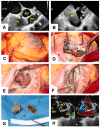
Illustration of intraoperative images of mass excision surgery. (A,B) Preoperative transesophageal echocardiogram: Image displaying the tumor mass in the RV obstructing the pulmonary artery outflow tract, as observed through transesophageal echocardiography (Image coding: RV = Right ventricle, AO = Aorta, PA = Pulmonary artery); (C) External view of tumor mass: This image shows an external perspective of the tumor mass, showing a greenish area on the surface, as observed from the surface of the RV (Image coding: RV = Right ventricle); (D,E) Tumor presentation prior to excision: (D) Image showing the tumor before its excision, revealing the presence of brown-green colored material on its surface; (E) Image demonstrating a widely opened RVOT, revealing that tumor cells were not adherent to the RVOT, enabling excision from the TV without accessing the RVOT; (F) Residual right ventricle wall: Image shows the aftermath of tumor excision. It displays the remaining wall of the RV with residual tumor cells attached to the RV free wall; (G) Surgical explant materials: These images show the materials extracted during surgery, for pathological examination. Due to the use of a throw away sucker, only a portion of the resected tumor cells is visible; (H) Postoperative transesophageal echocardiogram: Image from a postoperative view, displaying a relieved pulmonary outflow tract on the left-hand side and uninterrupted flow passing through without any obstruction, as observed in Doppler mode on the right-hand side (Image coding: RV = Right ventricle, AO = Aorta, PA = Pulmonary artery).
Figure 5
Figure 5
(A) Gross pathological examination revealed a well-defined mass with a brown-green, somewhat lobulated, cut surface. (B) Histologically, the mass consisted of sheets of malignant epithelioid hepatocytes, with areas showing the presence of brown bile pigment.

References

    1. Ahmedin J., Ward E.M., Johnson C.J., Cronin K.A., Ma J., Ryerson B., Mariotto A., Lake A.J., Wilson R., Sherman R.L., et al. Annual Report to the Nation on the Status of Cancer, 1975–2014, Featuring Survival. J. Natl. Cancer Inst. 2017;109:djx030. - PMC - PubMed
    1. Zhang X.T., Li Y., Ren S.H., Ren W.D., Song G., Xiao Y.J., Sun F.F., Sun L., Yang X.H., Tan X.Y. Isolated metastasis of hepatocellular carcinoma in the right ventricle. BMC Cardiovasc. Disord. 2019;19:287. doi: 10.1186/s12872-019-01290-6. - DOI - PMC - PubMed
    1. Kotani E., Kiuchi K., Takayama M., Takano T., Tabata M., Aramaki T., Kawamata H. Effectiveness of transcoronary chemoembolization for metastatic right ventricular tumor derived from hepatocellular carcinoma. Chest. 2000;117:287–289. doi: 10.1378/chest.117.1.287. - DOI - PubMed
    1. Gaurav J., Otto M., Abdul M.K.M., Chadha M., Sahajpal A. Cardiac Metastasis After Curative Treatment of Hepatocellular Carcinoma: Assessment of Risk Factors, Treatment Options, and Prognosis. J. Patient Centered Res. Rev. 2022;9:181–184. - PMC - PubMed
    1. Kawakami M., Koda M., Mandai M., Hosho K., Murawaki Y., Oda W., Hayashi K. Isolated metastases of hepatocellular carcinoma in the right atrium: Case report and review of the literature. Oncol. Lett. 2013;5:1505–1508. doi: 10.3892/ol.2013.1240. - DOI - PMC - PubMed

Publication types

LinkOut - more resources