Review
. 2023 Dec 15;12(24):7704.
doi: 10.3390/jcm12247704.

When Atrial Fibrillation Meets Cerebral Amyloid Angiopathy: Current Evidence and Strategies

Affiliations
Review

When Atrial Fibrillation Meets Cerebral Amyloid Angiopathy: Current Evidence and Strategies

Pierluigi Merella et al. J Clin Med. .

Abstract

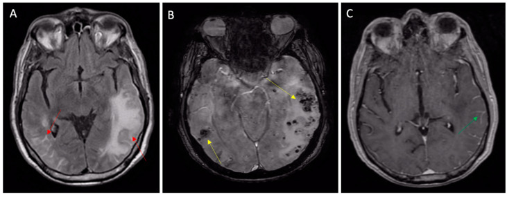
Non-valvular atrial fibrillation (AF) and cerebral amyloid angiopathy (CAA) are two common diseases in elderly populations. Despite the effectiveness of oral anticoagulant therapy in cardioembolic stroke prevention, intracranial hemorrhage represents the most serious complication of these therapies. Cerebral amyloid angiopathy is one of the main risk factors for spontaneous intracranial bleeding, and this risk is highly increased by age and concomitant antithrombotic therapies. Cerebral amyloid angiopathy can be silent for years and then manifest with clinical features simulating TIA (TIA-mimics) or stroke in AF patients, pushing clinicians to rapidly start VKAs or DOACs, thus increasing the risk of intracranial bleeding if the diagnosis of CAA was unknown. Because the cerebral amyloid angiopathy is easily diagnosed with non-contrast MRI, suspecting the disease can avoid catastrophic complications. In this review, we will provide physicians managing anticoagulant therapies with key tips to familiarize themselves with cerebral amyloid angiopathy, with a focus on the possible clinical presentations and on the diagnostic criteria.

Keywords: anticoagulation; atrial fibrillation; cerebral amyloid angiopathy; high bleeding risk; intracranial hemorrhage; left-atrial appendage occlusion.


Conflict of interest statement

We declare that we have no conflict of interest.

Figures

Figure 1
Figure 1
Final diagnoses of probable CAA according to the Boston Criteria 2.0. 84-year-old patient presenting with a TFNE (transient right hemiparesis and loss of speech for several minutes) and first detection of atrial fibrillation. (A) Apparently normal NECT. Oral anticoagulation was started for the secondary prevention of cardioembolism (the TFNE was misinterpreted as cardioembolic TIA). (B) MRI scan 10 days after the presentation showed signal loss in the left pre-central and central sulcus in T2* sequences (arrows), corresponding to “old” superficial siderosis. Oral anticoagulation was not interrupted based on CHA2DS2-VASc/HASBLED calculation. (C) Evolution with parenchymal left hematoma after few days (arrow). Abbreviations: NCET, non-enhanced computed tomography; CAA, cerebral amyloid angiopathy; TFNEs, transient focal neurological episodes; TIA, transient ischemic attack.
Figure 2
Figure 2
Hypertensive microangiopathy. Brain MRI, axial T2 gradient-echo images. Red arrows show microbleeds in the brain stem (A), left basal ganglia (B), and cerebellum (C). Deep localizations are typical features of hypertensive microbleeds.
Figure 3
Figure 3
Inflammatory CAA presenting with TIA-MIMIC (transient aphasia). (A) MRI FLAIR sequences showing vasogenic edema (left arrow) and leptomeningeal inflammatory hyperintensities (right arrow); (B) susceptibility-weighted imaging MRI showing subcortical microbleeds (yellow arrows) and (C) leptomeningeal post contrast enhancement (green arrow) on T1 sequences.
Figure 4
Figure 4
Cerebral involvement from confirmed bacterial endocarditis. (A,B) NECT showing left-temporal and right-frontal atypical hematomas linked with suspected CAA or cavernomas, according to radiologists. Same patient: second-line T2* brain MRI (C) with a further frontal lesion (yellow arrow) and subcortical microbleed (red arrow); the first two lesions are shown with signal loss in T2* sequences. The T1 post-contrast MRI (D) with ring enhancement (red arrow) raised the suspicion of an infective lesion. Control NECT after 3 weeks of antibiotic therapy (E,F): the complete disappearance of both frontal and temporal lesions, confirming cerebral emboli from endocarditis. Abbreviations: NCET, non-enhanced computed tomography; CAA, cerebral amyloid angiopathy.

Similar articles

Cited by

References

    1. Miyasaka Y., Barnes M.E., Gersh B.J., Cha S.S., Bailey K.R., Abhayaratna W.P., Seward J.B., Tsang T.S. Secular trends in incidence of atrial fibrillation in Olmsted County, Minnesota, 1980 to 2000, and implications on the projections for future prevalence. Circulation. 2006;114:119–125. doi: 10.1161/CIRCULATIONAHA.105.595140. - DOI - PubMed
    1. Heeringa J., van der Kuip D.A., Hofman A., Kors J.A., van Herpen G., Stricker B.H., Stijnen T., Lip G.Y., Witteman J.C. Prevalence, incidence and lifetime risk of atrial fibrillation: The Rotterdam study. Eur. Heart J. 2006;27:949–953. doi: 10.1093/eurheartj/ehi825. - DOI - PubMed
    1. Hindricks G., Potpara T., Dagres N., Arbelo E., Bax J.J., Blomström-Lundqvist C., Boriani G., Castella M., Dan G.-A., Dilaveris P.E., et al. 2020 ESC Guidelines for the diagnosis and management of atrial fibrillation developed in collaboration with the European Association of Cardio-Thoracic Surgery (EACTS): The Task Force for the diagnosis and management of atrial fibrillation of the European Society of Cardiology (ESC) Developed with the special contribution of the European Heart Rhythm Association (EHRA) of the ESC. Eur. Heart J. 2021;42:373–498. doi: 10.1093/eurheartj/ehaa612. - DOI - PubMed
    1. Hart R.G., Pearce L.A., Aguilar M.I. Meta-analysis: Antithrombotic therapy to prevent stroke in patients who have nonvalvular atrial fibrillation. Ann. Intern. Med. 2007;146:857–867. doi: 10.7326/0003-4819-146-12-200706190-00007. - DOI - PubMed
    1. Connolly S.J., Ezekowitz M.D., Yusuf S., Eikelboom J., Oldgren J., Parekh A., Pogue J., Reilly P.A., Themeles E., Varrone J., et al. Dabigatran versus Warfarin in Patients with Atrial Fibrillation. N. Engl. J. Med. 2009;361:1139–1151. doi: 10.1056/NEJMoa0905561. - DOI - PubMed

LinkOut - more resources