. 2023 Dec 12;11(12):2968.
doi: 10.3390/microorganisms11122968.

Characterization of Three New Outer Membrane Adhesion Proteins in Fusobacterium necrophorum

Affiliations

Characterization of Three New Outer Membrane Adhesion Proteins in Fusobacterium necrophorum

Prabha K Bista et al. Microorganisms. .

Abstract

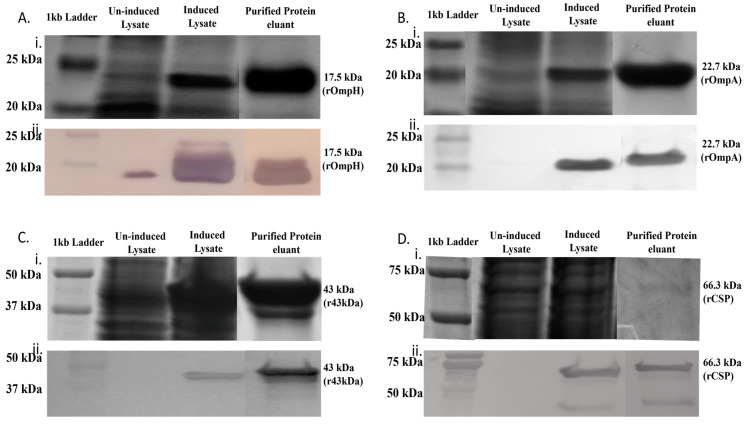
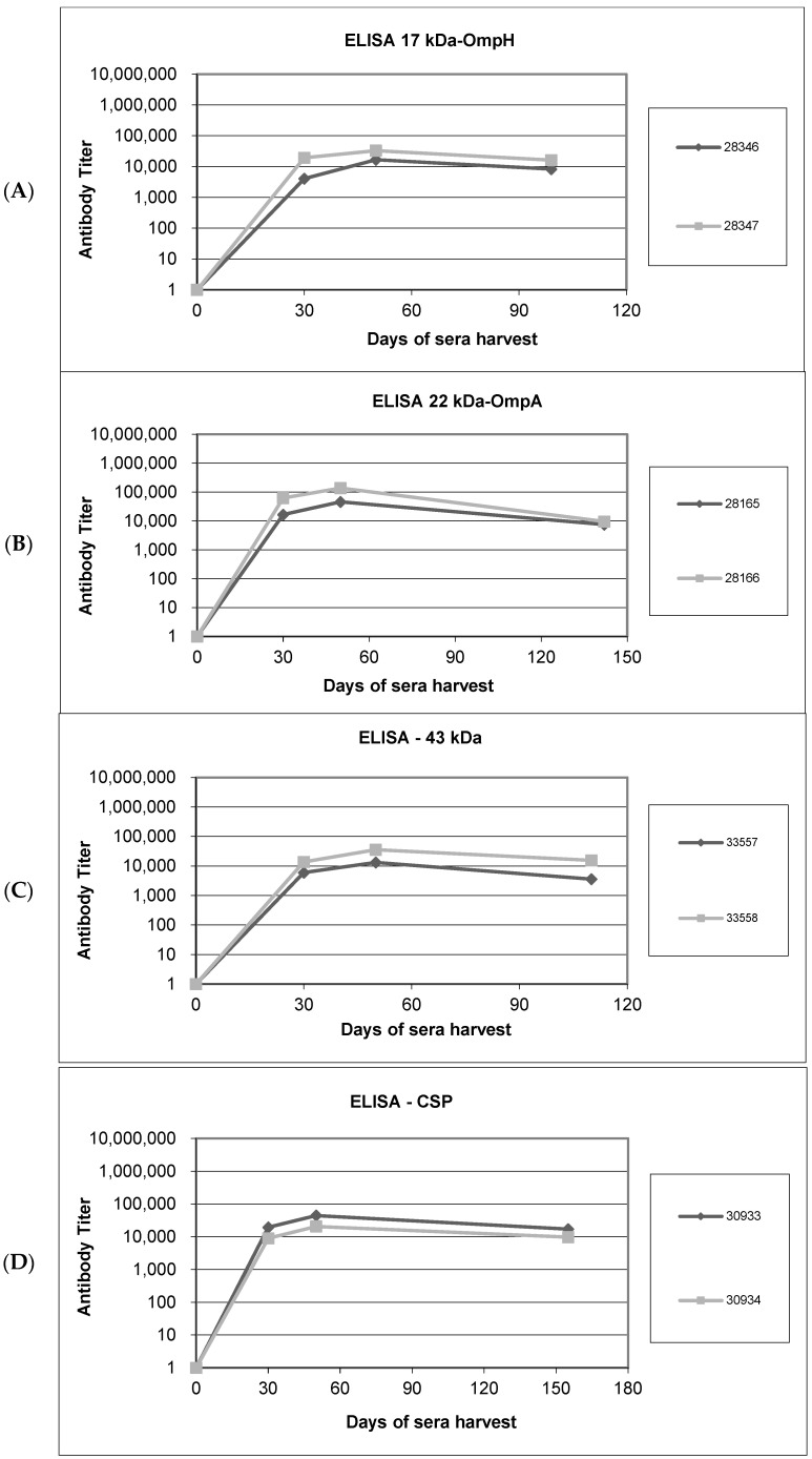
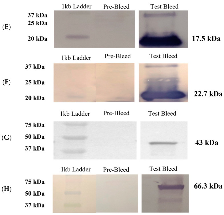
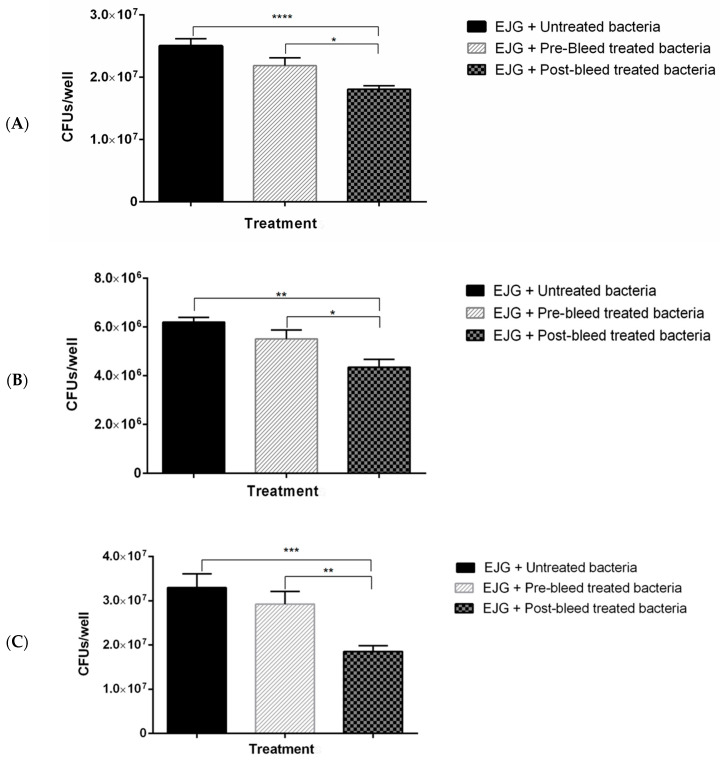
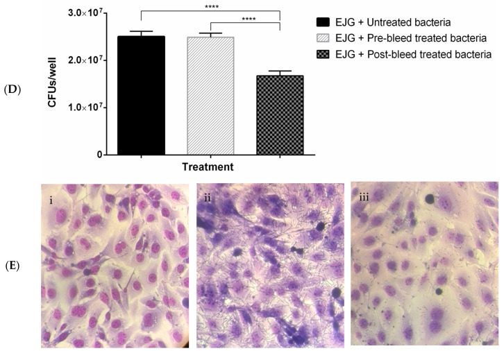
Fusobacterium necrophorum, an anaerobic Gram-negative pathogen, causes necrotic cattle infections, impacting livestock health and the US feedlot industry. Antibiotic administration is the mainstay for treating F. necrophorum infections, although resistance hampers their effectiveness. Vaccination, especially targeting outer membrane proteins (OMPs) due to their antigenic properties and host specificity, offers an alternative to antibiotics. This study identified high-binding-affinity adhesion proteins from F. necrophorum using binding and pull-down assays with bovine adrenal gland endothelial cells (EJG). Four OMP candidates (17.5 kDa/OmpH, 22.7 kDa/OmpA, 66.3 kDa/cell surface protein (CSP), and a previously characterized 43 kDa OMP) were expressed as recombinant proteins and purified. Rabbit polyclonal antibodies to recombinant OMPs were generated, and their ability to inhibit bacterial binding in vitro was assessed. The results show that treatment with individual polyclonal antibodies against 43 kDa significantly inhibited bacterial adhesion, while other antibodies were less potent. However, combinations of two or more antibodies showed a more prominent inhibitory effect on host-cell adhesion. Thus, our findings suggest that the identified OMPs are involved in fusobacterial attachment to host cells and may have the potential to be leveraged in combination for vaccine development. Future in vivo studies are needed to validate their roles and test the feasibility of an OMP-based subunit vaccine against fusobacterial infections.

Keywords: Fusobacterium necrophorum; OmpA; OmpH; cell surface protein; liver abscess; outer membrane proteins (OMPs).


Conflict of interest statement

The authors declare that the research was carried out in the absence of any personal, commercial, or financial relationships that could be construed as potential conflicts of interest.

Figures

Figure 1
Figure 1
Identification of high-binding-affinity adhesins from Fusobacterium necrophorum subspecies necrophorum. Results from binding assays (adapted from [17]) and pull-down assays with F. necrophorum outer membrane protein (OMP) extract and EJG bovine endothelial cells. OMPs bound to host cells were identified by Western blot analysis with anti-OMP primary antibody; Lane M indicates protein marker, and Lanes 1 and 2 show the OMPs identified from binding assays and pull-down assays, respectively.
Figure 2
Figure 2
Expression and purification of recombinant high-affinity-binding proteins from F. necrophorum in Escherichia coli. (AD) represent rOmpH, rOmpA, rCSP, and r43 kDa OMP, respectively. (i) depicts SDS-PAGE analysis, and (ii) corresponds to Western blot analysis. Western blots were incubated with anti-His primary antibody and anti-mouse IgG secondary antibody for rOmpH, rOmpA, r43kDa OMP, and rCSP, respectively. Lane 1: molecular marker; Lane 2: uninduced whole-cell lysates; Lane 3: induced whole-cell lysates; and Lane 4: purified recombinant OMPs.
Figure 3
Figure 3
Humoral responses in rabbits immunized with recombinant antigens and Western blot analyses to confirm antibody specificity in sera harvested after immunization. (AD). Enzyme-linked immunosorbent assays (ELISAs) to measure antibody titers in sera from rabbits immunized with recombinant OMPs: (A) rOmpH, (B) rOmpA, (C) r43 kDa OMP, and (D) rCSP. Pre-immunization antibody titers on day 0 and titers on days 30 and 50 post-immunization and on harvest day are shown. Diamond-shaped dots and square-shaped dots connecting lines in each graph represent the antibody titers from two rabbits (assigned label number) immunized with each protein. (EH). Western blot analysis to confirm the specificity of antibodies raised against the recombinant OMP antigens: (E) rOmpH, (F) rOmpA, (G) r43 kDa OMP, and (H) rCSP. The pre-bleed lanes were probed with control sera harvested before immunization (day 0), whereas day 30 sera were used as the primary antibody for analysis of the test bleed lanes.
Figure 3
Figure 3
Humoral responses in rabbits immunized with recombinant antigens and Western blot analyses to confirm antibody specificity in sera harvested after immunization. (AD). Enzyme-linked immunosorbent assays (ELISAs) to measure antibody titers in sera from rabbits immunized with recombinant OMPs: (A) rOmpH, (B) rOmpA, (C) r43 kDa OMP, and (D) rCSP. Pre-immunization antibody titers on day 0 and titers on days 30 and 50 post-immunization and on harvest day are shown. Diamond-shaped dots and square-shaped dots connecting lines in each graph represent the antibody titers from two rabbits (assigned label number) immunized with each protein. (EH). Western blot analysis to confirm the specificity of antibodies raised against the recombinant OMP antigens: (E) rOmpH, (F) rOmpA, (G) r43 kDa OMP, and (H) rCSP. The pre-bleed lanes were probed with control sera harvested before immunization (day 0), whereas day 30 sera were used as the primary antibody for analysis of the test bleed lanes.
Figure 4
Figure 4
Rabbit polyclonal antibodies to recombinant OMPs inhibit adhesion of F. necrophorum to bovine endothelial (EJG) cells. Adhesion inhibition assays with F. necrophorum and EJG cells using rabbit (1:100) polyclonal antibodies raised against (A) r43 kDa OMP; (B) rOmpH and rOmpA; (C) rOmpH, rOmpA, and rCSP; and (D) rOmpH, rOmpA, rCSP, and r43 kDa OMP. Significance was determined by unpaired two-tailed Student’s t-test, n = 3; * p < 0.05, ** p <0.01, *** p < 0.001, and **** p < 0.0001. The bars indicate the mean ± standard error of the mean (SEM) of data pooled from triplicate experiments. (E) Giemsa staining of EJG cells following adhesion inhibition assays: (i) uninfected (negative control), (ii) infected with F. necrophorum (positive control), and (iii) infected with F. necrophorum pre-treated with a combination of four polyclonal antibodies, as in panel (D).
Figure 4
Figure 4
Rabbit polyclonal antibodies to recombinant OMPs inhibit adhesion of F. necrophorum to bovine endothelial (EJG) cells. Adhesion inhibition assays with F. necrophorum and EJG cells using rabbit (1:100) polyclonal antibodies raised against (A) r43 kDa OMP; (B) rOmpH and rOmpA; (C) rOmpH, rOmpA, and rCSP; and (D) rOmpH, rOmpA, rCSP, and r43 kDa OMP. Significance was determined by unpaired two-tailed Student’s t-test, n = 3; * p < 0.05, ** p <0.01, *** p < 0.001, and **** p < 0.0001. The bars indicate the mean ± standard error of the mean (SEM) of data pooled from triplicate experiments. (E) Giemsa staining of EJG cells following adhesion inhibition assays: (i) uninfected (negative control), (ii) infected with F. necrophorum (positive control), and (iii) infected with F. necrophorum pre-treated with a combination of four polyclonal antibodies, as in panel (D).

Similar articles

Cited by

  • Pediatric Lemierre's Syndrome: A Comprehensive Literature Review.
    Lavalle S, Masiello E, Cocuzza S, Pavone P, Di Nora A, Calvo-Henriquez C, Lechien JR, Yanez MM, Praticò A, Ceccarelli M, Iannella G, Pace A, Parisi FM, Magliulo G, Maniaci A. Lavalle S, et al. Pediatr Rep. 2024 Mar 18;16(1):201-213. doi: 10.3390/pediatric16010018. Pediatr Rep. 2024. PMID: 38535514 Free PMC article. Review.

References

    1. Nagaraja T.G., Narayanan S.K., Stewart G.C., Chengappa M.M. Fusobacterium Necrophorum Infections in Animals: Pathogenesis and Pathogenic Mechanisms. Anaerobe. 2005;11:239–246. doi: 10.1016/j.anaerobe.2005.01.007. - DOI - PubMed
    1. Tadepalli S., Narayanan S.K., Stewart G.C., Chengappa M.M., Nagaraja T.G. Fusobacterium Necrophorum: A Ruminal Bacterium That Invades Liver to Cause Abscesses in Cattle. Anaerobe. 2009;15:36–43. doi: 10.1016/j.anaerobe.2008.05.005. - DOI - PubMed
    1. Clifton R., Giebel K., Liu N.L.B.H., Purdy K.J., Green L.E. Sites of Persistence of Fusobacterium Necrophorum and Dichelobacter Nodosus: A Paradigm Shift in Understanding the Epidemiology of Footrot in Sheep. Sci. Rep. 2019;9:14429. doi: 10.1038/s41598-019-50822-9. - DOI - PMC - PubMed
    1. Nyack B., Craig I., Padmore C., Bernard N. Necrotic Laryngitis in a Calf. Mod. Vet. Pr. 1981;62:937–938. - PubMed
    1. McCoy E.J., O’Quinn T.G., Schwandt E.F., Reinhardt C.D., Thomson D.U. Liver Abscess Severity at Slaughter Does Not Affect Meat Tenderness and Sensory Attributes in Commercially Finished Beef Cattle Fed Without Tylosin Phosphate. Kans. Agric. Exp. Stn. Res. Rep. 2017;3:33. doi: 10.4148/2378-5977.1367. - DOI - PMC - PubMed

LinkOut - more resources